Section III
Motor Learning and Performance
Copyright © 2005 CRC Press LLC

0-8493-1287-6/05/$0.00+$1.50
© 2005 by CRC Press LLC
10
The Arbitrary Mapping
of Sensory Inputs
to Voluntary and
Involuntary Movement:
Learning-Dependent
Activity in the Motor
Cortex and Other
Telencephalic Networks
Peter J. Brasted and Steven P. Wise
CONTENTS
10.1.1 Types of Arbitrary Mapping
10.1.1.1 Mapping Stimuli to Movements
10.1.1.2 Mapping Stimuli to Representations Other than
10.2 Arbitrary Mapping of Stimuli to Reflexes
10.2.1 Pavlovian Eye-Blink Conditioning
10.2.2 Pavlovian Approach Conditioning
10.2.2.1 Learning-Related Activity Underlying Pavlovian
10.2.2.2 Understanding Pavlovian Approach Behavior as a
10.3 Arbitrary Mapping of Stimuli to Internal Models
10.4 Arbitrary Mapping of Stimuli to Involuntary Response Habits
10.5 Arbitrary Mapping of Stimuli to Voluntary Movement
10.5.1 Learning Rate in Relation to Implicit and Explicit Knowledge
Copyright © 2005 CRC Press LLC

10.5.2.1 Premotor Cortex
10.5.2.2 Prefrontal Cortex
10.5.2.3 Hippocampal System
10.5.2.4 Basal Ganglia
10.5.2.5 Unnecessary Structures
10.5.2.6 Summary of the Neuropsychology
10.5.3.1 Premotor Cortex
10.5.3.2 Prefrontal Cortex
10.5.3.3 Basal Ganglia
10.5.3.4 Hippocampal System
10.5.3.5 Summary of the Neurophysiology
10.5.4.1 Methodological Considerations
10.5.4.2 Established Mappings
10.5.4.3 Learning New Mappings
10.5.4.4 Summary
10.6 Arbitrary Mapping of Stimuli to Cognitive Representations
10.7 Conclusion
References
ABSTRACT
Studies on the role of the motor cortex in voluntary movement usually focus on
standard sensorimotor mapping
, in which movements are directed toward sensory
cues. Sensorimotor behavior can, however, show much greater flexibility. Some
variants rely on an algorithmic transformation between a cue’s location and that of
a movement target. The well-known “antisaccade” task and its analogues in reaching
serve as special cases of such
transformational mapping
, one form of
nonstandard
mapping
. Other forms of nonstandard mapping differ from both of the above: they
are arbitrary. In
arbitrary sensorimotor mapping
, the cue’s location has no systematic
spatial relationship with the response. Here we explore several types of arbitrary
mapping, with emphasis on the neural basis of learning these behaviors.
10.1 INTRODUCTION
Many responses to sensory stimuli involve reaching toward or looking at them.
Shifting one’s gaze to a red traffic light and reaching for a car’s brake pedal exemplify
this kind of sensorimotor integration, sometimes termed
standard sensorimotor
mapping
.
1
Other behaviors lack any spatial correspondence between a stimulus and
a response, of which Pavlovian conditioned responses provide a particularly clear
example. The salivation of Pavlov’s dog follows a conditioned stimulus, the ringing
of a bell, but there is no response directed toward the bell or, indeed, toward anything
at all. Like braking at a red traffic light, Pavlovian learning depends on an arbitrary
Copyright © 2005 CRC Press LLC

relationship between a response and the stimulus that triggers it. That is, it depends
on
arbitrary sensorimotor mapping
.
1
Some forms of arbitrary mapping involve
choosing among goals or actions on the basis of color or shape cues. The example
of braking at a red light, but accelerating at a yellow one, serves as a prototypical
(and sometimes dangerous) example of such behavior. In the laboratory, this kind
of task goes by several names, including
conditional motor learning
,
conditional
discrimination
, and
stimulus–response
conditioning
. One stimulus provides the con-
text (or “instruction”) for a given response, whereas other stimuli establish the
contexts for different responses.
2
Arbitrary mapping enables the association of any
dimensions of any stimuli with any actions or goals.
The importance of arbitrary sensorimotor mapping is well recognized — a great
quantity of animal psychology revolves around stimulus–response conditioning —
but the diversity among its types is not so well appreciated. Take, once again, the
example of braking at a red light. On the surface, this behavior seems to depend on
a straightforward stimulus–response mechanism. The mechanism comprises an
input, the red light, a black box that relates this input to a response, and the response,
which consists of jamming on the brakes. This surface simplicity is, however,
misleading. Beyond this account lies a multitude of alternative neural mechanisms.
Using the mechanism described above, a person makes a braking response in the
context of the red light regardless of the predicted outcome of that action
3
and
without any consideration of alternatives.
2
Such behaviors are often called
habits
,
but experts use this term with varying degrees of rigor. Experiments on rodents
sometimes entail the assumption that all stimulus–response relationships are habits.
4,5
But other possibilities exist. Braking at a red light could reflect a voluntary decision,
one based on an attended decision among alternative actions
2
and their predicted
outcomes.
3
In addition, the same behavior might also reflect high-order cognition,
such as a decision about whether to follow the rule that traffic signals must be obeyed.
Because the title of this book is
Motor Cortex in Voluntary Movements
, this
chapter’s topic might seem somewhat out of place. However, the motor cortex —
construed broadly to include the premotor areas — plays a crucial role in arbitrary
sensorimotor mapping, which Passingham has held to be the epitome of voluntary
movement. In his seminal monograph, Passingham
2
defined a voluntary movement
as one made in the context of choosing among alternative, learned actions based on
attention to those actions and their consequences. We take up this kind of arbitrary
mapping in
, in which we discuss the premotor areas involved in this
kind of learning. In addition, we summarize evidence concerning the contribution
of other parts of the telencephalon — specifically the prefrontal cortex, the basal
ganglia, and the hippocampal system — to this kind of behavior. Because of the
explosion of data coming from neuroimaging methods, Section 10.5 also contains
a discussion of that literature and its relation to neurophysiological and neuropsy-
chological results. Before dealing with voluntary movement, however, we consider
arbitrary sensorimotor mapping in three kinds of involuntary movements — condi-
tioned reflexes (
).
Finally, we consider arbitrary mapping in relation to other aspects of response
selection, specifically those involving response rules (
). For a fuller
consideration of arbitrary mapping, readers might consult Passingham’s monograph
2
Copyright © 2005 CRC Press LLC

and previous reviews, which have focused on the changes in cortical activity that
accompany the learning of arbitrary sensorimotor mappings,
6
the role of the hippoc-
ampal system
7,8
and the prefrontal cortex
9
in such mappings, and the relevance of
arbitrary mapping to the life of monkeys.
10
10.1.1 T
YPES
OF
A
RBITRARY
M
APPING
10.1.1.1 Mapping Stimuli to Movements
Stimulus–Reflex Mappings.
Pavlovian conditioning is rarely discussed in the con-
text of arbitrary sensorimotor mapping. Also known as classical conditioning, it
requires the association of a stimulus, called the conditioned stimulus (CS), with a
different stimulus, called the unconditioned stimulus (US), which is genetically
programmed to trigger a reflex response, known as the unconditioned reflex (UR).
Usually, pairing of the CS with the US in time causes the induction of a conditioned
response (CR). For a CS consisting of a tone and an electric shock for the US, the
animal responds to the tone with a protective response (the CR), which resembles
the UR. The choice of CS is arbitrary; any neutral input will do (although not
necessarily equally well). The two types of Pavlovian conditioning differ slightly.
In one type, as described above, an initially neutral CS predicts a US, which triggers
a reflex such as eye blink or limb flexion. This topic is taken up in
.
In another form of Pavlovian conditioning, some neural process stores a similarly
predictive relationship between an initially neutral CS and the availability of sub-
stances like water or food that reduce an innate drive. Unlike the reflexes involved
in the former variety of Pavlovian conditioning, the latter involves the triggering of
consumatory behaviors such as eating and drinking. For example, animals lick a
water spout after a sound that has been associated with the availability of fluid from
that spout. This kind of behavior sometimes goes by the name Pavlovian-approach
behavior (a topic taken up in
). Both kinds of arbitrary sensorimotor
mapping rely on the fact that one stimulus predicts another stimulus, one that triggers
an innate, prepotent, or reflex response.
Stimulus–IM Mappings.
Stimuli can also be arbitrarily mapped to motor pro-
grams. For example, Shadmehr and his colleagues (this volume
11
) discuss the evidence
for internal models (IMs) of limb dynamics. These models involve predictions —
computed by neural networks — about what motor commands will be needed to
achieve a goal (and also about what feedback should occur). The IMs are not
examples of arbitrary sensorimotor mapping per se. Arbitrary stimuli can, however,
be mapped to IMs, a topic taken up in
Stimulus–Response Mappings in Habits.
When animals make responses in a
given stimulus context, that response is more likely to be repeated if a reinforcer,
such as water for a thirsty animal, follows the action. This fact lies at the basis of
instrumental conditioning. According to Pearce,
12
many influential learning theories
of the past 100 years or so
13–15
have held that after consistently making a response
in a given stimulus context, the expected outcome of the action no longer influences
an animal’s performance. The instrumental conditioning has produced an involuntary
movement, often known as a habit or simply as a stimulus–response (S–R) association.
Copyright © 2005 CRC Press LLC

Note, however, that many S–R associations are not habits. When used strictly, the
term “habit” applies only to certain learned behaviors, those that are so “overlearned”
that they have become involuntary in that they no longer depend on the predicted
outcome of the response.
3
It is also important to note that the response in an S–R
association is not a standard sensorimotor mapping. That is, it need not be directed
toward either the reinforcers, their source (such as water spouts and feeding trays),
or the conditioned stimuli. The response is spatially arbitrary. We take up this kind
of arbitrary mapping in
.
Stimulus–Response Mappings in Voluntary Movement.
takes
up arbitrary stimulus–response associations that are not habits, at least as defined
according to contemporary animal learning theory.
3
10.1.1.2 Mapping Stimuli to Representations Other
than Movements
Stimulus–Value Mappings.
Although we focus here on arbitrary sensorimotor map-
pings, there are many other kinds of arbitrary mappings. Stimuli can be arbitrarily
mapped to their biological value. For example, stimuli come to adopt either positive
or negative affective valence, i.e., “goodness” or “badness,” as a function of expe-
rience. This kind of arbitrary mapping is relevant to sensorimotor mapping because
stimulus–value mappings can lead to a response,
16–19
as discussed in
.
Stimulus–Rule Mappings.
In addition to stimulus–response and stimulus–value
mappings, stimuli can be arbitrarily mapping onto more general representations. For
example, a stimulus could evoke a response rule, a topic explored in Section 10.4.
Note that we focus here on the arbitrary mapping of stimuli to rules, not the
representation of a rule per se, as reported previously in both the spatial
20–22
and
nonspatial
22–24
domains.
Stimulus–Meaning Mappings.
In Murray et al.,
10
we argued that evolution co-
opted an existing arbitrary mapping ability for speech and language. Stimuli map
to their abstract meaning in an arbitrary manner. For example, the phonemes and
graphemes of language elicit meanings that usually have an arbitrary relationship
with those auditory and visual stimuli. And this kind of arbitrary mapping leads to
a type of response mapping not mentioned above. In speech production, the rela-
tionship between the meaning a speaker intends to express and the motor commands
underlying vocal or manual gestures that convey that meaning reflects a similarly
arbitrary mapping.
Given these several types of arbitrary mappings, what is known about the neural
mechanisms that underlie their learning?
10.2 ARBITRARY MAPPING OF STIMULI TO REFLEXES
Cells in a variety of structures show learning-related activity for responses that
depend upon Pavlovian conditioning, including the basal ganglia,
25–28
the
amygdala,
29–31
the motor cortex,
32
the cerebellum,
33
and the hippocampus.
34
Why are
there so many different structures involved? Partly, perhaps, because there are several
Copyright © 2005 CRC Press LLC

types of Pavlovian conditioning. One type relies mainly on the cerebellum and its
output mechanisms.
33
In response to potentially damaging stimuli, such as shocks,
taps, and air jets, this type of conditioned response involves protective movements
such as eye blinks and limb withdrawal. Another type, called Pavlovian approach
behavior, depends on parts of both the basal ganglia and the amygdala, and involves
consumatory behaviors such as eating and drinking. Although there are other types
of Pavlovian conditioning, such as fear conditioning and conditioned avoidance
responses, we will focus on these two.
10.2.1 P
AVLOVIAN
E
YE
-B
LINK
C
ONDITIONING
The many studies that describe learning-related activity in the cerebellar system
during eye-blink conditioning and related Pavlovian procedures have been well
summarized by Steinmetz.
33
The reader is referred to his review for that material.
In addition, a number of studies have shown that cells in the striatum, the principal
input structure of the basal ganglia, show learning-related activity during such
learning. For example, a specific population of neurons within the striatum, known
as tonically active neurons (TANs), have activity that is related in some way to
Pavlovian eye-blink conditioning. At first glance, this result seems curious: Pavlovian
conditioning of this type, which recruits protective reflexes, does not require the
basal ganglia but instead depends on cerebellar mechanisms.
33
TANs, which are
believed by many to correspond to the large cholinergic interneurons that constitute
~5% of the striatal cell population,
26,35,36
respond to stimuli that are conditioned by
association with either aversive stimuli
37,38
or with primary rewards.
25–28
TANs also
respond to rewarding stimuli.
37,39
However, studies that have recorded from TANs
while monkeys performed instrumental tasks
40
tend to report less selectivity for
reinforcers than in the Pavlovian conditioning tasks discussed above,
41,42
and it has
been suggested that reward-related responses may reflect the temporal unpredictab-
lility of rewards.
37
One current account of the function of TANs is that they serve
to encode the probability that a given stimulus will elicit a behavioral response.
Blazquez et al.
38
recorded from striatal neurons in monkeys during either appetitive
or aversive Pavlovian conditioning tasks. In addition to finding that responses to
aversive stimuli (air puffs) and reinforcers (water) can occur within individual TANs,
they also noted that as monkeys learned each association (CS-air puff or CS-water),
more TANs became responsive to the CS. Further analysis of the population
responses of TANs revealed that they were correlated with the probability of occur-
rence of the conditioned response.
Given that eye-blink conditioning depends on the cerebellum rather than the
striatum,
33
why would cells in the striatum reflect the probability of generating a
protective reflex response? The most likely possibility, according to Steinmetz,
33
is
that the basal ganglia uses information about the performance of these protective
reflexes in order to incorporate them into ongoing sequences of behavior. Thus,
recognizing the diversity of Pavlovian mechanisms can help us understand the
learning-dependent changes in striatal activity. As is always the case with neuro-
physiological data, a cell’s activity may be “related” to a behavior for many reasons,
only one of which involves causing that behavior.
Copyright © 2005 CRC Press LLC

Does this imply that structures mentioned above, such as the amygdala and the
basal ganglia, play no role in Pavlovian conditioning? Not at all. They participate
instead in other types of Pavlovian conditioning, such as Pavlovian approach behavior.
10.2.2 P
AVLOVIAN
A
PPROACH
C
ONDITIONING
10.2.2.1 Learning-Related Activity Underlying Pavlovian
Approach Conditioning
The properties of midbrain dopaminergic neurons are becoming reasonably well
characterized,
43
as is their importance in reward mechanisms.
44,45
Dopaminergic
neurons respond to unexpected rewards during the early stages of learning.
46,47
As
learning progresses and rewards become more predictable, neuronal responses to
reward decrease and neurons increasingly respond to conditioned stimuli associated
with the upcoming reward.
46,48
Furthermore, the omission of expected rewards can
phasically suppress the firing of these neurons.
49
In this context, Waelti et al.
50
predicted that dopaminergic neurons might reflect differences in reward expectancy
and tested this prediction in what is known as a
blocking
paradigm. Understanding
their experiment requires some background in the concepts underlying the
blocking
effect
, also known as the Kamin effect.
As outlined above, the paired presentation of a US such as food with a CS such
as a light or sound results in the development of an association between the repre-
sentation of the US and the CS. However, the simple co-occurrence of a potential
CS and the US does not suffice for the formation of such an association. Instead,
effective conditioning also depends upon a neural prediction: specifically, whether
the US is unexpected or surprising, and thus whether the CS can capture an animal’s
attention. Note that the concept of attention, in this sense, differs dramatically from
the concept of
top-down
attention. Top-down attention leads to an enhancement in
the neural signal of an object or place attended; it results from a stimulus (or aspect
of a stimulus) being predicted and its neural signal enhanced. By contrast, the kind
of “attention” studied in Pavlovian conditioning results from a stimulus, the US,
not
being predicted and its signal not cancelled by that prediction. Top-down attention,
which corresponds to attention in common-sense usage, is volitional: it results from
a decision and a choice among alternatives.
2
The other usage of the term refers to a
process that is completely involuntary. A number of prominent theories of learning
51–53
stress this aspect of expectancy and surprise, as demonstrated in a classic study by
Kamin.
54
In that study, one group of rats experienced, in a “pretraining” stage of
the experiment, pairings of a noise (a CS) and a mild foot shock (the US), whereas
a second group of rats received no such pretraining. Then, both groups subsequently
received an equal number of trials in which a compound CS composed of a noise
and a light was paired with shock. Finally, both groups were tested on trials in which
only the light was presented. The presentation of the light stimulus alone elicited a
conditioned response in rats that had received no pretraining, i.e., the group that had
never experienced noise–shock pairing. Famously, rats that had received pretraining
exhibited no such response. Kamin’s
blocking effect
indicated that the animals’
exposure to the noise–shock pairings had somehow prevented them from learning
about the light–shock pairings.
Copyright © 2005 CRC Press LLC
The study of Waelti et al.
50
tested the hypothesis that the activity of dopaminergic
midbrain neurons would reflect such blocking (Figure 10.1). As in the classic study
of Kamin, the paradigm comprised three stages. During a “pretraining” stage, mon-
keys were presented with one of two stimuli on a given trial (Figure 10.1A), one of
FIGURE 10.1
The response of dopaminergic neurons to conditioned stimuli in the Kamin
blocking paradigm. (A) In the pretraining stage one stimulus was paired with reward (A+)
and one stimulus (B-) was not. This dopaminergic neuron responded to the stimulus that
predicted the reward, A+. The visual stimuli presented to the monkey appear above the
histogram, which in turn appears above the activity raster for each presentation of that
stimulus. (B) During compound-stimulus training, stimulus A+ was presented in conjunction
with a novel stimulus (X), whereas stimulus B- was presented in conjunction with a second
novel stimulus (Y). Both compound stimuli were paired with reward at this stage (AX+,
BY+), and both stimulus pairs elicited firing. (C) The activity showed that compound-stimulus
training had prevented the association between stimulus X and reward but not that between
Y and reward. The association between stimulus X and reward was
blocked
because it was
paired with a stimulus (A) that already predicted reward. Stimulus X was thus redundant
throughout training. In contrast, the association between stimulus Y and reward was not
blocked because it was paired with a stimulus (B) that did not predict reward. (D) Other
dopamine neurons demonstrated a similar effect, but stimulus X elicited a weak increase in
firing rate rather than no increase at all (as in C). (E) Average population histograms for 85
dopamine cells that were tested with stimuli X and Y in the format of C and D. (From
Reference 50, with permission.)
AX+
BY+
X
−
D
Y
−
X
−
C
Y
−
B
A+
B–
A
20
X
−
E
Y
−
4
1.0
1.0
0
0
Stimulus
Stimulus
Stimulus
Stimulus
Stimulus
Stimulus
Stimulus
Stimulus
Stimulus
Stimulus
Copyright © 2005 CRC Press LLC

which was followed by a juice reward (designated A+, where + denotes reward) and
one of which was not paired with reward (B–, where – denotes the lack of reward).
Then, during compound stimulus conditioning (
), stimulus X was
presented in conjunction with the reward-predicting stimulus A+, whereas stimulus
Y was presented in conjunction with stimulus B–. Both compound stimuli were now
paired with reward (AX+, BY+), and trials of each type were interleaved. Because
of the Kamin blocking effect, learning the association of X with reward was pre-
vented because A already predicted reward, and thus rendered X redundant, whereas
the association of Y with reward was learned because B did not predict reward (and
therefore Y was not redundant). In the third stage, stimuli X and Y were presented
in occasional unrewarded trials as a probe to test this prediction (Figures 10.1C and
10.1D). (There were other trial types as well.) An analysis of anticipatory licking
was used as the measure of Pavlovian approach conditioning. This analysis demon-
strated that in the third stage of testing (Figures 10.1C and 10.1D), the monkeys did
not expect a reward when stimulus X was presented (i.e., learning had been blocked),
but were expecting a reward when stimulus Y appeared, as predicted by the Kamin
blocking effect.
Also as predicted, the Kamin blocking effect was faithfully reflected in the
activity of dopaminergic neurons in the midbrain.
50
A total of 85 presumptive
dopaminergic neurons were tested for the responses to probe-trial presentations of
stimuli X and Y (Figures 10.1C and 10.1D). Nearly half of these (39 cells) responded
to the nonredundant stimulus Y, but were not activated by the redundant stimulus X
(as in Figure 10.1C). No neuron showed the opposite result. Some cells showed the
same effects quantitatively (Figure 10.1D), rather than in an all-or-none manner
(Figure 10.1C). As a population, therefore, dopamine neurons responded much more
vigorously to stimulus Y than to X (Figure 10.1E). This finding demonstrated that
the dopaminergic cells had acquired stronger responses to the nonredundant stimulus
Y, compared to the redundant stimulus X, even though both stimuli had been equally
paired with reward during the preceding compound stimulus training. These cells
apparently predicted reward in the same way that the monkeys predicted reward.
10.2.2.2 Understanding Pavlovian Approach Behavior
as a Type of Arbitrary Mapping
The data reported by Waelti et al.
50
are consistent with contemporary learning
theories that posit a role for dopaminergic neurons in reward prediction.
55
This
system shows a close similarity to those involved in other forms of Pavlovian
conditioning, such as eye-blink conditioning. For eye-blink conditioning (and for
other protective reflexes), cells in the inferior olivary nuclei compare predicted and
received neuronal inputs, probably concerning predictions about the US.
33,56,57
The
outcome of this prediction then becomes a “teaching” signal, transmitted by climb-
ing-fiber inputs to the cerebellum, that induces the neural plasticity that underlies
this form of learning. Why should there be two such similar systems? One answer
is that the cerebellum subserves arbitrary stimulus–response mappings for protective
responses, whereas the dopamine system plays a similar role for appetitive responses.
The paradigmatic example of Pavlovian conditioning surely falls into the latter category:
Copyright © 2005 CRC Press LLC

the bell that triggered salivation in Pavlov’s dog did so because of its arbitrary
association with stimuli that triggered autonomic and other reflexes involved in
feeding.
What is the neural basis for this Pavlovian approach behavior? This issue has
been reviewed recently,
58,59
so we will only briefly consider this question here. The
central nucleus of the amygdala, the nucleus accumbens of the ventral striatum, and
the anterior cingulate cortex appear to be important components of the arbitrary
mapping system that underlies certain (but not all) types of Pavlovian approach
behavior in rats. Initially neutral objects, when mapped to a positive value, trigger
ingestive reflexes, such as those involved in procurement of food or water (licking,
chewing, salivation, etc.), and lesions of the central nucleus of the amygdala, the
nucleus accumbens, or the anterior cingulate cortex block such learning.
60,61
There are related mechanisms for arbitrary mapping of stimuli to biological
value that involve other parts of the amygdala, the basal ganglia, and the cortex, at
least in monkeys. As reviewed by Baxter and Murray,
59
these mechanisms involve
different parts of the frontal cortex and amygdala than the typical Pavlovian approach
behavior described above: the orbital prefrontal cortex (PF) instead of the anterior
cingulate cortex and the basolateral nuclei of the amygdala instead of the central
nucleus of the amygdala. These structures, very likely in conjunction with the parts
of the basal ganglia with which they are interconnected, underlie the arbitrary
mapping of stimuli to their value in a special and highly flexible way.
This flexibility is required when neutral stimuli map arbitrarily to food items
and the value of those food items changes over a short period of time. Stimuli that
map arbitrarily to specific food items can change their current value because of
several factors, for example, when that food item has been consumed recently in
quantity. Normal monkeys can use this information to choose stimuli that map to a
higher current value. This mechanism appears to depend on the basolateral nucleus
of the amygdala and the orbital PF: when these structures are removed or their
interconnections severed, monkeys can no longer use the stimuli to obtain the
temporarily more valued food item.
59
Separate analyses showed that the monkey
remembered the mapping of the arbitrary cue to the food item, so the deficit involved
mapping the stimulus to the food’s current value. Furthermore, monkeys with those
lesions remained perfectly capable of choosing the currently preferred food items.
(Presumably, the preserved food preference is due to other mechanisms, probably
hypothalamic ones, that are involved in foraging and food procurement.) Hence, the
lesioned monkeys seemed to know which arbitrary stimulus mapped to which food
item and they appeared to know which food they wanted. Their deficit — and
therefore the contribution of the basolateral amygdala’s interaction with orbital PF —
involved the arbitrary mapping of otherwise neutral stimuli to their current biological
value. The use of updated stimulus–value mappings allows animals to predict the
current, biologically relevant outcome of an action produced in the context of that
stimulus. This mechanism permits animals to make choices that lead to the best
possible outcome when several possible choices with positive outcomes are available,
and to choose appropriately in the face of changing values.
Copyright © 2005 CRC Press LLC

10.3 ARBITRARY MAPPING OF STIMULI
TO INTERNAL MODELS
The previous section deals with arbitrary mappings in Pavlovian conditioning. In
this section, we examine a different form of arbitrary mapping. Rao and Shadmehr
62
and Wada et al.
63
have recently shown that people can learn to map arbitrary spatial
cues and colors onto the motor programs needed to anticipate the forces and feedback
in voluntary reaching movements.
As summarized by Shadmehr and his colleagues in this volume,
11
in their
experiments people move a robotic arm from a central location to a visual target.
When, during the course of these movements, the robot imposes a complex pattern
of forces on the limb, the movement deviates from a straight line to the target. With
practice in countering a particular pattern of forces, the motor system learns to
produce a reasonably straight trajectory. The system is said to have learned (or
updated) an IM of the limb’s dynamics. There is nothing arbitrary about such IMs;
they reflect the physics of the limb and the forces imposed upon the limb.
People can, however, learn to map visual inputs arbitrarily onto such IMs. In
the experiments that first demonstrated this fact, Rao and Shadmehr
62
presented
participants with two different patterns of imposed force. They gave each person a
cue indicating which of these force patterns would occur on any given trial. This
cue could be either to the left or to the right of the target, and its location varied
randomly from trial to trial, but in neither case did the cue serve as a target of
movement or affect the trajectory of movement directly. Instead, the location of the
cue was arbitrary with respect to the forces imposed by the robot. The participants
in this experiment learned to use this arbitrary cue to call up the appropriate IM for
the pattern of imposed forces associated with that cue location. That is, they could
select the motor program needed to execute reasonably straight movements for either
of two different patterns of perturbations, as long as an arbitrary visual cue indicated
what the robot would do to the limb.
Having learned this mapping, the participants in these experiments could transfer
this ability to color cues. For example, a red cue indicated that the same forces
would occur as when the left cue appeared in the previous condition; a blue cue
indicated that the other pattern of forces would occur. Interestingly, in the experi-
ments of Shadmehr and his colleagues,
11,62
people could transfer the stimulus–IM
mapping from the arbitrary spatial cue to the color (nonspatial) cue, but not the
reverse. That is, if the color cues were presented first, participants were unable to
learn how to counteract the forces imposed by the robot, even after 3 days of practice.
Wada et al.
63
have recently shown, however, that color cues can be used to predict
the pattern of forces encountered during a movement. It takes extensive practice,
over days, not minutes, to learn this skill, and perhaps the people studied by Shad-
mehr and his colleagues would have learned, if given more time to do so. Although
the two studies do not fully agree about color–IM mappings, both show that people
can learn the mapping of arbitrary visual cues to internal models of limb dynamics.
Copyright © 2005 CRC Press LLC

10.4 ARBITRARY MAPPING OF STIMULI TO
INVOLUNTARY RESPONSE HABITS
Like the arbitrary mapping of stimuli to IMs, other types of arbitrary sensorimotor
mapping also involve involuntary aspects of movement. The finding that lesions of
the striatum impair performance guided by certain types of involuntary stimu-
lus–response associations
4
has encouraged neurophysiologists to examine learning-
related activity in that structure.
64
Jog et al.
5
trained rats in a T-maze. To receive
reinforcement, the rats were required to move through the left arm of the maze in
the presence of one auditory tone and to turn right in the presence of a tone of a
different frequency. Striatal cell activity was recorded using chronically implanted
electrodes in the dorsolateral striatum. This study reported that the proportion of
cells showing task relations increased over days as the rats gradually learned the
task. This increase was primarily the effect of more cells showing a relationship
with either the start of the trial or the end of the trial, when reward was gained.
Interestingly, relatively few cells were reported to respond to the stimuli (the tones)
per se, although the percentage of tone-related cells also increased with training. In
contrast, the number of cells that were related to the response decreased with training.
Jog et al.
5
also obtained activity data from a number of cells over multiple sessions,
as training progressed. These individual neurons showed the same changes that had
been noted in the population generally: the percentage of cells related to the task
increased as performance improved. Although the authors noted that such neuronal
changes could reflect the parameters of movement, a videotape analysis of perfor-
mance was used in an attempt to rule out such an account.
The results of Jog et al.
5
for the learning of arbitrary stimulus–response mappings
contrast somewhat with those of Carelli et al.,
65
who recorded neuronal activity from
what seems to be the same dorsolateral part of the striatum as rats learned the
instrumental response of pressing a lever in response to the onset of a tone. In
contrast to the rats studied by Jog et al., those of Carelli et al. were not required to
discriminate between different stimuli or make responses to receive a reward. Nev-
ertheless, rats required hundreds of trials on the task to become proficient. Carelli
et al.
65
reported the activity of 53 neurons that were both related to the lever-press
and also showed activity related to contralateral forepaw movement outside of the
task setting. However, the extent of the activity related to the conditioned lever-
pressing (compared to a premovement baseline) decreased with learning, leading
the authors to suggest that this population of cells in the dorsolateral striatum may
be necessary for the acquisition, but not the performance, of learned motor responses.
How can these apparently contrasting results be reconciled? It is always difficult
to compare studies performed in different laboratories with different behavioral
methods, but the results seem to be at odds. In the task of Jog et al., cells in the
dorsolateral striatum increased activity and task relationship during learning,
whereas in the task of Carelli et al., cells in much the same area decreased activity
and task relationship with learning. Much of the interpretation turns on the assump-
tion that what was learned in the task used by Jog et al. was a “habit,” as they
assumed. However, Jog et al. provided no evidence that their arbitrary sensorimotor
mapping task (two tones mapped arbitrarily to two responses, left and right) was
Copyright © 2005 CRC Press LLC

learned as a habit and, as we have seen, there are many types of arbitrary stimu-
lus–response relationships.
Any arbitrary sensorimotor mapping could be a habit, in the sense used in animal
learning theory,
3
but many are not. A commonly cited view concerning the functional
organization of the brain is that the basal ganglia, or more specifically the corticos-
triatal system, underlies the acquisition and performance of habits. This view remains
popular, but there is considerable weakness and ambiguity in the evidence cited in
support of it.
66,67
It remains an open question whether the basal ganglia plays the
central role in the performance of habits; it seems more likely that it plays a role in
the acquisition of such behaviors before they have become routine or relatively
automatic.
Taken together, the data of Jog et al.
5
and Carelli et al.
65
seem to support this
suggestion. It seems reasonable to presume that rats presented with only a few
hundred trials of an arbitrary sensorimotor mapping task, as in the study of Jog
et al.,
5
had not (yet) developed a stimulus–response habit, but rats presented with a
much larger number of trials pressing a bar in response to a single tone had done
so.
65
If one accepts this assumption, then their results can be interpreted jointly as
evidence that neuronal activity in the dorsolateral striatum reflects the acquisition
of learned instrumental behaviors and that this activity decreases once the learning
reaches the habitual stage in the overlearned condition. Note that this conclusion is
the reverse of the one most prominently asserted for this part of the striatum, namely
that the dorsolateral striatum subserves habits.
4,68,69
It is, however, consistent with
competing views of striatal function.
50,55,66,70
Thus, as with the other learning-related
phenomena considered in this chapter, the recognition that arbitrary sensorimotor
mappings come in many types provides interpretational benefits.
10.5 ARBITRARY MAPPING OF STIMULI
TO VOLUNTARY MOVEMENT
Up to this point, we have mainly considered arbitrary mapping for involuntary
movements and a limited amount of neurophysiological data on learning-related
activity during the acquisition of such mappings. The title of this book, however, is
Motor Cortex in Voluntary Movements, and consideration of that arbitrary mapping
for voluntary movement will consume most of the remainder of this chapter.
10.5.1 L
EARNING
R
ATE
IN
R
ELATION
TO
I
MPLICIT
AND
E
XPLICIT
K
NOWLEDGE
Arbitrary sensorimotor mappings clearly meet Passingham’s
2
definition of voluntary
action — learned actions based on context, with consideration of alternatives based
on expected outcome — but it is a definition that skirts the issue of consciousness.
Of course, it remains controversial whether nonhuman animals possess a human-
like consciousness,
71
and may always remain so. Regardless, the knowledge available
to consciousness is often called declarative or explicit. For example, if one is aware
of braking in response to a red traffic light, that would constitute explicit knowledge,
but one could also stop at the same red light in an automatic way, using implicit
Copyright © 2005 CRC Press LLC

knowledge. In a previous discussion of these issues, one of the authors presented
the case for considering arbitrary sensorimotor mappings — as observed under
certain circumstances — as explicit memories in monkeys.
7
We will not repeat that
discussion here, but in very abbreviated form we outlined two basic ways to approach
this problem: (1) identify the attributes of explicit learning that distinguish it from
implicit learning; or (2) assume that, when damage to a given structure in the brain
causes an inability to store new explicit memories in humans, damage to the homol-
ogous (and presumably analogous) structure in nonhuman brains does so as well.
We termed these alternatives the attribute approach and the ablation approach,
respectively.
The attribute approach is based partly on the speed of learning. Explicit knowl-
edge is said to be acquired rapidly, implicit knowledge slowly, over many repetitions
of the same input. But how rapid is rapid enough to earn the designation explicit?
As pointed out recently by Reber,
72
some implicit knowledge can be acquired very
rapidly indeed. What characterizes explicit knowledge in humans is the potential
for the information to be acquired after a single presentation. The learning rates
observed previously for arbitrary visuomotor mappings in rhesus monkeys
(
) are fast, but are they fast enough to warrant the term explicit?
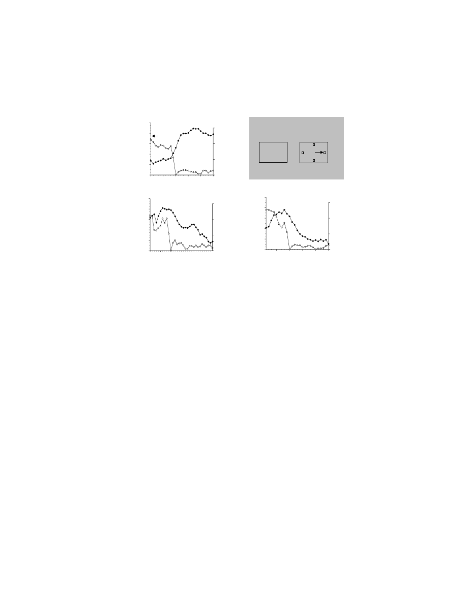
Figure 10.2 compares the learning rates for two different forms of motor learning.
Figure 10.2A shows some results for a traditional form of motor learning, described
in
, in which human participants adapt to forces imposed on their limbs
during a movement. Figure 10.2B presents a learning curve for arbitrary sensorim-
otor learning in rhesus monkeys. In that experiment, monkeys had to learn to map
three novel visual stimuli onto three spatially distinct movements of a joystick: left,
right, and toward the monkey. The stimulus presented on any given trial was ran-
domly selected from the set of three novel stimuli. Note that the learning rate
τ was
approximately 8 trials for both forms of learning. At first glance, this finding seems
odd: most experts would hold that traditional forms of motor learning are slower
than that shown in Figure 10.2A. In fact, under most experimental circumstances it
takes dozens if not hundreds of trials for participants to adapt to the imposed forces.
The curve shown in Figure 10.2A is unusual because it comes from a participant
performing a single out-and-back movement on every trial, rather than varying the
direction of movement among many targets, as is typically the case. When the
participants make movements in several directions, the learning that takes place for
a movement in one direction interferes to an extent with learning about movements
in other directions.
11
This interference slows learning. As Figure 10.2A shows,
traditional forms of motor learning need not be especially slow.
Figure 10.2B is also unusual, but in a different way than Figure 10.2A. Although
the learning rate is virtually identical, Figure 10.2B illustrates the concurrent learning
of three different sensorimotor mappings. In this task, each mapping can be consid-
ered a problem for the monkeys to solve. Thus, for a learning rate of ~8 trials, the
learning rate for any given problem less than 3 trials. Hence, to make the traditional
and arbitrary sensorimotor learning curves identical, the force adaptation problem
has to be reduced to one reaching direction, back and forth, and the arbitrary mapping
task has to be increased to three concurrently learned problems. This implies that
Copyright © 2005 CRC Press LLC
the learning of arbitrary visuomotor mappings in experienced rhesus monkeys is
faster than the fastest motor learning of the traditional kind.
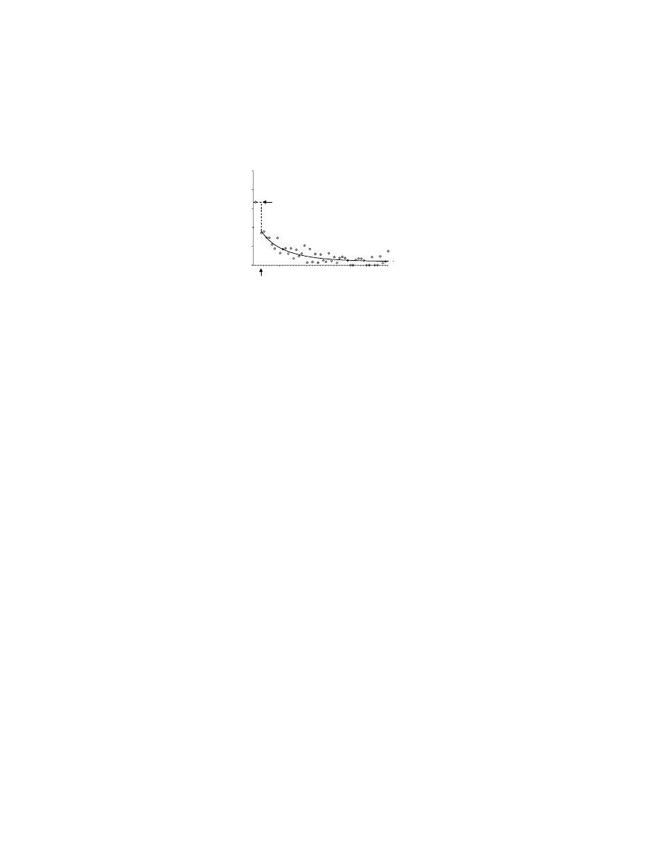
By one attribute, fast learning, a learning rate of less than 3 trials per problem
conforms reasonably well with the notion that arbitrary sensorimotor mappings in
monkeys, at least under certain circumstances, might be classed as explicit. But what
about one-trial learning, a hallmark of explicit learning?
72
, the average
error rate is plotted for four rhesus monkeys, each solving three-choice problems
concurrently and doing so many times. For reasons described in a previous review,
7
we plot only trials in which the stimulus on one trial has changed from that on the
previous trial. Then, we examine only responses to the stimulus (of the three) that
appeared on the first trial. For obvious reasons, the monkeys performed at chance
levels on the first trial of a 50-trial block. (Trial two is not illustrated because we
exclude all trials that repeat the stimulus of the previous trial.) On trial three (the
second presentation of the stimulus that had appeared on the first trial), one trial
learning is significant
10
and is followed by a gradual improvement in performance.
FIGURE 10.2 Learning curves for two forms of motor learning. (A) Adaptation to imposed
forces in human participants, in experiments similar to those described in the legend for
. In these experiments, participants adapted a novel pattern of imposed forces,
which perturbed their reaching movements. Motor learning is measured as a reduction in the
error — i.e., less deviation from a straight hand path to the target. For the data presented
here, participants moved back and forth to a single target trial after trial. (Data from
O. Donchin and R. Shadmehr, personal communication.) (B) Concurrent learning of three
arbitrary visuomotor mappings in rhesus monkeys. Three different, novel stimuli instructed
rhesus monkeys to make three different movements of a joystick. The plot shows the average
scores of four monkeys, each solving 40 sets of three arbitrary visuomotor mappings over
the course of 50 trials.
ττττ is a time constant that corresponds to learning rate, a reduction of
error to e
–1
. (Data from Reference 86.
)
Arbitrary sensorimotor mapping
Trial
Error (%)
Traditional motor learning: adaptation to imposed forces
Error (mm)
0
20
40
60
80
0
10
20
30
40
50
0
10
20
30
40
50
0
5
10
15
20
25
τ = 7.8 trials
τ = 7.8 trials
B
A
Copyright © 2005 CRC Press LLC
What about the ablation approach? Data reviewed in detail elsewhere
7,8
show
that ablations that include all of the hippocampus in both hemispheres abolish the
fast learning illustrated in
and Figure 10.3. Because it is thought that
the hippocampal system subserves the recording of new explicit knowledge in
humans,
73
these data also support the view that arbitrary sensorimotor mapping
represents explicit knowledge and that remaining systems, possibly neocortical,
remain intact to subserve the slower improvement.
Taking all of these data into account, one can argue that arbitrary sensorimotor
mappings of the type learned quickly by experienced animals differs, in kind, from
that learned slowly, and that this difference may correspond to the distinction
between explicit and implicit knowledge in humans. This understanding informs the
results obtained by lesion-, neurophysiological-, and brain-imaging methods for
studying arbitrary sensorimotor mapping. The next sections address the structures,
in addition to the hippocampal system, that support this kind arbitrary mapping.
10.5.2 N
EUROPSYCHOLOGY
Surgical lesions of a number of structures have produced deficits in arbitrary sen-
sorimotor mapping, either in learning new arbitrary mappings (acquisition) or in
performing according to preoperatively learned ones (retention).
10.5.2.1 Premotor Cortex
Severe deficits result from removal of the dorsal aspect of the premotor cortex (PM).
For instance, Petrides
74
demonstrated that monkeys with aspiration lesions that
primarily removed dorsal PM were unable to emit the appropriate response (choosing
FIGURE 10.3 Fast learning of a single arbitrary visuomotor mapping in rhesus monkeys.
The plot shows the average of four monkeys, each solving 40 sets of three arbitrary visuomotor
mappings over the course of 50 trials. For whichever of the three stimuli in the set that was
presented on trial one, the monkeys’ percent error is shown for all subsequent presentations
of the same stimulus. The plot shows only trials in which the stimulus changed from that on
the previous trial. Therefore, no trial-two data are shown: the stimulus on trial two could not
have both changed and been the same as that presented in trial one. (Data from Reference 86.)
0
20
40
60
80
100
chance
0
10
20
30
40
50
3
Fast Learning of Arbitrary Sensorimotor Mappings
Percent Error
Trial
Copyright © 2005 CRC Press LLC

to open either a lit or an unlit box) when instructed to do so, and never reached
criterion in this two-choice task, although they were given 1,020 trials. In contrast
to this poor performance, control monkeys mastered the same task in approximately
300 trials. The lesioned monkeys were able to choose the responses normally,
however, during sessions in which only one of the two responses was allowed,
showing that the monkeys were able to detect the stimuli and were able to make the
required movements.
Halsband and Passingham
75
produced a similarly profound deficit in monkeys
that had undergone bilateral, combined removals of both the dorsal and ventral PM.
Their lesioned monkeys could not relearn a preoperatively acquired arbitrary visuo-
motor mapping task in which a colored visual cue instructed whether to pull or turn
a handle. Unoperated animals relearned this task within 100 trials; lesioned monkeys
failed to reach criterion after 1,000 trials. However, lesioned monkeys were able to
learn arbitrary mappings between different visual stimuli. This pattern of results
confirms that the critical mapping function mediated by PM is that between a cue
and a motor response, rather than arbitrary mappings generally. Putting the results
of Petrides and Passingham together, the critical region for arbitrary sensorimotor
mapping appears to be dorsal PM. Subsequently, Kurata and Hoffman
76
confirmed
that injections of a GABAergic agonist, which transiently disrupts cortical informa-
tion processing, impair the performance of arbitrary visuomotor performance for
sites in the dorsal, but not the ventral, part of PM.
10.5.2.2 Prefrontal Cortex
There is also evidence indicating that the ventral and orbital aspects of the prefrontal
cortex (PF) are crucial for arbitrary sensorimotor mapping.
77
Compared to their
preoperative performance, monkeys were slower to learn arbitrary sensorimotor
mappings after disrupting the interconnections between these parts of PF and the
inferotemporal cortex (IT), either by the use of asymmetrical lesions
78,79
or by
transecting the uncinate fascicle,
80
which connects the frontal and temporal lobes.
These findings suggest that the deficits result from an inability to utilize visual
information properly in the formation of arbitrary visuomotor mappings.
Both Bussey et al.
81
and Wang et al.
82
have directly tested the hypothesis that
the ventral or orbital PF is integral to efficient arbitrary sensorimotor mapping. In
the study of Bussey et al.
81
monkeys were preoperatively trained to solve mapping
problems comprising either three or four novel visual stimuli, and then received
lesions of both the ventral and orbital aspects of PF. The rationale for this approach
was that both areas receive inputs from IT, which processes color and shape infor-
mation. Postoperatively, the monkeys were severely impaired both at learning new
mappings (
) and at performing according to preoperatively learned
ones. The same subjects were unimpaired on a visual discrimination task, which
argues against the possibility that the deficit resulted from an inability to distinguish
the stimuli from each other. A recent study by Rushworth and his colleagues
83
has
demonstrated that the learning impairment seen in monkeys with ventral PF lesions
reflects both the attentional demands inherent in the task and the acquisition of novel
arbitrary mappings.
Copyright © 2005 CRC Press LLC
Although this fast learning of arbitrary sensorimotor mappings was lost, and the
monkeys performed at only chance levels for the first few dozen trials for given sets
of stimuli, if given the same stimuli across days the monkeys slowly learned the
mappings (Figure 10.4B). This slow, across-session visuomotor learning after bilat-
eral lesions of ventral and orbital PF contrasts with the impairment that follows
lesions of PM. Recall that those monkeys could not learn (or relearn) a two-choice
task within 1,000 trials across several days.
74,75
This finding provides further evidence
that different networks subserve fast and slow learning of these arbitrary stimulus–
response mappings.
7
Whether this distinction between fast, within-session learning
and slow, across-session learning corresponds to explicit and implicit learning,
respectively, remains unknown.
In addition, Bussey et al. noted that lesioned monkeys lost the ability to employ
certain cognitive strategies, termed the repeat-stay and change-shift strategies
(Figure 10.4A). According to these strategies, if the stimulus changed from that on
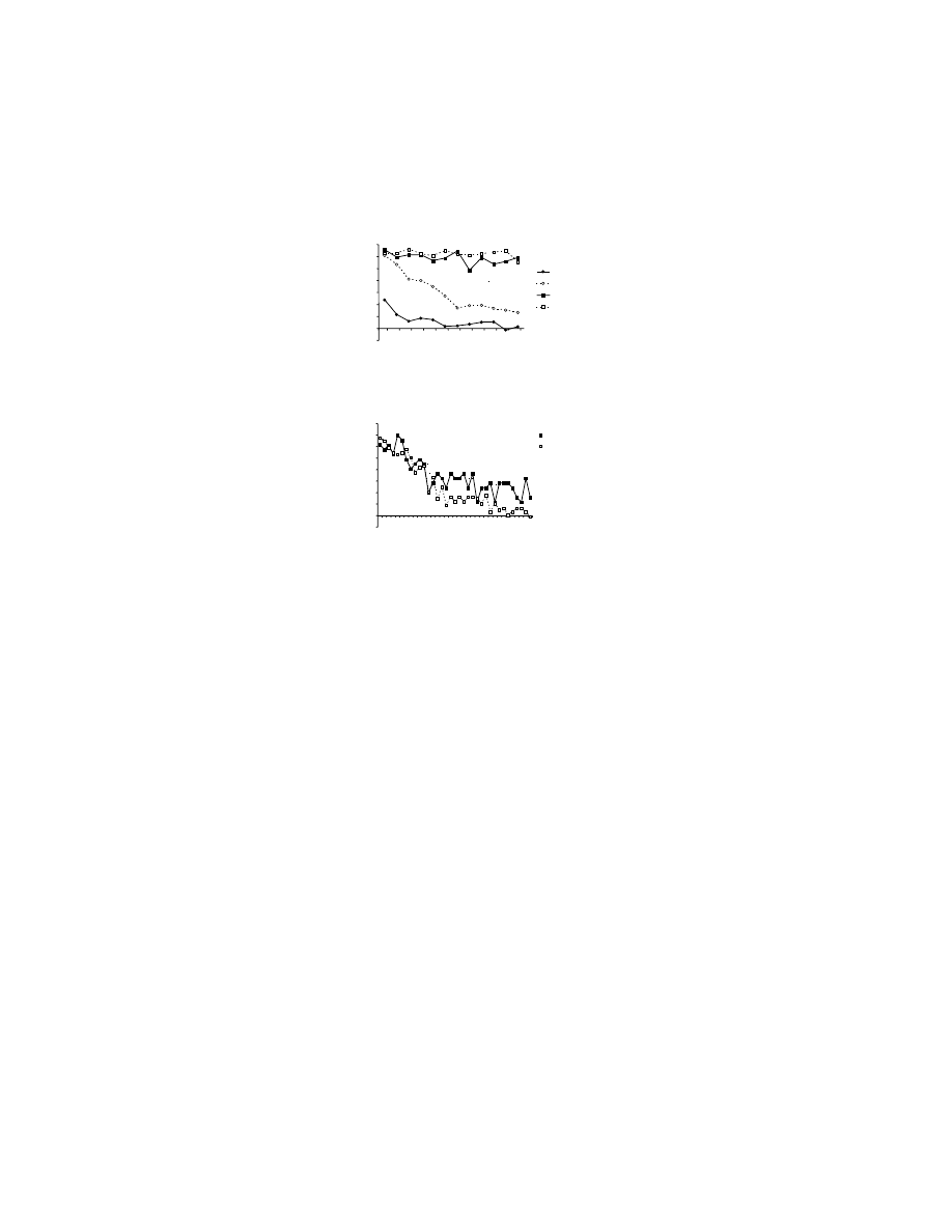
FIGURE 10.4 Effect of bilateral removal of the ventral and orbital prefrontal cortices on
arbitrary visuomotor mapping and response strategies. (A) Preoperative performance is shown
in the curves with circles for four rhesus monkeys. Note that over a small number of trials,
the monkeys improve their performance, choosing the correct response more frequently. Note
also that for repeat trials (filled circles, solid line), in which the stimulus was the same as the
immediately preceding trial, the monkeys performed better than for change trials (unfilled
circles, dashed line), in which the stimulus differed from that on the previous trial. The
difference between these curves is a measure of the application of repeat-stay and change-
shift strategies (see text); change-trial curve shows the learning rate. After removal of the
orbital and ventral prefrontal cortex (postoperative), the animals remain at chance levels for
the entire 48 trial session (curves with square symbols) and the strategies are eliminated.
(B) Two of those four monkeys could, postoperatively, learn the same arbitrary sensorimotor
mappings over the course of several days (sessions). (Data from Reference 81.)
70
60
50
40
30
20
10
0
8
16
24
32
40
48
Trial
Preoperative Repeat
Preoperative Change
Postoperative Repeat
Postoperative Change
70
60
50
40
30
20
10
0
80
8
16
24
32
Session
Percent Improvement
from Chance
Repeat
Change
Arbitrary sensorimotor mapping
across sessions
Arbitrary sensorimotor mapping
with a session
B
A
Percent Improvement
from Chance
Copyright © 2005 CRC Press LLC

the previous trial, then the monkey shifted to a different response; if the stimulus
was the same as on the previous trial, the monkey repeated its response. Application
of these strategies doubled the reward rate, as measured in terms of the percentage
of correct responses, prior to learning any of the sensorimotor mappings. Bilateral
ablation of the orbital and ventral PF abolished those strategies (
,
squares). Could the deficit shown in Figure 10.4A be due entirely to a disruption of
the monkeys’ high-order strategies? This possibility is supported by evidence that
strategies depend on PF function.
84,85
But the evidence presented by Bussey et al.
81
on familiar problem sets indicates otherwise. The repeat-stay and change-shift strat-
egies are relatively unimportant for familiar mappings, but the monkeys’ performance
was also impaired for them. Further, there was evidence — from studies that dis-
rupted the connection between ventral and orbital PF and IT in monkeys that did
not employ the high-order strategies — that learning across sessions was impaired.
79
Wang et al.
82
have also reported deficits in learning a two-choice arbitrary
sensorimotor mapping task after local infusions of the GABAergic antagonist bicu-
culline into the ventral PF. The monkeys in that study, however, showed no impairment
in performing the task with familiar stimuli, in contrast with the monkeys of Bussey
et al.,
81
which had permanent ventral and orbital PF lesions. This difference could
potentially reflect differences in task difficulty, in the temporary nature of the lesion
made by Wang et al.,
82
or both.
10.5.2.3 Hippocampal System
In addition to lesions of the dorsal PM and the ventral and orbital PF, which
substantially impair arbitrary sensorimotor mapping in terms of both acquisition and
performance, disruption of the hippocampal system (HS) also impairs this behav-
ior.
86–88
However, lesioned monkeys can perform mappings learned preoperatively.
This finding supports the idea that HS functions to store mappings in the intermediate
term, as opposed to the short term (seconds) or the long term (weeks or months).
The general idea
89
is that repeated exposure to these associations results in consol-
idation of the mappings in neocortical networks.
9
Impairments in learning new
arbitrary visuomotor mappings result from fornix transection, the main input and
output pathway for the HS, even when both the stimuli and responses are nonspatially
differentiated.
88
In contrast, monkeys with excitotoxic hippocampal lesions are not
impaired in learning these “nonspatial” visuomotor mappings.
90
This finding implies
that the impairment on this nonspatial task seen after fornix transection reflects either
the disruption of cholinergic inputs to areas near the hippocampus, such as the
entorhinal cortex, or dysfunction within those areas due to other causes.
10.5.2.4 Basal Ganglia
The ventral anterior nucleus of the thalamus (VA) receives input from a main output
nucleus of the basal ganglia, the internal segment of the globus pallidus, and projects
to PF and rostral PM. In an experiment reported by Canavan et al.,
91
radiofrequency
lesions were centered in VA. Monkeys in this experiment first learned a single, two-
choice arbitrary sensorimotor mapping problem to a learning criterion of 90%
correct. The experiment involved a lesion group and a control group. After the
Copyright © 2005 CRC Press LLC

“surgery,” the control group retained the preoperatively learned mappings; they made
only an average of ~20 errors to the learning criterion as they were retested on the
task. After lesions centered on VA, monkeys averaged ~1,340 errors in attempting
to relearn the mappings, and two of the three animals failed to reach criterion.
Nixon et al.
92
reported that disrupting the connections (within a hemisphere)
between the dorsal part of PM and the globus pallidus had little effect on the
acquisition of novel arbitrary mappings. This procedure, which involved lesions of
dorsal PM in one hemisphere and of the globus pallidus in the other, led instead to
a selective deficit in the retention and retrieval of familiar mappings. This finding
provides further evidence for the hypothesis that premotor cortex and the parts of
the basal ganglia with which it is connected play an important role in the storage
and retrieval of well-learned, arbitrary mappings.
10.5.2.5 Unnecessary Structures
Nixon and Passingham
93
showed that monkeys with cerebellar lesions are not
impaired on arbitrary sensorimotor mapping tasks. Similar observations have been
made in patients with cerebellar lesions,
94
but this conclusion remains somewhat
controversial. Lesions of the medial frontal cortex, including the cingulate motor
areas and the supplementary and presupplementary motor areas, also fail to impair
arbitrary sensorimotor mapping.
95,96
Similarly, lesions of the dorsolateral PF have
been shown to have either mild impairments in arbitrary sensorimotor mapping,
97,98
or no effects.
99
Arbitrary sensorimotor mapping also does not require an intact
posterior parietal cortex.
100
Along the same lines, a patient with a bilateral posterior
parietal cortex lesion has been reported to have nearly normal timing for correcting
reaching movements when these corrections were instructed by changes in the color
of the targets.
101
Two issues of connectivity arise from the lesion literature. First, it is interesting
to note that the most severe deficits in arbitrary sensorimotor mapping are apparent
after dorsal PM lesions and ventral PF lesions, and yet there is said to be little in
the way of direct cortical connectivity between these two regions. Second is the
issue of how and where the nonspatial information provided by a sensory stimulus
is associated with distinct responses within the motor system. Perhaps the informa-
tion underlying arbitrary sensorimotor mappings is transmitted via a third cortical
region, for which the dorsal PF would appear to be a reasonable candidate. However,
preliminary data indicated that lesions of dorsal PF do not cause the predicted
deficit.
102
Similarly, the medial frontal cortex and the posterior parietal cortex would
appear to be ruled out by the data presented in the preceding paragraph. It is possible
that the basal ganglia play a pivotal role, as suggested by Passingham,
2
but the
precise anatomical organization of inputs and outputs through the basal ganglia and
cortex militates against this interpretation. The parts of basal ganglia targeted by IT
and PF do not seem to overlap much with those that involve PM. Specific evidence
that high-order visual areas project to the parts of basal ganglia that target PM —
via the dorsal thalamus, of course — would contribute significantly to understanding
the network underlying arbitrary visuomotor mapping. Unfortunately, clear evidence
for this connectivity has not been reported.
Copyright © 2005 CRC Press LLC

10.5.2.6 Summary of the Neuropsychology
The hippocampal system, ventral and orbital PF, premotor cortex, and the associated
part of the basal ganglia are involved in the acquisition, retention, and retrieval of
arbitrary sensorimotor mappings.
10.5.3 N
EUROPHYSIOLOGY
10.5.3.1 Premotor Cortex
There is substantial evidence for premotor neurons showing learning-related changes
in activity,
105–107
and these data have been reviewed previously.
6,8–10
Electrophysio-
logical evidence for the role of dorsal PM in learning arbitrary mappings derived
from a study by Mitz et al.
105
in which monkeys were required to learn which of
four novel stimuli mapped to four possible joystick responses (left/right/up/no-go).
In more than half of the cells tested, there was shown to be learning-dependent
activity. Typically, but not exclusively, these learning-related changes were the result
of increases in activity that correlated with an improvement in performance. More-
over, 46% of all learning-related changes were observed in cells that demonstrated
directional selectivity, which would argue against such changes reflecting nonspecific
factors such as reward expectancy. One finding of particular interest was that the
evolution of neuronal activity during learning appeared to lag improved performance
levels, at least slightly. This raised the possibility that the arbitrary mappings may
be represented elsewhere in the brain prior to neurons in dorsal PM reflecting this
sensorimotor learning. This idea is consistent with the findings, mentioned above,
that HS damage disrupts the fastest learning of arbitrary sensorimotor (and other)
associations, but slower learning remains possible. It is also consistent with models
suggesting that the neocortex underlies slow learning and consolidation of associa-
tions formed more rapidly elsewhere. However, it should be noted that, as illustrated
in
, PF damage also disrupts the fastest arbitrary visuomotor mapping,
while allowing across-session learning to continue,
81
albeit at a slower rate.
79
Accord-
ingly, fast mapping is not the exclusive province of the HS.
Chen and Wise
106
used a similar experimental approach to demonstrate learning-
related changes in other parts of the premotor cortex, specifically the supplementary
eye field (SEF) and the frontal eye field (FEF). In their experiment, some results of
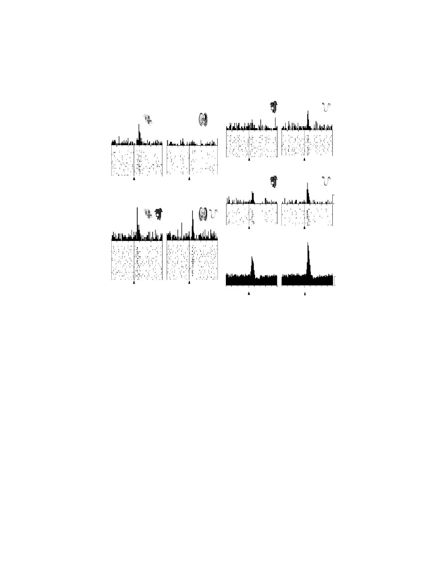
which are illustrated in
, the monkeys were required to fixate a novel
visual stimulus, which was an instruction for an oculomotor response to one of four
targets. The suggestion that subsequent changes in neuronal activity could reflect
changes in motor responses could now be rejected with more confidence than in the
earlier study of learning-dependent activity:
105
saccades do not vary substantially as
a function of learning. Changes in activity during learning were common in SEF,
but less so in FEF.
10.5.3.2 Prefrontal Cortex
Asaad et al.
108
recorded activity from the cortex adjacent and ventral to the principal
sulcus (the ventral and dorsolateral PF) as monkeys learned to make saccades to
Copyright © 2005 CRC Press LLC
one of two targets in response to one of two novel stimuli. In addition to recording
cells that demonstrated stimulus and/or response selectivity (80%), they observed
many cells (44%) in which activity for a specific stimulus–response association was
greater than the additive effects of stimulus and response selectivity. Such “nonlinear”
cells could therefore represent the sensorimotor mapping per se, and the occurrence
of such nonlinearity was essentially constant as a trial progressed: the percentage
of cells showing this effect was 34% during the cue period, 35% during the delay
FIGURE 10.5 Three subpopulations of cells in the supplementary eye field (SEF), showing
their change in activity modulation during learning (filled circles, right axis). Also shown is
the monkeys’ average learning rate over the same trials (unfilled circles, left axis). In the
upper right part of the figure is a depiction of the display presented to the monkeys. The
monkeys fixated the center of a video screen, and at that fixation point an initially novel
stimulus (?) appeared. Later, four targets were presented, and the monkey had to learn — by
trial and error — which of the four targets was to be fixated in order to obtain a reward on
that trial in the context of that stimulus. The arrow illustrates a saccade to the right target.
(A) The average activity (filled circles) of a population of neurons showing learning-dependent
activity that increases with learning, normalized to the maximum for each neuron in the
population. Learning-dependent activity was defined as significant modulation, relative to
baseline activity, for responses to both novel and familiar stimuli. Unfilled circles show mean
error rate (for a moving average of three trials), aligned on the first occurrence of three
consecutive correct responses. Note the close correlation between the improvement in per-
formance and increase in population activity. (B) Learning-dependent activity that decreases
during learning. (C) Learning-selective activity, defined as neuronal modulation that was only
significant for responses to novel stimuli. (Data from Reference 129.)
-10
-5
0
5
10
15
Performance
(percent error)
0
20
40
60
80
100
0.0
0.2
0.4
0.6
Chance
Normalized A
ctivity
Learning-
dependent
(increasing)
Activity
Errors
-10
-5
0
5
10
15
20
Learning-
dependent
(decreasing)
B
A
Normalized Trial Number
-5
0
5
10
15
Learning-
selective
C
Stimulus
Response
?
Normalized Trial Number
Copyright © 2005 CRC Press LLC

period, and 33% during the presaccadic period. In contrast, cells showing cue
selectivity decreased during the trial from 45% during the cue period to 32% and
21% during the delay and presaccadic periods, respectively; and cells showing
response selectivity increased during the trial, from 14% during the cue period to
21% and 34% during the delay and presaccadic periods, respectively. Neuronal
changes during learning were reported for these directional-selective cells in the
delay period, with such selectivity becoming apparent at earlier time points within
the trial as learning progressed. Also of note was the fact that the activity for novel
stimuli typically exceeded that shown for familiar stimuli, even during the delay
period.
10.5.3.3 Basal Ganglia
Tremblay et al.
109
studied the activity of cells in the anterior portions of the caudate
nucleus, the putamen, and the ventral striatum while animals performed an arbitrary
visuomotor mapping task using either familiar or novel stimuli. In this task, there
were three trial types, signaled by one of three stimuli: a rewarded movement trial,
in which a lever touch would result in reward; a rewarded nonmovement trial, in
which the monkey maintained contact with a resting key (and thus did not move
toward the lever) and consequently gained reward; or an unrewarded movement trial,
in which a lever touch would result in the presentation of an auditory conditioned
reinforcer (which also signaled that the next trial would be of the rewarded variety).
Thus reward-related activity and movement-related activity could be compared
across trials to demonstrate the specificity of the cell’s activity modulations.
When the task was performed using familiar stimuli, 17% of neurons showed
task-related activity.
110
When the activity between novel and familiar stimuli was
compared,
109
44% of neurons (90/205) exhibited significant decreases in task-related
activation, while 46% of cells (95/205) demonstrated significant increases in task-
related activity. These increases and decreases in activity were either transient in
nature or were sustained for long after the association had been learned. This pattern
of activity is reminiscent of changes observed in the neocortex,
105,106
and neurons
that showed such task relations were distributed nonpreferentially over the caudate
nucleus, the putamen, and the ventral striatum. Recent data from Brasted and Wise
111
not only confirm the presence of learning-related activity in striatal (putamen)
neurons, but also showed that the time course of these changes in striatal neurons
is similar to that seen in the dorsal premotor cortex, with changes in activity typically
occurring in close correspondence with the learning curve.
Finally, there is evidence for learning-related changes in neuronal activity in
cells in the globus pallidus.
112
Monkeys learned to perform a three-choice arbitrary
visuomotor mapping task in which one of three cues presented on a monitor could
instruct subjects to push, pull, or rotate a manipulator. Monkeys were required to
maintain a center hold position with the manipulator until the cue appeared in the
center of the screen. The cue was then replaced by a neutral stimulus for a variable
delay period before the appearance of a trigger cue instructed monkeys to make
Copyright © 2005 CRC Press LLC

their response. In a control condition, monkeys performed the task using three
familiar stimuli that instructed well-learned associations. In a learning condition
performed in a separate blocks of trials, one of the familiar stimuli was replaced by
a novel stimulus, which required the same response as the replaced familiar stimuli.
Inase et al. focused their efforts on delay-period activity and found about one-third
of cells (49/157) to have delay-period activity, about half of which reflected a
decrease in firing (inhibited neurons) and half an increase in firing (excited neurons)
during the delay period. A difference between learning and control conditions was
seen for 17/23 inhibited neurons, and for 10/26 excited neurons. The majority of
the cells (21/27) that were sensitive to novel stimuli were located in the dorsal medial
aspect of the internal segment of the globus pallidus, which projects indirectly to
the dorsolateral PF according to Middleton and Strick.
113
10.5.3.4 Hippocampal System
Cahusac et al.
114
reported changes in cell activity in the hippocampus and parahippo-
campal gyrus while monkeys learned arbitrary visuomotor tasks. Animals were
presented with one of two visual stimuli on a monitor and were required either to
tap the screen three times or to withhold such movement. Both responses were
rewarded if performed appropriately. Cahusac et al.
114
reported similar types of
learning as seen in the dorsal PM and in the SEF. Thus, 22% (19/87) of neurons
demonstrated differential changes in activity for the two trial types as learning
progressed, while 45% (39/87) of neurons showed only a transient difference
between the two trial types, akin to the learning-selective neurons reported in SEF,
106
More recently, Wirth et al.
115
have also presented reports on arbitrary visual
motor mapping. In their experiment, a complex scene filled a video monitor and
four potential eye-movement targets were superimposed on that scene. Each scene
instructed an eye movement to one and only one of the four targets. They describe
their task as a scene–location association, but it does not differ from the tasks
described above for studies of the premotor cortex. Wirth et al. report that 47% of
hippocampal cells sampled showed activity related to the stimulus or delay period,
and 36% of these cells showed learning-related changes in activity. On average,
changes in neural activity were shown to lag behind changes in behavior. Neverthe-
less, Wirth et al. report that ~38% of their learning cases show changes in neural
activity prior to behavioral changes. A more precise comparison of those data with
the learning-related changes in activity observed in other parts of the brain, using
similar analytical procedures, remains to be undertaken.
10.5.3.5 Summary of the Neurophysiology
There appears to be a close correspondence between the parts of the brain in which
learning-related changes in activity are observed during the acquisition of arbitrary
sensorimotor mappings and the areas necessary for those mappings. How, then, does
this network correspond to that observed for comparable tasks in humans, as their
Copyright © 2005 CRC Press LLC

brains are imaged with positron emission tomography (PET) or functional magnetic
resonance imaging (fMRI)?
10.5.4 N
EUROIMAGING
10.5.4.1 Methodological Considerations
A number of neuroimaging studies have sought to identify the neural network
involved in arbitrary sensorimotor mapping in our species. These reports provide an
interesting parallel to the neurophysiological and neuropsychological work summa-
rized above, although any comparison requires assumptions about the homologies
between cortical areas. In addition to potential species differences, there are sub-
stantial differences between examining single-cell activity and changes in blood flow
rates or other local hemodynamic events. The relationships between the signals
obtained in PET and fMRI studies to neural discharge rates are becoming better
understood,
116,117
and we believe that a consensus is emerging that neuroimaging
signals mainly reflect synaptic events rather than neural discharge rates. Further,
difficult as it might be to resist doing so, negative results in neuroimaging cannot
be interpreted in any meaningful way.
118
Take the example of learning-related activity
changes during the acquisition of arbitrary sensorimotor mappings. As illustrated in
, some cells increase activity during learning, but others decrease, and
they are so closely intermingled that the synaptic inputs causing these changes must
contribute to a single fMRI or PET voxel. So it is likely that no change in PET and
fMRI signals would be observed, despite the occurrence of important changes in
information processing. In addition, of course, there are always issues about whether
human and nonhuman participants approach learning in the same way.
Putting such general methodological issues aside for the sake of discussion,
there remains the issue of how to identify the processes related to the formation of
stimulus–response mappings, as opposed to its many covariants. Three approaches
seem most popular. It is possible (1) to compare activation at different stages of
arbitrary sensorimotor mapping;
119,120
(2) to compare arbitrary sensorimotor mapping
with other kinds of learning, such as sequence learning;
121
or (3) to compare the
learning of novel associations with performance controlled by established associa-
tions.
122
As for the first and third approaches, comparing activation in the early stages
of learning with later stages or with established associations demonstrates that the
experimental and control conditions are well matched for factors relating to stimulus
processing, response preparation, selection, and execution. However, it is reasonable
to assume that the early stages of learning may be associated with greater demands
on attention, novelty detection, motivation levels, and task difficulty. As for the
second approach, comparing arbitrary sensorimotor mapping to other kinds of learn-
ing with similar attentional demands and difficulty, the differences between the tasks
create other interpretational difficulties.
Taking these interpretational problems into account, can we make sense of the
brain-imaging data bearing on arbitrary sensorimotor mapping and its relation to
the neurophysiological and neuropsychological data summarized here?
Copyright © 2005 CRC Press LLC

10.5.4.2 Established Mappings
One of the first studies to examine activity related to arbitrary sensorimotor mapping
was a PET study of Deiber et al.
123
Participants were required to make one of four
joystick movements, with each movement arbitrarily associated with a distinct tone.
In a separate block of trials, the task was performed using the same auditory stimuli
but with the contingencies reversed. Thus, both of these conditions involved arbitrary
sensorimotor mappings. Participants received ~100 trials of training prior to the
scanning in the former task and about 13 trials in the latter. Activity in these two
conditions was compared with a condition in which subjects always moved the
joystick in the same direction in response to a tone. The arbitrary mapping task with
reversal showed some activity increases in the dorsal and dorsolateral PF, and both
arbitrary mapping conditions resulted in significant increases in superior parietal
areas, but neither showed activity increases in PM. There were no reported changes
in other structures associated with arbitrary sensorimotor learning and performance,
such as the basal ganglia and the hippocampal region.
Grafton et al.
124
examined arbitrary visuomotor associations by requiring sub-
jects in a PET scanner to make one of two different grasping actions in response to
the presentation of a red or green light. This condition was compared to an average
of two control conditions in which subjects had to perform only one of the two
grips, with the color light providing only response execution (“go”) rather than
response selection instructions. Thus, although the control task lacked the element
of response selection that was required in the arbitrary mapping task, neither the
stimuli nor the responses could be readily described as spatially differentiated. The
arbitrary mapping task produced greater activation in the posterior parietal cortex
and also resulted in significant increases in the rostral extent of dorsal PM contralat-
eral to the arm used.
Toni et al.
125
conducted a PET study to compare activity in two tasks in which
one of four objects instructed one of four movements. In one task, the stimuli instructed
a spatially congruent grasping movement, while in the other task, stimuli cued an
arbitrarily associated hand movement. The task that used arbitrary visuomotor asso-
ciations was associated with significantly differential regional cerebral blood flow
(rCBF) in the ventral PF, the dorsal PM, and the putamen and/or globus pallidus. It
should be noted, however, that this dorsal PM activity was located in its medial
aspect, which lesion studies suggest may not be necessary for effecting arbitrary
mappings.
95,96
Such activity thus may reflect a more general role in monitoring
visuomotor transformations.
Ramnani and Miall
126
found a selective increase in dorsal PM activity in an
fMRI study of arbitrary visuomotor mapping. In their study, stimulus shape indicated
which of four buttons to depress (and therefore which finger to move) and stimulus
color indicated whether the participant, another person in the room, or a computer
should perform the task. Their findings not only indicated that dorsal PM showed
a significant hemodynamic response during performance of the arbitrary mapping
task, but also that it was selective for a specific instruction (as opposed to a non-
specific warning stimulus) and for the scanned participant performing the task (as
opposed to the other person in the room). Ramnani and Miall suggested that predictions
Copyright © 2005 CRC Press LLC

about the actions of another person rely on a different brain system, one commonly
activated when people attribute mental states to others. This brain system included
part of Broca’s area, among other regions of medial PF and the superior temporal
cortex.
The above studies seem to provide supportive evidence for PF and basal ganglia
involvement in arbitrary sensorimotor mappings, especially ventral PF and puta-
men.
125
There is, by contrast, a notable lack of consistent activation in PM in such
tasks. In the studies outlined above, only the findings of Toni et al.
125
and Ramnani
and Miall
126
provided support for the neuropsychological results, which have indi-
cated that dorsal PM is necessary for performing arbitrary mappings. The PM activity
reported in the study of Grafton et al.
124
(see also Sweeney et al.
127
) could simply
have reflected greater response-selection demands. The potential reasons for false-
negative PET or fMRI results for dorsal PM include the methodological issues
mentioned above, as well as one important additional problem. PM mediates a
diverse range of response-related functions in addition to arbitrary sensorimotor
mappings. Thus, many control tasks may fail to yield contrasts because dorsal PM
is involved in those tasks as well. For example, Deiber et al.
123
reported no PM
activity at all during performance of arbitrary mappings, even when the mappings
were reversed. But there is evidence from monkeys that PM might be involved in
the control condition of their task: repetitive movement. Indeed, cells in dorsal PM
show movement-related modulation in activity for arm movements in total dark-
ness,
128
so a genuine control task might be difficult to devise.
10.5.4.3 Learning New Mappings
A number of imaging studies have also attempted to identify the neural substrates
of arbitrary sensorimotor mapping during learning. Deiber et al.
119
measured rCBF
as participants performed two kinds of arbitrary mapping tasks. One required them
to perform a joystick movement that depended on arbitrary cues, and the other
required them to report whether an arrow matched the arbitrary stimulus-to-place
mapping for the same cues. Increases in rCBF during learning were reported for the
putamen in the latter condition and in ventrocaudal PM in the former. Decreases in
rCBF were more extensive: ventral PF, dorsal PF, and dorsal PM showed rCBF
decreases for the reporting task.
Toni and his colleagues have undertaken a series of imaging studies designed
to identify the neural network involved in arbitrary visuomotor mapping. Toni and
Passingham
121
conducted a PET study in which they compared arbitrary sensorimotor
learning with motor sequence learning. The responses were the same in the two
tasks (one of four finger movements) and the stimuli also were of the same type,
though not identical, for each task. Thus, although the two tasks were matched for
sensory and motor components, the stimulus pattern only instructed the correct
response in the arbitrary sensorimotor mapping task. (In a baseline task, subjects
passively viewed four categorically similar stimuli.) The critical comparison between
arbitrary visuomotor mapping and sequential motor learning revealed learning-
related increases near the cingulate sulcus and in the body of the caudate nucleus
in the left hemisphere and in orbital PF in the right hemisphere. The two tasks also
Copyright © 2005 CRC Press LLC

showed differential patterns of activity in the left superior parietal cortex, with
activity decreasing during arbitrary sensorimotor mapping and increasing during
sequence learning. Increases in the parahippocampal gyrus and the putamen and
globus pallidus, as well as decreases in the dorsolateral PF, were only seen when
arbitrary sensorimotor mapping was compared to the passive, baseline condition.
These data seem to confirm a role for the ventral PF and basal ganglia in arbitrary
sensorimotor mapping, but the evidence for hippocampal involvement in this study
remains weak because it was revealed only in comparison with a passive baseline
condition. The lack of PM activity in the arbitrary sensorimotor learning condition,
although seemingly inconsistent with the neuropsychological and neurophysiologi-
cal literature on monkeys, may reflect the involvement of dorsal PM in the visuo-
motor transformations underlying both arbitrary visuomotor mapping and motor
sequence learning.
In a subsequent fMRI study, Toni et al.
122
compared the learning of arbitrary
sensorimotor mappings with performance using already established mappings. There
were four stimuli in each condition, which mapped to the four finger movement
responses mentioned above. Blocks of familiar and novel stimuli were mixed. Toni
et al.
122
reported hemodynamic events in many regions in the frontal and temporal
lobes, as well as in the HS and basal ganglia. Their results included signal increases
in the ventral PF, the ventral PM, the dorsal PF, the orbital PF, the parahippocampal
and hippocampal gyri, and the caudate nucleus, among other structures. Interestingly,
whereas most time-related changes consisted of the hemodynamic signal for learning
and control conditions converging over time, this was not true of increases in the
caudate nucleus, in which the signal became greater in the learning than in the
control condition as the task progressed. Subsequent structural equation modeling
of the same data set led the authors to suggest that corticostriatal interactions
strengthen when arbitrary sensorimotor mappings are learned.
130
An analysis of
“effective connectivity” suggested that variation in the fMRI signal in the striatum
showed a stronger correlation, as learning progressed, with changes seen in PM, the
inferior frontal cortex, and the medial temporal lobe. On the basis of these analyses,
the investigators inferred that the learning of arbitrary mappings was the result of
an increase in activity in corticostriatal connections, although it should be noted that
such inferences assume a level of corticostriatal convergence that remains to be
shown neuroanatomically.
As in the previous section, the results summarized here also show a surprising
lack of consistency regarding dorsal PM. Although Toni et al.
122
report learning-
associated increases in ventral PM, Toni and Passingham
121
did not report any
changes in PM activity when subjects learn arbitrary sensorimotor mappings that
require skeletomotor responses (see also Paus et al.
131
). Similarly, while Deiber
et al.
119
reported learning-associated increases in ventral PM during learning in one
of their arbitrary mapping tasks, they also found no changes in PM activation in the
other version of the task. Such negative results are at variance with neurophysiolog-
ical (
) studies. As mentioned
earlier, this lack of correspondence may reflect interpretational problems with neuro-
imaging techniques. As Toni and Passingham
121
suggest, “[I]t could be that the
learning-related signal measured with PET is diluted by the contribution of a
Copyright © 2005 CRC Press LLC

neuronal subpopulation that is related to the execution of movements” (p. 29).
Alternatively, it may be that dorsal PM mediates both the learning of new mappings
and the retrieval of established mappings, thus nullifying any contrast between the
two conditions. In this regard, Toni et al.
122
state that “our results suggest that, at
the system level, dorsal premotor regions were similarly involved in both the
[retrieval] and [learning] tasks,” and that “the discrepancy between these results and
those obtained at the single-unit level … calls for further investigation of the specific
contribution of ventral and dorsal premotor cortex to visuomotor association.” (p.
1055) That such caution is warranted is confirmed by the observation that in the
PF,
108
SEF, and FEF
129
of monkeys, a number of cells discharge preferentially for
either familiar or novel mappings. Also, to repeat the argument presented above,
some cells increase activity, but others decrease activity during learning. Given that
a myriad of synaptic signals that must have driven these changes, which also
increased or decreased during learning, and given that these synaptic signals (rather
than neuronal discharge rate) probably dominated the neuroimaging results,
116,117
one can argue that nothing specific can be predicted for either PET or fMRI learning-
related activity. Another possibility is that human participants approach these learn-
ing tasks differently than do monkeys. Specifically, it seems possible that participants
in neuroimaging studies may preferentially employ only fast-learning mechanisms,
and for that reason fail to show PM activation.
79
There is, however, a greater consensus in the neuroimaging literature that ventral
PF and the basal ganglia mediate arbitrary visuomotor mapping. Toni et al.
122
and
Toni and Passingham
121
(see also Paus et al.
131
) all reported learning-related changes
in the ventral PF. Although Deiber et al.
119
did not find such changes in the visuo-
motor version of their learning task, they did see learning-related decreases in ventral
PF in the version in which participants reported such relations. It has been suggested
that the ventral PF plays a critical role in mediating arbitrary sensorimotor mapping,
since it is in a position to represent knowledge about stimuli, responses, and out-
comes,
77
a view supported by lesion studies.
81,82
Regarding the role of the basal
ganglia, Toni et al.
122
reported increases in caudate as learning progresses, whereas
Deiber et al.
119
reported learning-related increases in the putamen (see also Toni and
Passingham
121
and Paus et al.
131
).
10.5.4.4 Summary
This section has focused on the neural mechanisms for learning arbitrary mappings
in voluntary movement. Neurophysiological, neuropsychological, and neuroimaging
findings appear to agree that ventral prefrontal cortex and parts of the basal ganglia
play an important role in such learning. Neuroimaging findings are less consistently
supportive of the neurophysiological and neuropsychological evidence that dorsal
premotor cortex and the hippocampal system also play necessary roles in this kind
of learning. Although others will surely disagree, we think that the limitations of
neuroimaging methods make negative results of this kind uninterpretable. Accord-
ingly, we conclude that the hippocampal system is necessary for the rapid learning
of arbitrary mappings, but not for slow learning and not for the retention or retrieval
of familiar mappings. Ventral and orbital PF are necessary for fast learning and the
Copyright © 2005 CRC Press LLC

application of at least certain strategies, if not strategies in general. These parts of
PF also contribute, in part through their interaction with IT, to the slow learning of
arbitrary visuomotor mappings. The dorsal premotor cortex and the associated part
of the basal ganglia are involved in the retention and retrieval of familiar mappings,
not the learning of new ones. It seems likely that the role of the basal ganglia is
diverse,
104
with some parts involved in fast learning, much like the hippocampal
system and the prefrontal cortex, and other parts involved in slow learning and long-
term retention, as postulated here for the premotor cortex.
10.6 ARBITRARY MAPPING OF STIMULI TO
COGNITIVE REPRESENTATIONS
In addition to the many specific stimulus–response associations, it is also possible
that a stimulus can map to a response rule. The arbitrary mapping of stimuli to rules
is relevant to this chapter for two reasons: not only does the relationship between
the stimulus and the rule represent an example of arbitrary mapping in its own right,
but such rules also allow correct responses to be chosen even if the stimulus that
will cue the response has never been encountered.
Wallis et al.
23
examined the arbitrary mapping of stimuli to response rules, and
showed that abstract rules are encoded in PF (
). They trained two monkeys
to switch flexibly between two rules: a matching-to-sample rule and a nonmatching-
to-sample rule. Monkeys were presented with a sample stimulus and then, after a
delay, were shown a test stimulus, and were required to judge whether the sample
stimulus matched the test stimulus, and to respond (i.e., release or maintain bar
press) accordingly. The rule applicable to each trial was indicated by a cue that was
presented at the same time as the sample stimulus. In order to rule out the sensory
properties of the cue as a confounding factor, each rule type could be signaled by
one of two distinct cues from two different modalities (e.g., juice or low tone for
the matching rule, no juice or high tone for the nonmatching rule). Thus, the specific
event that occurred at the same time that the sample appeared served as an arbitrary
cue that mapped onto one of the two rules. The authors reported the presence of
rule-selective cells, such as those that were preferentially active during match trials,
regardless of whether the match rule had been signaled a drop of juice or a low
tone. Of 492 cells recorded from in dorsolateral or ventral PF, 200 cells (41%)
showed such selectivity for either the match rule (101 cells) or the nonmatch rule
(99 cells). There was no obvious segregation of rule cells in any one area of PF for
either of the rule types, and rule specificity was recorded in both the stimulus and
delay task periods, although there was a higher incidence of rule-selective neurons
in the dorsolateral PF (29%) than in the ventrolateral PF (16%) and the orbitofrontal
PF (18%). The same authors have subsequently shown such abstract rules to be
encoded in premotor areas during the sample period.
134
These results are consistent
with those of Hoshi et al.,
24
who also recorded from cells in the prefrontal cortex
that were related to the abstract rules that govern responding.
While such studies demonstrate the ability of PF to encode abstract rules, Strange
et al.
133
conducted an fMRI study during which subjects were required to learn rules.
Copyright © 2005 CRC Press LLC
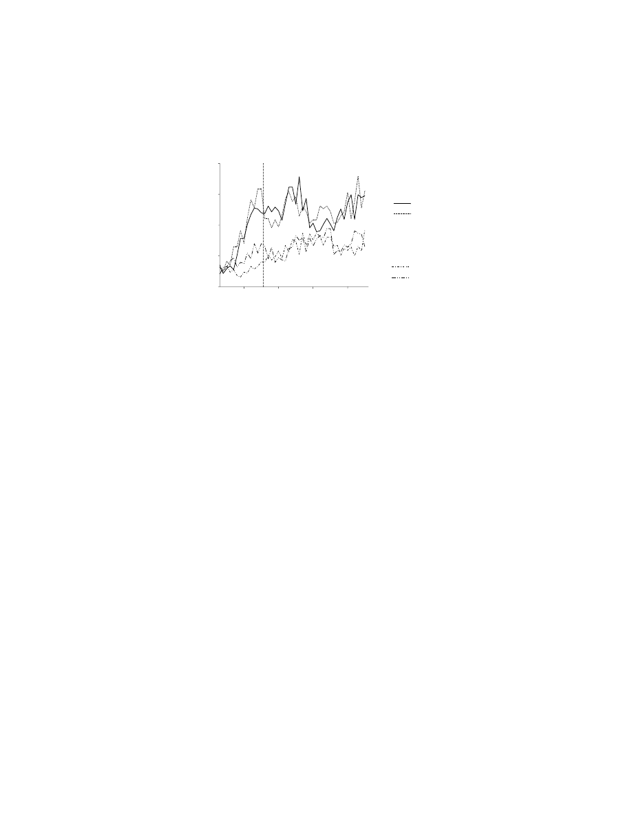
Subjects would be presented with a string of four letters, and would have to respond
according to whether the letters conformed or not with an unstated rule, such as
“the second and fourth letters must match.” While subjects were given examples of
such rules prior to scanning, they were not informed of the exact rule. During
scanning, the experimenters could then change either the exemplars, or the rule, or
both the rule and the exemplars. A factorial analysis revealed that, after a rule change,
increases in activity were observed in rostral aspects of PF, including dorsal, ventral,
and polar PF. A slightly different pattern of activity was seen in the left hippocampal
region: a decrease in activity was observed and it was greater when the rule changed
than when only the exemplars changed.
Thus, although research on learning to map stimuli onto arbitrary response rules
has only recently begun, it seems likely that it has much the same character and involves
many of the same neural structures as the other kinds of arbitrary sensorimotor
mapping outlined in this chapter.
10.7 CONCLUSION
Arbitrary sensorimotor mapping occurs in many types, which appear to be special
cases of a more general arbitrary mapping capacity. Advanced brains can map stimuli
FIGURE 10.6 Neuronal activity in the prefrontal cortex, reflecting arbitrary stimulus–rule
mappings. This cell’s modulation was relatively high when a high-pitched tone or no additional
stimulus appeared at the same time as a sample stimulus. The monkey later, after a delay
period, was required to respond to a stimulus other than the sample in order to receive a
reward. This response rule is called a nonmatching-to-sample rule. The cell showed much
less modulation when the low-pitched tone or a reward occurred at the same time as the
sample. In that eventuality, the monkey was required to respond to the sample when it
reappeared later in the trial (called a matching-to-sample rule). Note that the cells firing rate
was greater when the arbitrary cue signaled the nonmatching-to-sample rule, regardless of
which cue mapped to that rule. (Data from Reference 23.)
Firing rate (impulses/s)
high ton
e
no IS
juice
low ton
e
0.5
1.0
1.5
2.0
0
5
10
15
20
Sample
Delay
Matching Rule
Nonmatching Rul
Time (s)
Rule-Related Activity
Copyright © 2005 CRC Press LLC

arbitrarily to (1) reflex-like responses, (2) internal models of limb dynamics, and
response choices that either (3) habitually follow a stimulus or (4) follow a stimulus
based on a prediction about response outcome. They can also map stimuli arbitrarily
to biological value, response rules, and abstract meaning, which, in turn, can be
mapped to the four kinds of action listed above. The recurrent nature of such arbitrary
mappings provides much of their power to enable the behavioral flexibility charac-
teristic of advanced animals.
REFERENCES
1. Wise, S.P., di Pellegrino, G., and Boussaoud, D., The premotor cortex and nonstandard
sensorimotor mapping, Can. J. Physiol. Pharmacol., 74, 469, 1996.
2. Passingham, R.E., The Frontal Lobes and Voluntary Action, Oxford University Press,
Oxford, 1993.
3. Dickinson, A. and Balleine, B., Motivational control of goal-directed action., Animal
Learn. Behav., 22, 1, 1994.
4. McDonald, R.J. and White, N.M., A triple dissociation of memory systems: hippoc-
ampus, amygdala, and dorsal striatum, Behav. Neurosci., 107, 3, 1993.
5. Jog, M.S. et al., Building neural representations of habits, Science, 286, 1745, 1999.
6. Wise, S.P., Evolution of neuronal activity during conditional motor learning, in
Acquisition of Motor Behavior in Vertebrates, Bloedel, J.R., Ebner, T.J., and Wise,
S.P., Eds., MIT Press, Cambridge, MA, 1996, 261.
7. Wise, S.P. and Murray, E.A., Role of the hippocampal system in conditional motor
learning: mapping antecedents to action, Hippocampus, 9, 101, 1999.
8. Wise, S.P. and Murray, E.A., Arbitrary associations between antecedents and actions,
Trends Neurosci., 23, 271, 2000.
9. Murray, E.A., Bussey, T.J., and Wise, S.P., Role of prefrontal cortex in a network for
arbitrary visuomotor mapping, Exp. Brain Res., 133, 114, 2000.
10. Murray, E.A., Brasted, P.J., and Wise, S.P., Arbitrary sensorimotor mapping and the
life of primates, in Neuropsychology of Memory, Squire, L.R. and Schacter, D.L.,
Eds., Guilford, New York, 2002, 339.
11. Shadmehr, R. et al., Learning dynamics of reaching, in Motor Cortex in Voluntary
Movements, Riehle, A. and Vaadia, E., Eds., CRC Press, Boca Raton, FL, 2005.
12. Pearce, J.M., Animal Learning and Cognition: An Introduction, Psychology Press,
UK, 1997.
13. Thorndike, E.L., Animal Intelligence, Macmillan, New York, 1911.
14. Guthrie, E.R., The Psychology of Learning, Harper, New York, 1935.
15. Hull, C.L., Principles of Behavior, Appleton-Century-Crofts, New York, 1943.
16. Adams, C.D. and Dickinson, A., Actions and habits: variations in associative repre-
sentations during instrumental learning, in Information Processing in Animals: Mem-
ory Mechanisms, Spear, N.E. and Miller, R.R., Eds., Lawrence Erlbaum Associates,
Inc., Hillsdale, NJ, 1981, 143.
17. Colwill, R.M. and Rescorla, R.A., Instrumental responding remains sensitive to
reinforcer devaluation after extensive training, J. Exp. Psychol. Animal Behav. Proc.,
11, 520, 1985.
18. Balleine, B.W. and Dickinson, A., Goal-directed instrumental action: contingency and
incentive learning and their cortical substrates, Neuropharmacology, 37, 407, 1998.
Copyright © 2005 CRC Press LLC

19. Corbit, L.H. and Balleine, B.W., The role of the hippocampus in instrumental con-
ditioning, J. Neurosci., 20, 4233, 2000.
20. di Pellegrino, G. and Wise, S.P., Visuospatial vs. visuomotor activity in the premotor
and prefrontal cortex of a primate, J. Neurosci., 13, 1227, 1993.
21. Riehle, A., Kornblum, S., and Requin, J., Neuronal coding of stimulus-response
association rules in the motor cortex, NeuroReport, 5, 2462, 1994.
22. White, I.M. and Wise, S.P., Rule-dependent neuronal activity in the prefrontal cortex,
Exp. Brain Res., 126, 315, 1999.
23. Wallis, J.D., Anderson, K.C., and Miller, E.K., Single neurons in prefrontal cortex
encode abstract rules, Nature, 411, 953, 2001.
24. Hoshi, E., Shima, K., and Tanji, J., Neuronal activity in the primate prefrontal cortex
in the process of motor selection based on two behavioral rules, J. Neurophysiol., 83,
2355, 2000.
25. Kimura, M., The role of primate putamen neurons in the association of sensory stimuli
with movement, Neurosci. Res., 3, 436, 1986.
26. Aosaki, T. et al., Responses of tonically active neurons in the primate’s striatum
undergo systematic changes during behavioral sensorimotor conditioning, J. Neurosci.,
14, 3969, 1994.
27. Aosaki, T., Graybiel, A.M., and Kimura, M., Effect of the nigrostriatal dopamine
system on acquired neural responses in the striatum of behaving monkeys, Science,
265, 412, 1994.
28. Raz, A. et al., Neuronal synchronization of tonically active neurons in the striatum
of normal and parkinsonian primates, J. Neurophysiol., 76, 1996.
29. Rosenkranz, J.A. and Grace, A.A., Dopamine-mediated modulation of odour-evoked
amygdala potentials during Pavlovian conditioning, Nature, 417, 282, 2002.
30. Schafe, G.E. et al., Memory consolidation of Pavlovian fear conditioning: a cellular
and molecular perspective, Trends Neurosci., 24, 540, 2001.
31. Repa, J.C. et al., Two different lateral amygdala cell populations contribute to the
initiation and storage of memory, Nat. Neurosci., 4, 724, 2001.
32. Aou, A., Woody, C.D., and Birt, D., Increases in excitability of neurons of the motor
cortex of cats after rapid acquisition of eye blink conditioning, J. Neurosci., 12, 560, 1992.
33. Steinmetz, J.E., Brain substrates of classical eyeblink conditioning: a highly localized
but also distributed system, Behav. Brain Res., 110, 13, 2000.
34. McEchron, M.D. and Disterhoft, J.F., Hippocampal encoding of non-spatial trace
conditioning, Hippocampus, 9, 385, 1999.
35. Kimura, M., Rajkowski, J., and Evarts, E., Tonically discharging putamen neurons
exhibit set-dependent responses, Proc. Nat. Acad. Sci. U.S.A., 81, 4998, 1984.
36. Aosaki, T., Kimura, M., and Graybiel, A.M., Temporal and spatial characteristics of
tonically active neurons of the primate’s striatum, J. Neurophysiol., 73, 1234, 1995.
37. Ravel, S., Legallet, E., and Apicella, P., Tonically active neurons in the monkey
striatum do not preferentially respond to appetitive stimuli, Exp. Brain Res., 128,
531, 1999.
38. Blazquez, P.M. et al., A network representation of response probability in the striatum,
Neuron, 33, 973, 2002.
39. Apicella, P., Legallet, E., and Trouche, E., Responses of tonically discharging neurons
in the monkey striatum to primary rewards delivered during different behavioral states,
Exp. Brain Res., 116, 456–466.
40. Ravel, S. et al., Reward unpredictability inside and outside of a task context as a
determinant of the responses of tonically active neurons in the monkey striatum,
J. Neurosci., 21, 5730, 2001.
Copyright © 2005 CRC Press LLC

41. Lebedev, M.A. and Nelson, R.J., Rhythmically firing neostriatal neurons in monkey:
activity patterns during reaction-time hand movements, J. Neurophysiol., 82, 1832, 1999.
42. Shimo, Y. and Hikosaka, O., Role of tonically active neurons in primate caudate in
reward-oriented saccadic eye movement, J. Neurosci., 21, 7804, 2001.
43. Schultz, W. and Dickinson, A., Neuronal coding of prediction errors, Annu. Rev.
Neurosci., 23, 473, 2000.
44. Robbins, T.W. and Everitt, B.J., Neurobehavioral mechanisms of reward and moti-
vation, Curr. Opin. Neurobiol., 6, 228, 1996.
45. Schultz, W., Dopamine neurons and their role in reward mechanisms, Curr. Opin.
Neurobiol., 7, 191, 1997.
46. Ljungberg, T., Apicella, P., and Schultz, W., Responses of monkey dopamine neurons
during learning of behavior reactions, J. Neurophysiol., 67, 145, 1992.
47. Hollerman, J.R. and Schultz, W., Dopamine neurons report an error in the temporal
prediction of reward during learning, Nat. Neurosci., 1, 304, 1998.
48. Schultz, W., Apicella, P., and Ljungberg, T., Responses of monkey dopamine neurons
to reward and conditioned stimuli during successive steps of learning a delayed
response task, J. Neurosci., 13, 900, 1993.
49. Schultz, W., Predictive reward signal of dopamine neurons, J. Neurophysiol., 80, 1,
1998.
50. Waelti, P., Dickinson, A., and Schultz, W., Dopamine responses comply with basic
assumptions of formal learning theory, Nature, 412, 43, 2001.
51. Mackintosh, N.M., A theory of attention: variations in the associability of stimuli
with reinforcement, Psychol. Rev., 82, 276, 1975.
52. Pearce, J.M. and Hall, G., A model for Pavlovian learning: variations in the effec-
tiveness of conditioned but not of unconditioned stimuli, Psychol. Rev., 87, 532, 1980.
53. Rescorla, R.A. and Wagner, A.R., A theory of Pavlovian conditioning: variations in
the effectiveness of reinforcement and non-reinforcement, in Classical Conditioning:
Current Research and Theory, Black, A.H. and Prokasy, W.F., Eds., Appleton-Century-
Crofts, New York, 1972, 64.
54. Kamin, L.J., Predictability, surprise, attention, and conditioning, in Punishment and
Aversive Behavior, Campbell, B.A. and Church, R.M., Eds., Appleton-Century-
Crofts, New York, 1969, 279.
55. Suri, R.E. and Schultz, W., Learning of sequential movements by neural network
model with dopamine-like reinforcement signal, Exp. Brain Res., 121, 350–354.
56. Kim, J.J., Krupa, D.J., and Thompson, R.F., Inhibitory cerebello-olivary projections
and blocking effect in classical conditioning, Science, 279, 570, 1998.
57. Medina, J.F. et al., Parallels between cerebellum- and amygdala-dependent condi-
tioning, Nat. Rev. Neurosci., 3, 122, 2002.
58. Everitt, B.J., Dickinson, A., and Robbins, T.W., The neuropsychological basis of
addictive behaviour, Brain Res. Rev., 36, 129, 2001.
59. Baxter, M.G. and Murray, E.A., The amygdala and reward, Nat. Rev. Neurosci., 3,
563, 2002.
60. Cardinal, R.N. et al., Effects of selective excitotoxic lesions of the nucleus accumbens
core, anterior cingulate cortex, and central nucleus of the amygdala on autoshaping
performance in rats, Behav. Neurosci., 116, 553, 2002.
61. Parkinson, J.A. et al., Disconnection of the anterior cingulate cortex and nucleus
accumbens core impairs Pavlovian approach behavior: further evidence for limbic
cortical-ventral striatopallidal systems, Behav. Neurosci., 114, 42, 2000.
62. Rao, A.K. and Shadmehr, R., Contextual cues facilitate learning of multiple models
of arm dynamics, Soc. Neurosci. Abstr., 26, 302, 2001.
Copyright © 2005 CRC Press LLC

63. Wada, Y. et al., Acquisition and contextual switching of multiple internal models for
different viscous force fields, Neurosci. Res., 46, 319, 2003.
64. Packard, M.G., Hirsh, R., and White, N.M., Differential effects of fornix and caudate
nucleus lesions on two radial maze tasks: evidence for multiple memory systems,
J. Neurosci., 9, 1465, 1989.
65. Carelli, R.M., Wolske, M., and West, M.O., Loss of lever press-related firing of rat
striatal forelimb neurons after repeated sessions in a lever pressing task, J. Neurosci.,
17, 1804, 1997.
66. Wise, S.P., The role of the basal ganglia in procedural memory, Sem. Neurosci., 8,
39, 1996.
67. Gaffan, D., Memory, action and the corpus striatum: current developments in the
memory-habit distinction, Sem. Neurosci., 8, 33, 1996.
68. Mishkin, M. and Petri, H.L., Memories and habits: some implications for the analysis
of learning and retention, in Neuropsychology of Memory, Squire, L.R. and Butters,
N., Eds., Guilford Press, New York, 1984, 287.
69. Knowlton, B.J., Mangels, J.A., and Squire, L.R., A neostriatal habit learning system
in humans, Science, 273, 1399, 1996.
70. Houk, J.C. and Wise, S.P., Distributed modular architectures linking basal ganglia,
cerebellum, and cerebral cortex: their role in planning and controlling action, Cereb.
Cortex, 5, 95, 1995.
71. Tulving, E., Episodic memory and common sense: how far apart? Phil. Trans. R. Soc.
Lond. B Biol. Sci., 356, 1505, 2001.
72. Reber, P.J., Attempting to model dissociations of memory, Trends Cog. Sci., 6, 192, 2002.
73. Squire, L.R. and Zola, S.M., Structure and function of declarative and nondeclarative
memory systems, Proc. Nat. Acad. Sci. U.S.A., 93, 13515, 1996.
74. Petrides, M., Deficits in non-spatial conditional associative learning after periarcuate
lesions in the monkey, Behav. Brain Res., 16, 95, 1985.
75. Halsband, U. and Passingham, R.E., Premotor cortex and the conditions for a move-
ment in monkeys, Behav. Brain Res., 18, 269, 1985.
76. Kurata, K. and Hoffman, D.S., Differential effects of muscimol microinjection into
dorsal and ventral aspects of the premotor cortex of monkeys, J. Neurophysiol., 71,
1151, 1994.
77. Passingham, R.E., Toni, I., and Rushworth, M.F., Specialisation within the prefrontal
cortex: the ventral prefrontal cortex and associative learning, Exp. Brain Res., 133,
103, 2000.
78. Gaffan, D. and Harrison, S., Inferotemporal-frontal disconnection and fornix transec-
tion in visuomotor conditional learning by monkeys, Behav. Brain Res., 31, 149, 1988.
79. Bussey, T.J., Wise, S.P., and Murray, E.A., Interaction of ventral and orbital prefrontal
cortex with inferotemporal cortex in conditional visuomotor learning, Behav. Neuro-
sci., 116, 703, 2002.
80. Eacott, M.J. and Gaffan, D., Inferotemporal-frontal disconnection: the uncinate fas-
cicle and visual associative learning in monkeys, Eur. J. Neurosci., 4, 1320, 1992.
81. Bussey, T.J., Wise, S.P., and Murray, E.A., The role of ventral and orbital prefrontal
cortex in conditional visuomotor learning and strategy use in rhesus monkeys
(Macacca mulatta), Behav. Neurosci., 115, 971, 2001.
82. Wang, M., Zhang, H., and Li, B.M., Deficit in conditional visuomotor learning by
local infusion of bicuculline into the ventral prefrontal cortex in monkeys, Eur. J.
Neurosci., 12, 3787, 2000.
83. Rushworth, M.F. et al., Attentional selection and action selection in the ventral
prefrontal cortex, Soc. Neurosci. Abstr., 28, 722.11, 2003.
Copyright © 2005 CRC Press LLC

84. Gaffan, D., Easton, A., and Parker, A., Interaction of inferior temporal cortex with
frontal cortex and basal forebrain: double dissociation in strategy implementation and
associative learning, J. Neurosci., 22, 7288, 2002.
85. Collins, P. et al., Perseveration and strategy in a novel spatial self-ordered sequencing
task for nonhuman primates: effects of excitotoxic lesions and dopamine depletions
of the prefrontal cortex, J. Cog. Neurosci., 10, 332, 1998.
86. Murray, E.A. and Wise, S.P., Role of the hippocampus plus subjacent cortex but not
amygdala in visuomotor conditional learning in rhesus monkeys, Behav. Neurosci.,
110, 1261, 1996.
87. Rupniak, N.M.J. and Gaffan, D., Monkey hippocampus and learning about spatially
directed movements, J. Neurosci., 7, 2331, 1987.
88. Brasted, P.J. et al., Fornix transection impairs conditional visuomotor learning in
tasks involving nonspatially differentiated responses, J. Neurophysiol., 87, 631, 2002.
89. McClelland, J.L., McNaughton, B., and O’Reilly, R., Why there are complementary
learning systems in the hippocampus and neocortex: Insights from the successes and
failures of connectionist models of learning and memory, Psychol. Rev., 102, 419,
1995.
90. Brasted, P.J. et al., Bilateral excitotoxic hippocampal lesions do not impair nonspatial
conditional visuomotor learning in a task sensitive to fornix transection, Soc. Neuro-
sci. Abstr., 28, 129.4, 2003.
91. Canavan, A.G.M., Nixon, P.D., and Passingham, R.E., Motor learning in monkeys
(Macaca fascicularis) with lesions in motor thalamus, Exp. Brain Res., 77, 113, 1989.
92. Nixon P.D. et al., Corticostriatal pathways in conditional visuomotor learning, Soc.
Neurosci. Abstr., 27, 282.1, 2002.
93. Nixon, P.D. and Passingham, R.E., The cerebellum and cognition: cerebellar lesions
impair sequence learning but not conditional visuomotor learning in monkeys, Neuro-
psychologia, 38, 1054, 2000.
94. Tucker, J. et al., Associative learning in patients with cerebellar ataxia, Behav. Neuro-
sci., 110, 1229, 1996.
95. Thaler, D. et al., The functions of the medial premotor cortex (SMA) I. Simple learned
movements, Exp. Brain Res., 102, 445, 1995.
96. Chen, Y.-C. et al., The functions of the medial premotor cortex (SMA) II. The timing
and selection of learned movements, Exp. Brain Res., 102, 461, 1995.
97. Gaffan, D. and Harrison, S., A comparison of the effects of fornix transection and
sulcus principalis ablation upon spatial learning by monkeys, Behav. Brain Res., 31,
207, 1989.
98. Petrides, M., Motor conditional associative-learning after selective prefrontal lesions
in the monkey, Behav. Brain Res., 5, 407, 1982.
99. Petrides, M., Conditional learning and the primate frontal cortex, in The Frontal Lobes
Revisited, Perecman, E., Ed., IRBN Press, New York, 1987, 91.
100. Rushworth, M.F.S., Nixon, P.D., and Passingham, R.E., Parietal cortex and movement.
I. Movement selection and reaching, Exp. Brain Res., 117, 292, 1997.
101. Pisella, L. et al., An ‘automatic pilot’ for the hand in human posterior parietal cortex:
toward reinterpreting optic ataxia, Nat. Neurosci., 3, 729, 2000.
102. Passingham, R.E., From where does the motor cortex get its instruction? in Higher
Brain Functions, Wise, S.P., Ed., John Wiley & Sons, New York, 1987, 67.
103. Wise, S.P., Murray, E.A., and Gerfen, C.R., The frontal cortex–basal ganglia system
in primates, Crit. Rev. Neurobiol., 10, 317, 1996.
104. Hikosaka, O. et al., Parallel neural networks for learning sequential procedures, Trends
Neurosci., 22, 464, 1999.
Copyright © 2005 CRC Press LLC

105. Mitz, A.R., Godschalk, M., and Wise, S.P., Learning-dependent neuronal activity in
the premotor cortex of rhesus monkeys, J. Neurosci., 11, 1855, 1991.
106. Chen, L.L. and Wise, S.P., Neuronal activity in the supplementary eye field during
acquisition of conditional oculomotor associations, J. Neurophysiol., 73, 1101, 1995.
107. Germain, L. and Lamarre, Y., Neuronal activity in the motor and premotor cortices
before and after learning the associations between auditory stimuli and motor
responses, Brain Res., 611, 175, 1993.
108. Asaad, W.F., Rainer, G., and Miller, E.K., Neural activity in the primate prefrontal
cortex during associative learning, Neuron, 21, 1399, 1998.
109. Tremblay, L., Hollerman, J.R., and Schultz, W., Modifications of reward expectation-
related neuronal activity during learning in primate striatum, J. Neurophysiol., 80,
964, 1998.
110. Hollerman, J.R., Tremblay, L., and Schultz, W., Influence of reward expectation on
behavior-related neuronal activity in primate striatum, J. Neurophysiol., 80, 947, 1998.
111. Brasted, P.J. and Wise, S.P., Comparison of learning-related neuronal activity in the
dorsal premotor cortex and striatum, Eur. J. Neurosci., 19, 721, 2004.
112. Inase, M. et al., Pallidal activity is involved in visuomotor association learning in
monkeys, Eur. J. Neurosci., 14, 897, 2001.
113. Middleton, F.A. and Strick, P.L., Anatomical evidence for cerebellar and basal ganglia
involvement in higher cognitive function, Science, 266, 458, 1994.
114. Cahusac, P.M. et al., Modification of the responses of hippocampal neurons in the
monkey during the learning of a conditional spatial response task, Hippocampus, 3,
29, 1993.
115. Wirth, S. et al., Single neurons in the monkey hippocampus and learning of new
associations, Science, 300, 1578, 2003.
116. Logothetis, N.K. et al., Neurophysiological investigation of the basis of the fMRI
signal, Nature, 412, 150, 2001.
117. Logothetis, N.K., MR imaging in the non-human primate: studies of function and of
dynamic connectivity, Curr. Opin. Neurobiol., 13, 630, 2003.
118. Aguirre, G.K. and D’Esposito, M., Experimental design for brain fMRI, in Functional
MRI, Moonen, C.T.W. and Bandettini, P.A., Eds., Springer-Verlag, Berlin, 2000, 369.
119. Deiber, M.P. et al., Frontal and parietal networks for conditional motor learning: a
positron emission tomography study, J. Neurophysiol., 78, 977, 1997.
120. Eliassen, J.C., Souza, T., and Sanes, J.N., Experience-dependent activation patterns
in human brain during visual-motor associative learning, J. Neurosci., 23, 10540,
2003.
121. Toni, I. and Passingham, R.E., Prefrontal-basal ganglia pathways are involved in the
learning of arbitrary visuomotor associations: a PET study, Exp. Brain Res., 127, 19,
1999.
122. Toni, I. et al., Learning arbitrary visuomotor associations: temporal dynamic of brain
activity, NeuroImage, 14, 1048, 2001.
123. Deiber, M.P. et al., Cortical areas and the selection of movement: a study with positron
emission tomography, Exp. Brain Res., 84, 393, 1991.
124. Grafton, S.T., Fagg, A.H., and Arbib, M.A., Dorsal premotor cortex and conditional
movement selection: a PET functional mapping study, J. Neurophysiol., 79, 1092, 1998.
125. Toni, I., Rushworth, M.F., and Passingham, R.E., Neural correlates of visuomotor
associations. Spatial rules compared with arbitrary rules, Exp. Brain Res., 141, 359,
2001.
126. Ramnani, N. and Miall, R.C., A system in the human brain for predicting the action
of others, Nat. Neurosci., 7, 85, 2004.
Copyright © 2005 CRC Press LLC

127. Sweeney, J.A. et al., Positron emission tomography study of voluntary saccadic eye
movements and spatial working memory, J. Neurophysiol., 75, 454, 1996.
128. Wise, S.P., Weinrich, M., and Mauritz, K.-H., Movement-related activity in the pre-
motor cortex of rhesus macaques, Prog. Brain Res., 64, 117, 1986.
129. Chen, L.L. and Wise, S.P., Supplementary eye field contrasted with the frontal eye
field during acquisition of conditional oculomotor associations, J. Neurophysiol., 73,
1122, 1995.
130. Toni, I. et al., Changes of cortico-striatal effective connectivity during visuomotor
learning, Cereb. Cortex, 12, 1040, 2002.
131. Paus, T. et al., Role of the human anterior cingulate cortex in the control of oculo-
motor, manual, and speech responses: a positron emission tomography study,
J. Neurophysiol., 70, 453, 1993.
132. Dolan, R.J. and Strange, B.A., Hippocampal novelty responses studied with functional
neuroimaging, in Neuropsychology of Memory, Squire, L.R. and Schacter, D.L., Eds.,
Guilford, New York, 2002, 204.
133. Strange, B.A. et al., Anterior prefrontal cortex mediates rule learning in humans,
Cereb. Cortex, 11, 1040, 2001.
134. Wallis, J.D. and Miller, E.K., From rule to response: neuronal processes in the
premotor and prefrontal cortex, J. Neurophysiol., 90, 1790, 2003.
Copyright © 2005 CRC Press LLC
Document Outline
- Motor Cortex in Voluntary Movements
- Table of Contents
- Section III Motor Learning and Performance
- Chapter 10: The Arbitrary Mapping of Sensory Inputs to Voluntary and Involuntary Movement: Learning-Dependent Activity in the Motor Cortex and Other Telencephalic Networks
- ABSTRACT
- 10.1 INTRODUCTION
- 10.2 ARBITRARY MAPPING OF STIMULI TO REFLEXES
- 10.3 ARBITRARY MAPPING OF STIMULI TO INTERNAL MODELS
- 10.4 ARBITRARY MAPPING OF STIMULI TO INVOLUNTARY RESPONSE HABITS
- 10.5 ARBITRARY MAPPING OF STIMULI TO VOLUNTARY MOVEMENT
- 10.6 ARBITRARY MAPPING OF STIMULI TO COGNITIVE REPRESENTATIONS
- 10.7 CONCLUSION
- REFERENCES
- Chapter 10: The Arbitrary Mapping of Sensory Inputs to Voluntary and Involuntary Movement: Learning-Dependent Activity in the Motor Cortex and Other Telencephalic Networks
- Section III Motor Learning and Performance
- Table of Contents
Wyszukiwarka
Podobne podstrony:
(eBook PL,matura, kompedium, nauka ) Matematyka liczby i zbiory maturalne kompedium fragmid 1287
Ch10 Q3
ch10
Dz U 2008 nr 206 poz 1287 Tekst aktu
1287
Ch10 E2
Ch10 Q1
BW ch10
Ch10 Q5
1287
1287
ch10 012604
ch10
ch10
ch10
czestosciomierz radiowy id 1287 Nieznany
Ch10 Placed Features
Ch10 Standard Parts
Ch10 E3
więcej podobnych podstron