netter49

I
I
Cardiac Mustlt*: Structure
MUSCLE PHYSIOLOGY


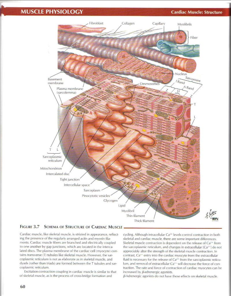
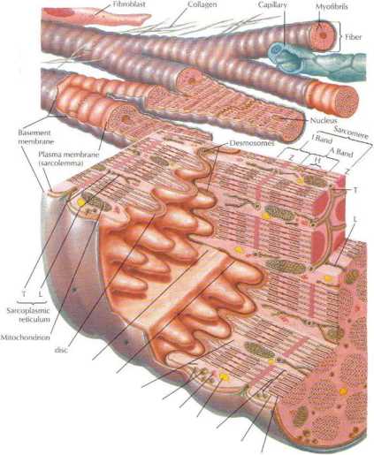
Cardiac musde, like skeletal musde, is slriated in appearance, reflect mg the presence o i Ihe regularly arrangcd actin and myosin fila-mcnts. Cardiac musde fibers are branched and olectrically coupled to one another by gap junctions, which are locatcd in the intercalated discs. Tłu* plasma membranę of the cardiac celi Imyocyte) con-tains transverse (T) tubules like skeletal musde. However, the sar-coplasmic reticulum is not as elaborat? as in skeletal musde, and dyads (rather than triads) are formed between the T tubules and sar-coplasmic reticulum.
bccitation-contraction coupling in cardiac musde is similar to that oł skeletal musde, as is the process of cross-bridge formation and cycling. Although intracdlular Ca;* levels control contraction in both skeletal and cardiac musde, there are sonie important differences. Skeletal musde contraction Ls dependent on the release of Ca '* from the sarcoplasinit reticulum, and changes in extraccllular |Ca-' | do not apprecinbly alter the strength of the skeletal musde contraction. In contrast. Ca entry into the cardiac myocyte from the extiaccllular fluid is necessary for the release of Ca-'* from the sarcoplasmic reticulum, and removal of extraccllular Ca- * will decrease the force of contraction. The ratę and force of contraction of cardiac myocytes can be im reased by 0-adrenergic agonists.
/3-Adrenergic agonists do not have these effec ts on skeletal musde.
Wyszukiwarka
Podobne podstrony:
netter49 I I Cardiac Mustlt*: StructureMUSCLE PHYSIOLOGYFlCURF 3.7 SCHEMA OF STRUCFURt OF CARDIAC MU
38936 netter50 Smooth Musde: StructureMUSCLE PHYSIOLOGY J. Pcrkins Ms. MFA°^
netter109 Renal Clearance: IIRENAL PHYSIOLOGY PRINCIPIE OF TUBULAR SECRETION LIMITATION (Tm) USINC P
netter136 Gaslric Secretion: IIGASTROINTESTINAL PHYSIOLOGY Secrelions of gastric acid (H* ) by parie
16523 netter73 Rrsponse to txeras»CARDIOVASCULAR PHYSIOLOGY Anticipation of exercise stimulates
netter73 Rrsponse to txeras»CARDIOVASCULAR PHYSIOLOGY Anticipation of exercise stimulates
41783 netter168 Thyroid Gland: StructureENDOCRINE PHYSIOLOGY Hyoid bonę Lymph node Phrenic nerve Sup
16523 netter73 Rrsponse to txeras»CARDIOVASCULAR PHYSIOLOGY Anticipation of exercise stimulates
netter52 Fxcitation-Conlraclion CouplingMUSCLE PHYSIOLOGY HART 3.1 COMPARISION OF MUSCLE STRUCTURE A
11871 netter148 Pancreas Structuri-GASTROINTESTINAL PHYSIOLOGY V / Root of Superio
netter135 Gastric Secretion: IGASTROINTESTINAL PHYSIOLOGY Neurocrine Regulalion of Acid
netter152 Intrahepatii Biliary SystemGASTROINTESTINAL PHYSIOLOGY Noto. The figurę shows bile canalic
więcej podobnych podstron