netter32

NEUROPHYSIOLOCY
Sensory Pathways: III
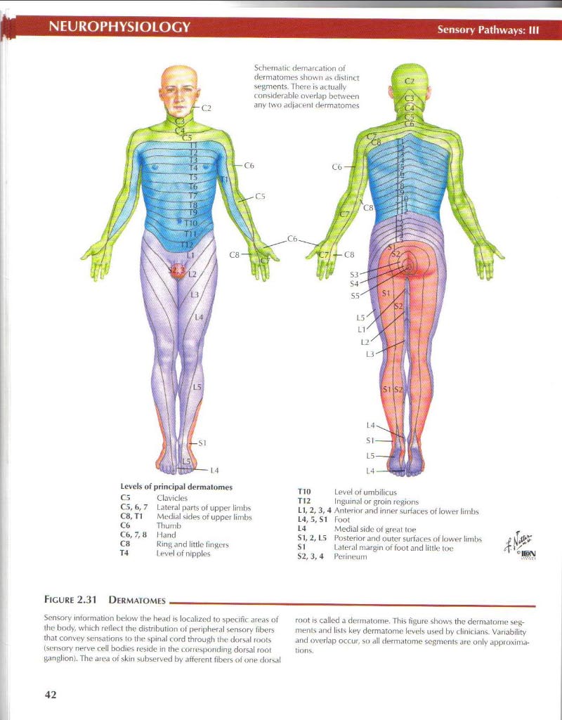
Schematic demarcation of dermatomes shown as distinct segments. Thcre is actually conśiderable overlap between any two adjacenl dermatomes
|
T10 |
Level of umbilicus |
|
TI2 |
Inguinal or groin region* |
|
LI, 2, 3,4 |
Anterior and inner surfaces of lowei limbs |
|
L4, 3, SI |
Coot |
|
L4 |
Medial side of greal toe |
|
SI, 2,13 |
Posterior and outer surfaces of lower limbs |
|
SI |
Lateral margin of foot and little toe |
|
$2, 3, 4 |
Pciinetim |
Lcvcls of principal dermatomes
C3 Clavides
CS, 6, 7 Latoral partS oł upper liml is
C8, Tl Mediał sides of uppoi limbs
C6 Tliurnb
C6,7,8 Hand
C8 Ring and little fingers
T4 Level of nipples
Figurę 2.31 Dermatomes
Sensory intormation bel<nv the head is localized to specific areas of the body, which refled the distribution of peripheral sensory fibers that convey sensations to the spinał cord through the dorsal roots (sensory ner\'e celi bodies residc in the corres|>onding dorsal root ganglion}. The aiea of skin subserved by afferent fibers of one dorsal
root is called a dennatonte. This figurę shows the dermatome segment* and lists key dermatome levels used by clinicians. Variability and overlap occur. su all dermatome segments are only approxima-
42
Wyszukiwarka
Podobne podstrony:
netter30 NEUROPHYSIOLOGY Sensory Pathways: I Spinothałamic iract lower part of medulla
netter24 NEUROPHYSIOLOGYCutaneous Sensory Receplors Melanoryte Arrectot muscle ofliair, Sebaceous gl
netter22 NEUROPHYSIOLOGYCerebellum: Afferent Pathways Cortical input uperior cerebellar peduncłe Mid
netter40 NEUROPHYSIOLOGYGustatory (Taste) System: Pathways Sensory cortex łjust below face area) Lat
netter2 NEUROPHYSIOLOGY Central sulcus (Rotando)Organizalion of Ihe Brain: Cerebrum Postcentral gyru
netter20 NEUROPHYSIOLOGYThe Cerebral Cortex Sm!,} Spn4,,,v Sensory Promotor; orientation, eye and
netter36 NEUROPHYSIOLOGY Audilory System: Pathways temporal lobe cortex Medial geniculate body High
netter49 I I Cardiac Mustlt*: StructureMUSCLE PHYSIOLOGYFlCURF 3.7 SCHEMA OF STRUCFURt OF CARDIAC MU
więcej podobnych podstron