CHAPTER 13
Fibrohistiocytic Tumours
The concept of fibrohistiocytic tumours in all locations is cur-
rently being challenged. Stout was the first pathologist to sug-
gest that some of the very pleomorphic sarcomas, especially
those containing cells with foamy cytoplasm, represent neo-
plasms arising from histiocytes or at least have the potential of
histiocytic differentiation.
Benign fibrous histiocytomas have the histological features of
the common metaphyseal fibrous defect but occur in adults and
in unusual locations. Some giant cell tumours may have areas
simulating benign fibrous histiocytoma.
The diagnosis of malignant fibrous histiocytoma is made when
a highly malignant spindle cell tumour is arranged in a storiform
pattern or if the tumour cells have abundant cytoplasm suggest-
ing a histiocytic origin. This tumour is rare and it does not seem
reasonable to subclassify it any further.
bb5_21.qxd 13.9.2006 13:19 Page 291
Benign fibrous histiocytoma of bone
M. Kyriakos
Definition
A benign lesion of bone composed of
spindle-shaped fibroblasts, arranged in
a storiform pattern, with a variable
admixture of small, multinucleated osteo-
clast-like giant cells. Foamy cells (xan-
thoma), chronic inflammatory cells, stro-
mal haemorrhages and haemosiderin
pigment are also commonly present.
ICD-O code
8830/0
Synonyms
Fibroxanthoma, fibrous xanthoma, xan-
thofibroma, xanthogranuloma.
Epidemiology
Benign fibrous histiocytoma is rare, with
less than 100 reported cases. Patients
have ranged in age from 6 to 74 years at
diagnosis {110,180}, 60% being older
than age 20 years, with a slight female
prevalence.
Sites of involvement
Approximately 40% of benign fibrous
hisiocytomas occur in the long bones,
with femur and tibia most frequently
involved. As many as 25% of cases
involve the pelvic bones, in particular the
ilium. However, this tumour may involve
virtually any bone. In the long bones,
benign fibrous histiocytoma is centred in
the epiphysis or diaphysis.
Clinical features / Imaging
Although some patients (~15%) are
asymptomatic {180,583,1639}, in most
(65%) the lesion causes pain which may
be present for days {877} up to several
years {364,1308,1468,1781}. Occasional
patients present because of pathological
fracture {365,506,939}.
Roentgenographically, benign fibrous
histiocytoma (BFH) appears as a well
defined, benign appearing, radiolucent
medullary defect without matrix forma-
tion; internal trabeculation or pseu-
doseptations, may be evident {365}.
Approximately two-thirds of the lesions
have sclerotic margins, at times best
seen with computed tomography {100,
180,723,877,959}. The lesion may thin
and expand the cortex, however, a
periosteal reaction is lacking in the
absence of fracture {180,364,365,506,
723,765,959,1639,1781,2155}. Soft tis-
sue extension is not present. Rarely, the
lesion is less well defined, with indistinct
borders, having a pattern suggestive of
malignancy {939,2155}. At the end of a
long bone it may be central or eccentric
and be indistinguishable from a giant cell
tumour (GCT) {1355,1875}. The diagno-
sis of benign fibrous histiocytoma should
be considered in cases in which the clin-
ical radiographic setting is also compat-
ible with diagnoses of: metaphyseal
fibrous cortical defect, non-ossifying
fibroma or giant cell tumour of bone.
Macroscopy
Most lesions are 3.0 cm in diameter or
smaller {100,110,364,583,1308,1484,
1639}, although cases up to 7.0 cm have
been reported {723,2155}. The tumour
tissue is usually firm, grey-white, and fre-
quently contains irregular yellow to red-
dish brown foci.
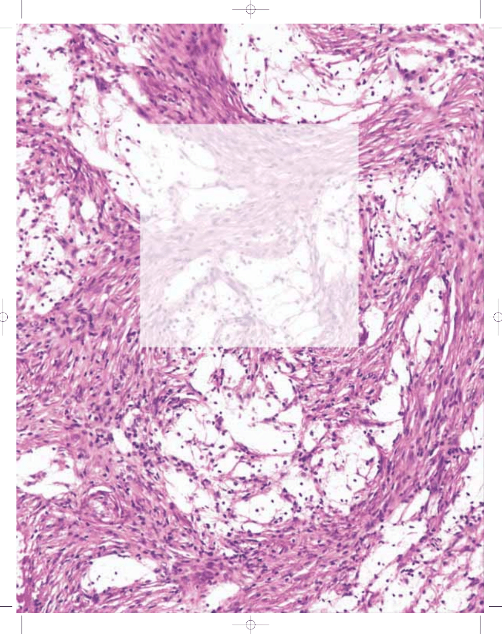
Histopathology
The basic pattern of BFH consists of a
stroma of spindle-shaped fibroblasts,
arranged, at least focally, in a whorled,
storiform pattern, among which a variable
number of small, multinucleated, osteo-
clast-type giant cells are scattered. The
spindle cell nuclei may be dark, thin and
elongate, or round to oval and vesicular
with a micronucleolus. In rare cases the
stromal cells exhibit mild nuclear atypia
justifying the term "atypical fibrous histio-
cytoma". There is no consensus as to
how extensive or severe the degree of
atypia should be to consider a low grade
malignancy {1587,1840}. Foam (xan-
thoma) cells, with small, dark nuclei are
frequently, but not always, found inter-
spersed among the stromal cells either
individually, or in small clusters or sheet-
like masses. Scattered inflammatory
cells, mainly lymphocytes, are present,
occasionally situated in small, loose clus-
ters. Mitotic figures may be evident but
atypical forms are not present. Small stro-
mal haemorrhages are common as are
deposits of haemosiderin either as fine
cytoplasmic granules within the stromal
cells or small macrophages, or as large,
extracellular clumps. Zones of ischaemic
necrosis may occur secondary to frac-
ture. The lesion is sharply demarcated
from the adjacent uninvolved bone, scal-
loping it without permeation.
BFH must be distinguished from fibrohis-
tiocytic degenerative or repair tissue that
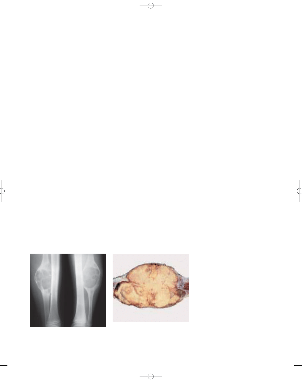
Fig. 13.01
Benign fibrous histiocytoma. A well
defined, lytic lesion involves the mid- diaphysis of the
fibula in a 10-year- old boy. The lesion expands and
scallops the bone.
Fig. 13.02 Benign fibrous histiocytoma. Resection
specimen of the same lesion (Fig. 13.01) shows a
pale, cream-yellow cut surface with focal rust brown
areas along its periphery. Marked cortical thinning
and endosteal scalloping are evident.
292
Fibrohistiocytic tumours
bb5_21.qxd 13.9.2006 13:19 Page 292
293
Benign fibrous histiocytoma
occurs in other bone lesions, most
notably and frequently in GCT of long
bones {180,365,1468}. In an adult with a
lytic, destructive lesion at the end of a
long bone, careful search for residual
foci of GCT must be made before making
a diagnosis of BFH {180}.
BFH is histologically indistinguishable
from non-ossifying fibroma (NOF), being
separated from the latter only on clinical
and radiological grounds {1875,2286},
i.e., its location in non-long bones, or lack
of metaphyseal involvement if in a long
bone; its usual occurrence in older
patients; the presence of pain even in the
absence of pathological fracture; and a
radiological pattern that may lack the
well defined, sclerotic, bubble-type mar-
gins typical of NOF.
Immunophenotype
There are no specific marker proteins.
Prognostic factors
The prognosis is excellent, surgical curet-
tage / resection usually being curative.
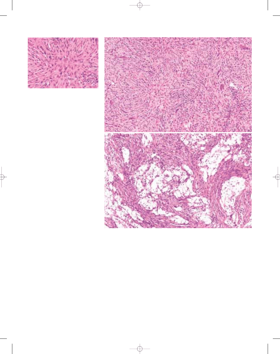
Fig. 13.04 Benign fibrous histiocytoma. A Storiform arrangement of spindle cells admixed with small, mult-
inucleated osteoclast-like giant cells. Loose clusters of lymphoid cells are also present. B Clusters of foam
cells with pale cytoplasm and small, dark nuclei are seen interspersed among whorled spindle cells. Such
foam cells may be absent or so extensive as to dominate the lesion.
A
B
Fig. 13.03 Benign fibrous histiocytoma. Centre of
storiform focus shows spindle cells, whose nuclei
are elongated or oval with a fine chromatin pattern.
Note intracytoplasmic haemosiderin.
bb5_21.qxd 13.9.2006 13:19 Page 293
Definition
Malignant fibrous histiocytoma (MFH)
defines a malignant neoplasm com-
posed of fibroblasts and pleomorphic
cells with a prominent storiform pattern.
ICD-O code
8830/3
Synonyms
MFH was initially described in the bone
in 1972 by Feldman and Norman {647},
although similar tumours were earlier
described in the soft tissue by Stout and
co-workers in 1963 {1632} and 1964
{1589}. MFH has also been termed
malignant histiocytoma, xanthosarcoma,
malignant fibrous xanthoma and fibrox-
anthosarcoma.
Epidemiology
Males are more frequently affected than
females. MFH of bone is a relatively rare
tumour which represents less than 2% of
all primary malignant bone lesions. The
age of patients at the time of diagnosis is
broad and usually varies from the 2nd to
8th decades, with a higher incidence in
adults over 40 years of age. Approxi-
mately 10-15% of cases occur in patients
less than 20 years of age. MFH can arise
as a primary bone tumour or may devel-
op secondary to pre-existing bone con-
ditions such as Paget disease or bone
infarct, or at the site of bone which was
irradiated for the treatment of osseous or
extraosseous tumours {503,997,1367}.
Secondary MFH accounts for approxi-
mately 28% of all MFH {305,538,990,
1571,1648}.
Sites of involvement
Primary MFH predilects the long bones
of the lower extremities, particularly the
femur (30-45%), followed by tibia and
humerus. The knee is a common loca-
tion, with concurrent involvement of the
distal femur and proximal tibia {193}.
Among the trunk bones, the pelvis is
most frequently involved. Almost all MFH
are solitary lesions, but rare multifocal
tumours have been reported {1367}.
Clinical features / Imaging
Clinically, most patients complain of pain
and, less frequently, swelling that varies
from 1 week to 3 years (average 7-9
months). Rarely, a pathological fracture
may be the initial presenting symptom.
MFH in the long bones predilects the
metaphyseal region with epiphyseal
extension in some cases. Diaphyseal
location is infrequent. The tumours are
essentially osteolytic lesions, but sclerot-
ic areas may be present. The margins
are usually ill defined and a moth-eaten
or permeative pattern of bone destruc-
tion can be observed. Some tumours
have well defined borders. The cortex is
commonly involved and destroyed by the
tumour with often soft tissue extension.
Periosteal reaction is not a frequent find-
ing {1272,1522}.
G.C. Steiner
G. Jundt
J.A. Martignetti
Malignant fibrous histiocytoma of bone
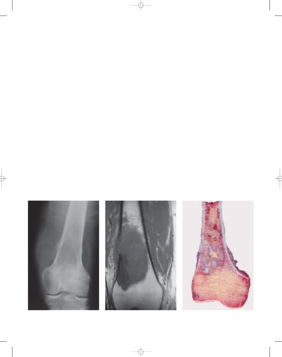
Fig. 13.05 Malignant fibrous histiocytoma of the dis-
tal femur, presenting as large, lytic lesion with ill
defined margins and focal periosteal reaction.
Fig. 13.07 Malignant fibrous histiocytoma. Greyish-
white, circumscribed tumour with yellowish necrotic
area, focally destroying the cortex (left).
Fig. 13.06 Malignant fibrous histiocytoma. Sagittal
T1-weighted MR image showing a large medullary
lesion with focal soft tissue extension.
294
Fibrohistiocytic tumours
bb5_21.qxd 13.9.2006 13:19 Page 294
MFH occurring in metaphyseal location
can be very aggressive {1522}. The radi-
ographic features of primary MFH are
nonspecific and in older patients can
mimic lymphoma, myeloma or osteolytic
metastases. In younger patients, osteo-
sarcoma and Ewing sarcoma are includ-
ed in the differential diagnosis. MRI usu-
ally helps to demonstrate the intra- and
extraosseous extent of the tumour, but
the imaging features are not specific to
differentiate MFH from other tumours.
However, the presence of an eccentric
lytic and diaphyseal lesion with cortical
destruction and soft tissue extension, or
a metaphyseal lytic lesion that extends to
the epiphysis but not to the subchondral
bone, should raise suspicion of MFH
{1272,1522}.
In secondary MFH arising in Paget dis-
ease and bone infarct, the radiographs
indicate the presence of an underlying
bone process in most cases.
Macroscopy
The gross appearance of this tumour is
not characteristic. It varies in colour from
tan to greyish-white, soft to firm in con-
sistency. Areas of yellowish discoloura-
tion, necrosis and haemorrhage are fre-
quently seen. The margins are irregular
and often cortical destruction and soft
tissue infiltration are present.
Histopathology
Microscopically, MFH consists mainly of a
mixed population of spindle cells, histio-
cytoid and pleomorphic cells. Varying
amounts of multinucleated giant cells of
the osteoclast type are seen as well as
foamy cells and chronic inflammatory
cells. The nuclei of the tumour cells may
be quite atypical, particularly in the malig-
nant giant cells. Typical and atypical
mitoses are present. There is a variability
of cellular patterns within these tumours.
A characteristic storiform pattern is com-
monly seen in the fibroblastic areas, in
which bundles of spindle cells are
arranged in a storiform or pinwheel pat-
tern.
Different histological subtypes have
been described in MFH of soft tissue and
bone: storiform–pleomorphic, histiocytic,
myxoid, giant cell, and inflammatory. The
storiform-pleomorphic is the most com-
mon histological subtype in bone. The
myxoid pattern is rare. Most MFH are
high grade tumours, but a few low grade
lesions have been reported {305,538,
990,1571,1648}.
Immunophenotype
Immunomarkers are of limited value in
the diagnosis of MFH of bone. They are
useful to rule out other malignant neo-
plasms that may resemble MFH such as
leiomyosarcomas, metastatic carcino-
mas and melanomas {675}. Vimentin is
strongly positive in tumour cells. Smooth
muscle actin, indicative of myofibroblas-
tic differentiation, may be focally positive.
The presence of cytokeratin immunore-
activity in MFH is nonspecific. CD68
reactivity may be present in the tumour
cells but is not a specific marker for histi-
ocytes and therefore of no diagnostic
significance {69,2051}.
Differential diagnosis
MFH may have foci of osteoid or primitive
bone formation at the periphery of the
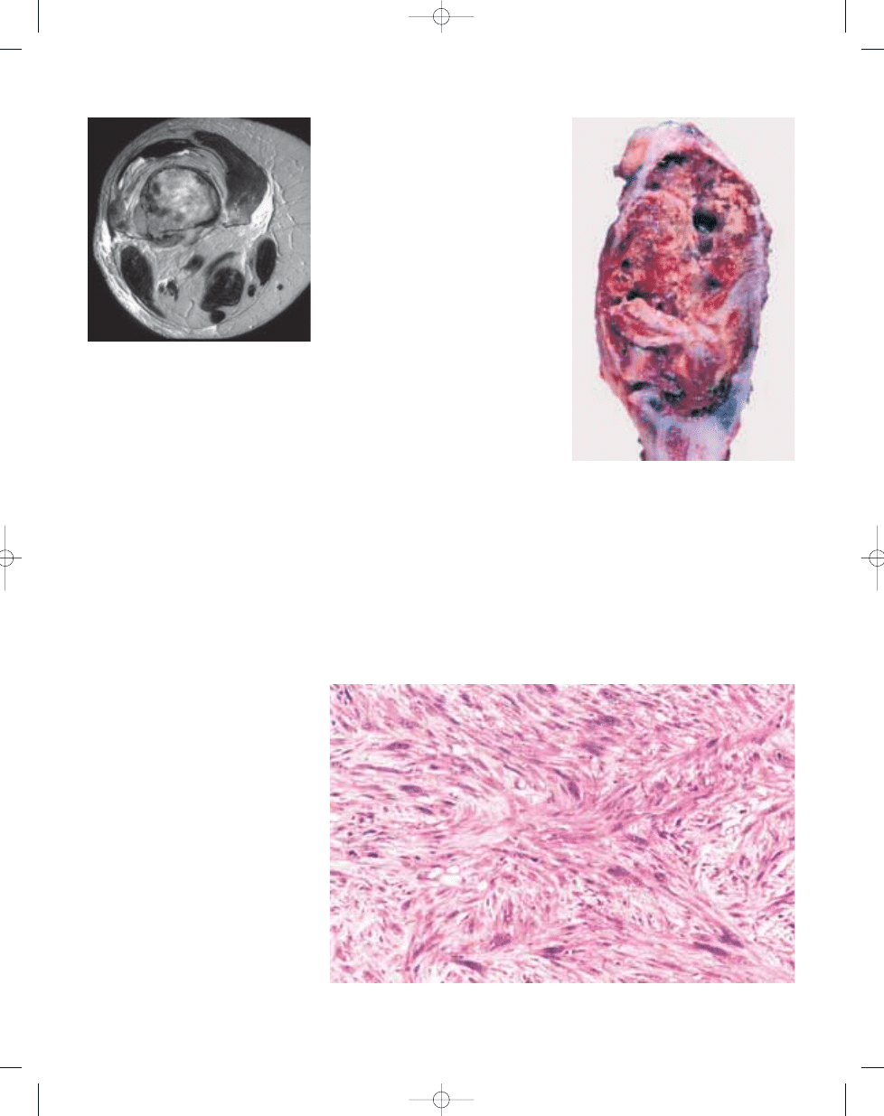
Fig. 13.08 Malignant fibrous histiocytoma of bone.
Axial T2-weighted MR image demonstrating an inho-
mogeneous lesion with cortical destruction and soft
tissue extension.
Fig. 13.09 Malignant fibrous histiocytoma of bone.
Gross photograph of resected specimen shows
yellowish brown, partly cystic tumour tissue.
Fig. 13.10 Malignant fibrous histiocytoma of bone. High power view of storiform pattern containing large atyp-
ical tumour cells.
295
Malignant fibrous histiocytoma of bone
bb5_21.qxd 13.9.2006 13:19 Page 295
tumours in the areas of soft tissue
involvement. This usually represents
periosteal reactive bone and not
osteosarcoma {990}. Also, foci of irregu-
lar and coarse collagen fibres within the
tumour, which are present in some
cases, can be misinterpreted as neo-
plastic osteoid. In these instances,
detailed histological evaluation will help
to rule out osteosarcoma, particularly the
MFH-like variant of osteosarcoma, which
shows unequivocal evidence of miner-
alised osteoid and bone {1528}. Fibro-
sarcoma often overlaps histologically,
clinically and radiologically with MFH. In
contrast to MFH, which contains pleo-
morphic cells and a storiform pattern,
fibrosarcoma consists of bundles of spin-
dle cells with a herringbone pattern
{538}. However, histological distinction
between one tumour and the other can
be arbitrary.
Metastatic carcinoma with a spindle cell
component and melanoma should be
distinguished from MFH by the use of
appropriate immunomarkers.
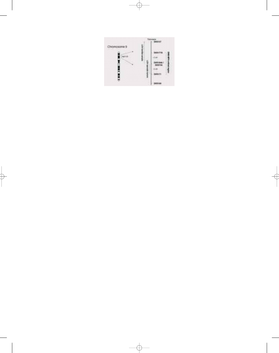
Genetics
In 5/7 sporadic MFHs, LOH was found for
markers within the 9p21-22 region, and
the minimally defined region of LOH
could be narrowed down even further
{1338}. Loss of the 9p21-22 region in
bone MFH has previously also been
noted using comparative genomic
hybridization {1957}. The LOH results are
in accord with mutation studies which
suggest that
CDKN2A is not the critical
gene {2096}.
Genetic susceptibility
Diaphyseal medullary stenosis with
malignant fibrous histiocytoma (DMS-
MFH) is a rare, autosomal dominant
bone dysplasia / cancer syndrome of
unknown aetiology {85,886}. The skele-
tal phenotype is characterized by cor-
tical growth abnormalities, including
diffuse diaphyseal medullary stenois
with overlying endosteal cortical thick-
ening, metaphyseal striations, and
scattered infarctions and sclerotic
areas throughout the long bones.
Notably, malignant transformation has
occurred in 13 of 40 patients in the five
reported DMS-MFH families {85,886,
1337,1521,1583}. Malignant fibrous
histiocytoma has been the consistent
diagnosis in all the tumours studied.
Using a positional cloning strategy,
the DMS-MFH gene was localized in
three unrelated families to chromo-
some bands 9p21-22 with a maximal
two-point LOD score of 5.49 {1337}.
Haplotype analysis narrowed the
boundaries of the gene locus to an ~3
cM region. These results were inde-
pendently corroborated in another
DMS-MFH family {1521}.
Prognostic factors
MFH is a highly malignant neoplasm
with frequent tendency to metastasis,
particularly to the lungs (45-50%). The
recommended treatment is wide
surgical excision. In those patients
with histologically high grade and res-
ectable lesions, preoperative chemo
therapy appears to be the standard of
care. The chemotherapy regimen is
similar to that used in osteosarcoma.
The degree of tumour necrosis in the
resected specimen after chemothera-
py is apparently an important prognos-
tic factor, as in the management of
osteosarcoma {193,1648}. In patients
with localized disease, the 5-year
disease-free survival has been
reported to be over 50% {246,1648}.
Radiotherapy is used particularly in
patients with inadequate surgical treat-
ment.
Favourable prognostic factors are:
younger age at manifestation (under
40 years); adequate surgical margins
and histological low grade. Some
authors report that a prominent chronic
inflammatory infiltrate is associated
with a better prognosis, as opposed to
the presence of prominent desmopla-
sia with hyalinization {2325}. The histo-
logical subtype of the lesion does not
affect the prognosis.
296
Fibrohistiocytic tumours
Fig. 13.11 Malignant fibrous histiocytoma of bone.
Minimal region of deletion on chromosome arm 9p.
bb5_21.qxd 13.9.2006 13:19 Page 296
Wyszukiwarka
Podobne podstrony:
chap13
bb5 chap3
bb5 chap8
bb5 chap1
BB5 BOX
bb5 chap16
bb5 chap15
bb5 contents
bb5 chap12
bb5 chap4
bb5 references
bb5 chap6
bb5 chap17
bb5 chap20
bb5 chap5
Lista wszystkich dostępnych polskich Product Code dla telefonów platformy BB5
bb5 chap21
bb5 source
więcej podobnych podstron