CHAPTER 20
Tumours of Undefined Neoplastic Nature
There are many conditions of bone that are generally consi-
dered non-neoplastic, but often constitute important lesions to
be considered in the differential diagnosis of bone tumours.
Some feature the appearance and cytogenetic characteristics of
neoplasms, although the clinical behaviour rather supports a
non-neoplastic nature. Only the most important conditions are
included in this chapter.
bb5_28.qxd 13.9.2006 14:16 Page 337
Aneurysmal bone cyst
A.E. Rosenberg
G.P. Nielsen
J.A. Fletcher
Definition
Aneurysmal bone cyst (ABC) is a benign
cystic lesion of bone composed of blood
filled spaces separated by connective
tissue septa containing fibroblasts,
osteoclast-type giant cells and reactive
woven bone. ABC may arise de novo
(primary ABC), or secondarily compli-
cate other benign and malignant bone
tumours (secondary ABC) that have
undergone haemorrhagic cystic change.
Synonyms
Multilocular haematic cyst, giant cell
reparative granuloma.
Epidemiology
ABC affects all age groups, but is most
common during the first two decades of
life (median age approximately 13 years)
and has no sex predilection {1345,
2200}. The estimated annual incidence is
0.15 per million individuals {1239}.
Sites of involvement
ABC can affect any bone but usually aris-
es in the metaphysis of long bones espe-
cially the femur, tibia and humerus, and
the posterior elements of vertebral bodies.
Rare tumours whose morphology is iden-
tical to primary ABC of bone have also
been described in the soft tissues {53}.
Clinical features / Imaging
The most common signs and symptoms
are pain and swelling, which are rarely
secondary to fracture. In the vertebrae it
can compress nerves or the spinal cord
and cause neurological symptoms.
Radiographically, ABC presents as a lytic,
eccentric, expansile mass with well
defined margins. Most tumours contain a
thin shell of subperiosteal reactive bone.
Computed tomography and magnetic
resonance imaging studies show internal
septa and characteristic fluid-fluid levels
created by the different densities of the
cyst fluid caused by the settling of red
blood cells {1173,2200}. In secondary
ABC, CT and MRI may show evidence of
an underlying primary lesion.
Macroscopy
ABC is a well defined and multiloculated
mass of blood filled cystic spaces
separated by tan white gritty septa. More
solid areas can be seen which may rep-
resent either a solid portion of the ABC or
a component of a primary tumour that has
undergone secondary ABC-like changes.
Histopathology
ABC may arise de novo (primary ABC),
or secondarily complicate other benign
and malignant bone tumours (secondary
ABC) that have undergone haemorrhagic
cystic change {1281,1557,1699,1849,
1926}.
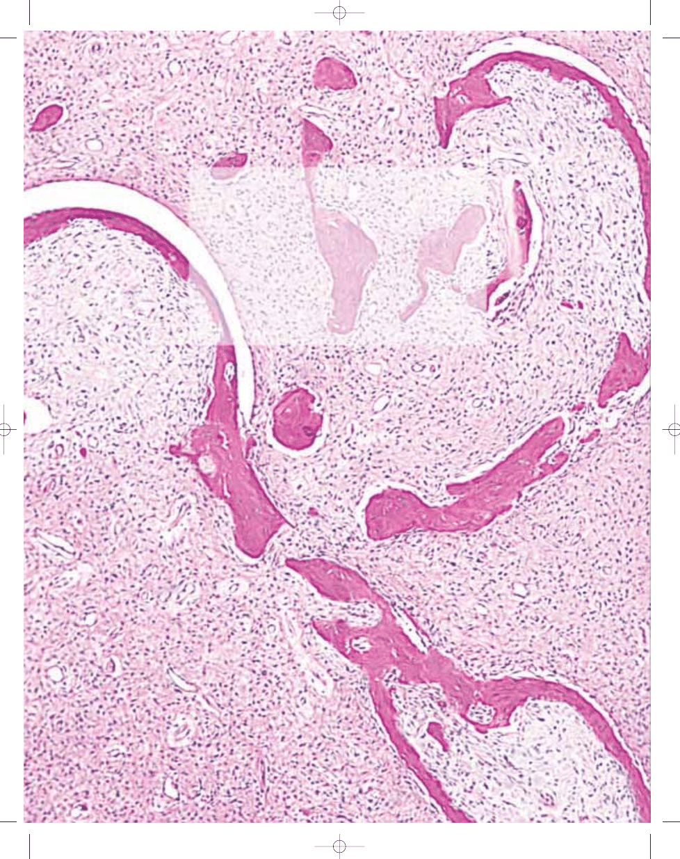
Primary ABC is well circumscribed and
composed of blood filled cystic spaces
separated by fibrous septa. The fibrous
septa are composed of a moderately
dense cellular proliferation of bland
fibroblasts, with scattered multinucleated
osteoclast-type giant cells and reactive
woven bone rimmed by osteoblasts. The
woven bone frequently follows the con-
tours of the fibrous septa. In approxi-
mately 1/3 of cases the bone is
basophilic and has been termed "blue
bone", however, its presence is not diag-
nostic as it can be seen in other entities.
Mitoses are commonly present and can
be numerous, however, atypical forms
are absent. Necrosis is rare unless there
has been a pathological fracture. The
solid variant of ABC has the same
components as the septa and is very
similar, if not identical, to giant cell repar-
ative granuloma. Primary ABC accounts
for approximately 70% of all cases
{177,1859}.
The majority of secondary ABC develop
in association with benign neoplasms,
most commonly giant cell tumour of
bone, osteoblastoma, chondroblastoma
and fibrous dysplasia {1173,1345, 2200}.
However, ABC-like changes may also
omplicate sarcomas, especially
osteosarcoma.
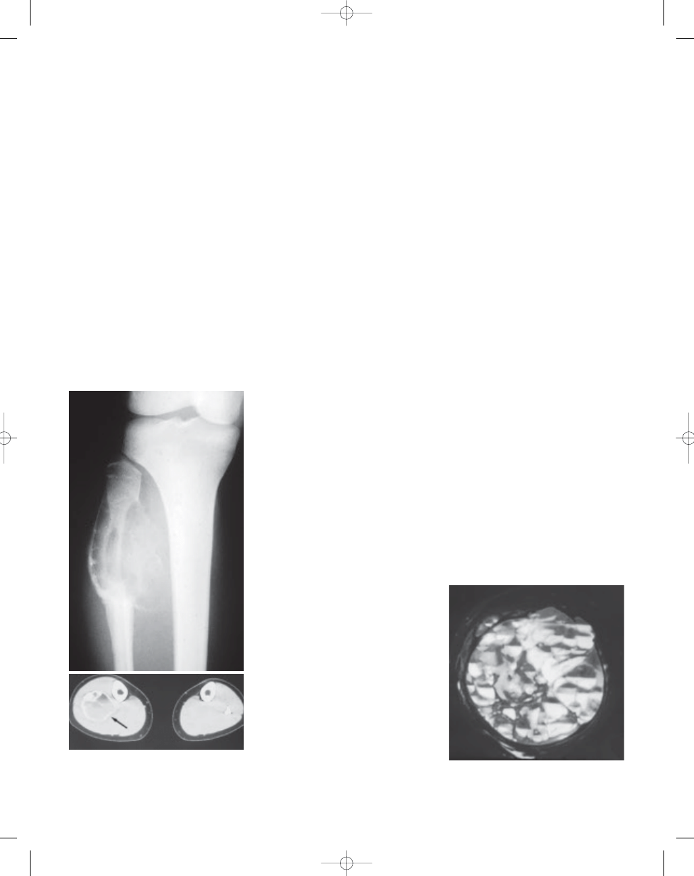
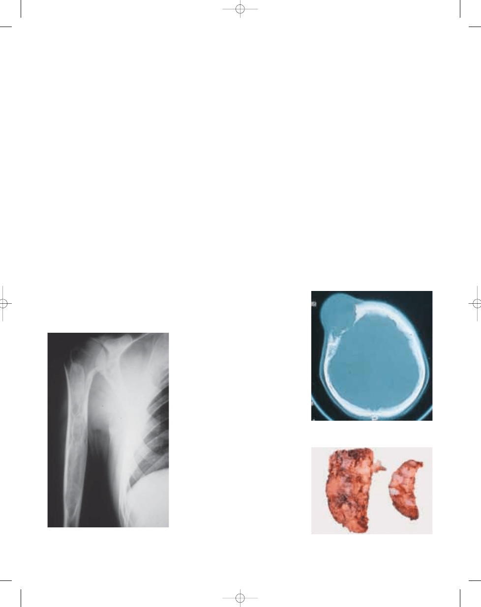
Fig. 20.01 Aneurysmal bone cyst. A Plain X-ray of
an eccentric lytic mass of the proximal fibula. Note
the peripheral shell of reactive bone.
B CT of the
same lesion (arrow).
Fig. 20.02
Aneurysmal bone cyst. MRI of large
destructive lesion of distal femur. Note numerous
fluid-fluid levels.
338
Tumours of undefined neoplastic nature
A
B
bb5_28.qxd 13.9.2006 14:16 Page 338
339
Aneurysmal bone cyst
Genetics
The most notable genetic feature is the
characteristic rearrangement of the
chromosome 17 short arm {1645}. The
chromosome 17 rearrangements are
often in the form of balanced transloca-
tions, in which material is exchanged
with the long arm of chromosome 16.
However, there are many variations on
this theme, and at least five different
chromosomes can serve as transloca-
tion partners with chromosome 17
{435,938,1645,1909,2281,2311}. The
cytogenetic analyses invariably reveal
normal metaphases along with those
bearing the translocations. Therefore,
the translocations can be assumed to
result from acquired aberrations, arising
in cytogenetically normal precursor
cells. The cytogenetic findings provide
compelling evidence that many aneursy-
mal bone cysts are clonal proliferations,
with activation of a 17p oncogene play-
ing a key role in their tumourigenesis.
The mechanisms of oncogene activation
appear to be heterogeneous, as shown
by the different types of 17p rearrange-
ment, and as evidenced by the absence
of 17p rearrangement in some cyto-
genetically abnormal aneursymal bone
cysts {135,435,938,1645,1909,2281,
2311}. It is also striking that these
varied, but related, cytogenetic abnor-
malities have been reported across the
entire clinicopathological spectrum of
aneursymal bone cysts. Chromosome
16 rearrangement was identified in a
solid variant aneursymal bone cyst,
whereas chromosome 17 rearrangement
was found in an extra-osseous case
{435}. Hence, it appears that there are
generalisable transforming mecha-
nisms, that are utilised irrespective of
histological subtype or site of origin.
Prognostic factors
ABC is a benign potentially locally
recurrent lesion. The recurrence rate fol-
lowing curettage is variable (20-70%).
Spon- taneous regression following
incomplete removal is very unusual.
Rare cases of apparent malignant trans-
formation of ABC have been reported
{1197}.
B
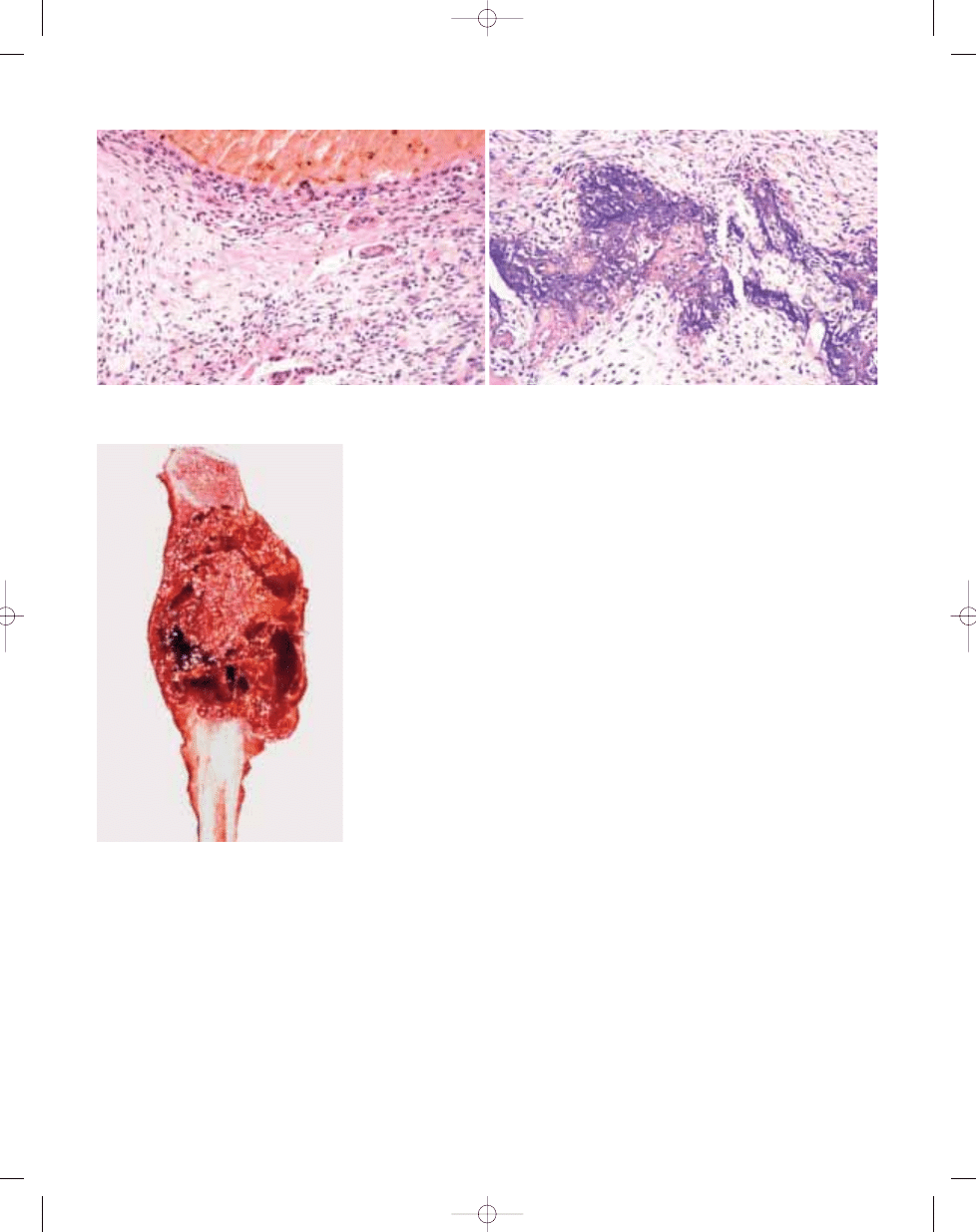
A
Fig. 20.03 Aneurysmal bone cyst. A Septa composed of reactive woven bone, fibroblasts, and scattered osteoclast-like giant cells. B So-called 'blue bone" in wall of
the lesion.
Fig. 20.04 Aneurysmal bone cyst of proximal fibula.
The well-defined haemorrhagic multicystic mass
has a prominent solid component in the centre.
bb5_28.qxd 13.9.2006 14:16 Page 339
Definition
An intramedullary, usually unilocular,
bone cyst (cavity) filled with serous or
sero-sanguineous fluid.
Synonyms
Solitary bone cyst; unicameral bone cyst;
juvenile bone cyst; essential bone cyst.
Epidemiology
Males predominate in a ratio of 3:1.
About 85% of patients are in the first two
decades of life.
Sites of involvement
There is a predilection for long bones,
proximal humerus, proximal femur and
proximal tibia accounting for up to 90%
of cases. Pelvis and calcaneus are also
common locations in older patients.
Clinical features / Imaging
Simple bone cyst can produce pain and
swelling but, more frequently, patients
present with a pathological fracture.
Roentgenograms show a metaphysio-
diaphyseal lucency, extending up to
epiphyseal plate, with little or no expan-
sion of bone; marginal sclerosis is
absent or very thin. The cortex is usual-
ly eroded and thin, but is intact unless
pathological fracture has occurred.
There can be partial or complete septa-
tions of the cavity. MRI usually confirms
its fluid content, that can be bloody in
fractured lesions {1328}.
Aetiology
Growth defect at the epiphyseal plate
has been postulated, or that a venous
blood flow obstruction causes the sim-
ple cyst {342}.
Macroscopy
The cystic cavity is usually filled with
serous or sero-sanguineous fluid. The
inner surface of the cyst shows ridges
separating depressed zones covered
by a layer of thin membrane. Partial
septae may be seen.
The occasionally curetted specimen
consists of fragments of a usually thin,
whitish membrane that may be
attached at one surface to bone
spicules.
Histopathology
The inner lining and septae of the cyst
consist of connective tissue that can,
occasionally, contain foci of reactive
new bone formation, haemosiderin pig-
ment and scattered giant cells.
Fibrinous deposits are often seen.
Some of these are mineralized, resem-
bling cementum. Occasionally, histo-
logical features of fracture callus may
be prominent. Rare "solidified" cases of
simple bone cyst have been described
in older subjects.
Genetics
A highly complex clonal structural
rearrangement involving chromosomes
4, 6, 8, 16, 21 and both chomosomes
12 has been described in a surgically
resected solitary bone cyst in an 11-
year-old boy {2195}.
Prognostic factors
Recurrence is reported at 10-20% of
cases, especially in children. Growth
arrest of the affected bone and avascular
necrosis of the head of the femur after
pathological fracture can occur {2022}.
Spontaneous healing after fracture has
been described {52}.
R.K. Kalil
E.S. Araujo
Simple bone cyst
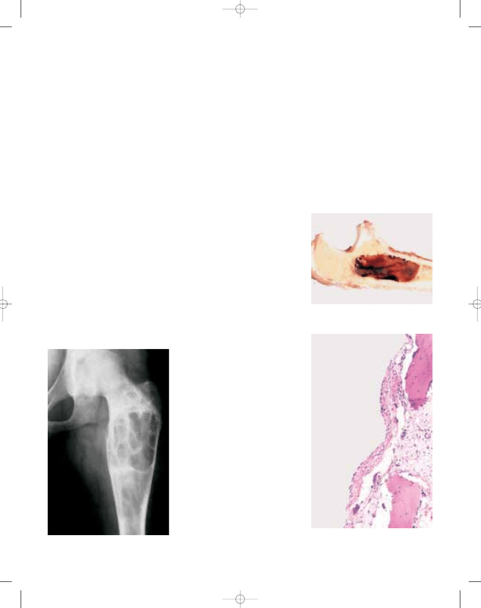
Fig. 20.05 Simple bone cyst of proximal femur. The
lesion does not expand the bone.
Fig. 20.06 Simple bone cyst of proximal ulna. A
unilocular cyst contains fibrin clot.
Fig. 20.07 Simple bone cyst. The lining is usually
inconspicuous and contains scattered spindle
cells and giant cells.
340
Tumours of undefined neoplastic nature
bb5_28.qxd 13.9.2006 14:16 Page 340
Definition
Fibrous dysplasia (FD) is a benign
medullary fibro-osseous lesion which
may involve one or more bones.
Synonyms
Fibrocartilagenous dysplasia, general-
ized fibrocystic disease of bone.
Epidemiology
Fibrous dysplasia occurs in children and
adults world-wide and affects all racial
groups with an equal sex distribution.
The monostotic form is six times more
common than polyostotic fibrous dys-
plasia.
Sites of involvement
The gnathic (jaw) bones are the most
common site of involvement in surgical
series (because they are often sympto-
matic) {1596}. In women, long bones are
more often involved, whereas ribs and
the skull are favoured sites in men
{2154}. In the monostotic form, about
35% of cases involve the head, a sec-
ond 1/3 occur in the femur and tibia, and
an additional 20% in the ribs. In the
polyostotic form, the femur, pelvis, and
tibia are involved in the majority of cases
{890}.
Clinical features / Imaging
Fibrous dysplasia may present in a
monostotic or polyostotic form, and in
the latter case, can be confined to one
extremity or one side of the body or be
diffuse. The polyostotic form often mani-
fests earlier in life than the monostotic
form {890}. The lesion is often asympto-
matic but pain and fractures may be part
of the clinical spectrum {333}. Fibrous
dysplasia may also be associated with
oncogenic osteomalacia {1660}.
The polyostotic form of fibrous dysplasia
is intimately associated with McCune-
Albright syndrome, in which there are
endocrine abnormalities and skin pig-
mentation. There is also a relationship
between fibrous dysplasia and intramus-
cular myxomas (Mazabraud syndrome)
{630}.
Rontgenographic studies often show a
non-aggressive geographic lesion with a
ground glass matrix. There is generally
no soft-tissue extension, and a
periosteal reaction is not seen unless
there is a complicating fracture. CT
scans and MRI further delineate these
features and better define the extent
{422,1035,2118}.
Aetiology
Activating mutations of the G proteins
have been identified in both the mono-
stotic and polyostotic forms and may be
aetiologically important.
Macroscopy
The bone is often expanded and the
lesional tissue has a tan grey colour with
a firm-to-gritty consistency. There may
be cysts, which may contain some yel-
low-tinged fluid {1948}. When cartilage
is present, it often stands out as sharply
circumscribed of blue-tinged translucent
material {2154}.
Histopathology
The lesion is generally well circum-
scribed and composed of fibrous and
osseous components; which are present
in varying proportions from lesion to
lesion and also within the same lesion.
The fibrous component is composed of
cytologically bland spindle cells with a
low mitotic rate. The osseous component
is comprised of irregular curvilinear tra-
beculae of woven (or rarely lamellar)
bone. Occasionally, the osseous compo-
nent may take the form of rounded
psammomatous or cementum-like bone.
Secondary changes such as foam cells,
G. Siegal
P. Dal Cin
E.S. Araujo
Fibrous dysplasia
Fig. 20.08 X-ray of a polyostotic form of fibrous dys-
plasia. There is a well defined lucency with scle-
rotic margins.
Fig. 20.09 CT of skull with fibrous dysplasia. In flat
bones the process is often expansile.
Fig. 20.10 Fibrous dysplasia with gross cartilaginous
components.
341
Fibrous dysplasia
bb5_28.qxd 14.9.2006 8:19 Page 341
multinucleate giant cells, a secondary
aneurysmal bone cyst or myxoid change
may occur.
Genetics
Activating mutations in the
GNAS1
gene, encoding the alfa subunit of stim-
ulatory G protein, has been demonstrat-
ed in monostotic as well as polyostotic
fibrous dysplasia {382} (see also chap-
ter on McCune-Albright syndrome).
Clonal chromosome aberrations have
been reported in eight of eleven in-
vestigated cases, suggesting that this
entity is neoplastic in nature {439}. The
only recurrent changes described so
far are structural rearrangements in-
volving 12p13 and trisomy 2 (three
cases each).
Prognostic factors
The prognosis of patients with FD is
good. Malignant transformation occurs,
but rarely.
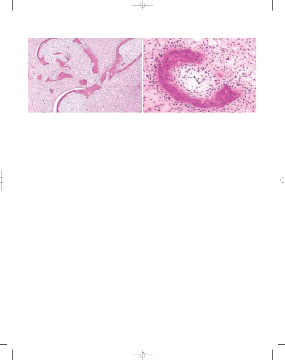
B
A
Fig. 20.11 Fibrous dysplasia. A Characteristic C shaped bony spicules with hypocellular spindle cell stroma. B High power appearance showing the typical appear-
ance of bone which seems to be dissected by spindle cell proliferation. Note that there is no osteoblastic rimming.
342
Tumours of undefined neoplastic nature
bb5_28.qxd 13.9.2006 14:16 Page 342
Definition
Osteofibrous dysplasia (OFD) is a self-
limited benign fibro-osseous lesion of
bone characteristically involving cortical
bone of the anterior mid-shaft of the tibia
during infancy and childhood.
Synonyms
Kempson-Campanacci lesion, cortical
fibrous dysplasia.
Epidemiology
The lesion is more commonly seen in
boys during the first two decades of life
with a precipitous drop-off thereafter,
OFD has been reported in neonates, but
is extremely rare after skeletal matura-
tion.
Sites of involvement
The proximal or middle-third of the tibia is
the most frequent site of involvement
{301}. Lesions can be bilateral with ipsi-
lateral or contralateral involvement of the
fibula. Other sites include the ulna and
radius {1055}. Multifocal or large conflu-
ent lesions oriented longitudinally along
the cortical axis are not unusual.
Clinical features / Imaging
The lesion is rare after the age of 15. The
most common presenting symptoms are
swelling or a painless deforming bowing
of the involved segment of the limb. OFD
is typically epicentered in the cortical
bone but may involve the medullary cavi-
ty by extension. Although slow growth is
characteristic of OFD, some lesions are
aggressive and may involve the entire
bone with significant bowing deformity.
Often well demarcated, it is associated
with a thinning, expanding or even miss-
ing cortex. The expanding cortex is often
sclerotically rimmed near the medullary
bone. Separate or confluent oval-shaped,
scalloped, saw-toothed or bubbly multi-
loculated lytic lesions are often noted.
Perilesional sclerosis may be consider-
able. The radiodensity of the interior of
the lytic foci are typically more radio-
dense than soft tissue. Periosteal reac-
tions and soft tissue extensions are
unusual. Bone scans are typically hot. CT
scans classically delineate a cortical epi-
centre to the lesion not breaking through
into the soft tissue and demarcated from
medullary bone by sclerosis. MRI find-
ings show high intensity lesions on T2
weighted images and mixed signals on
T1 and fat suppressed images.
Aetiology
The occurrence of so-called OFD-like
adamantinoma, to be distinguished from
classic epithelium-rich adamantinoma
but differentiated from OFD with difficulty,
raises the possibility of an association
between OFD and adamantinoma {112,
918,1188}. Some cases of OFD may
arise de novo and are not related to
adamantinoma.
Macroscopy
OFD is solid with a whitish, yellowish or
reddish colour and soft or gritty texture
blending into the surrounding host
bone. The periosteum often appears
intact but the cortex is thin or absent.
The medullary extension is usually
demarcated by a sclerotic rim.
Histopathology
The histopathologic findings in OFD
are irregular fragments of woven bone
often rimmed by lamellar layers of bone
laid down by well defined osteoblasts.
Osteoclasts may be present. The fibrous
component consists of bland spindle
cells with collagen production and a
matrix that varies from a myxoid com-
ponent to one that is moderately fibrous.
Mitoses are extremely rare. A zonal
architecture has been delineated with
thin spicules and woven bone or even
fibrous tissue predominating in the cen-
tre of the lesion with more abun-
dant anastomosing and lamellar bone
peripherally, the latter often blending
V.J. Vigorita
B. Ghelman
P.C.W. Hogendoorn
Osteofibrous dysplasia
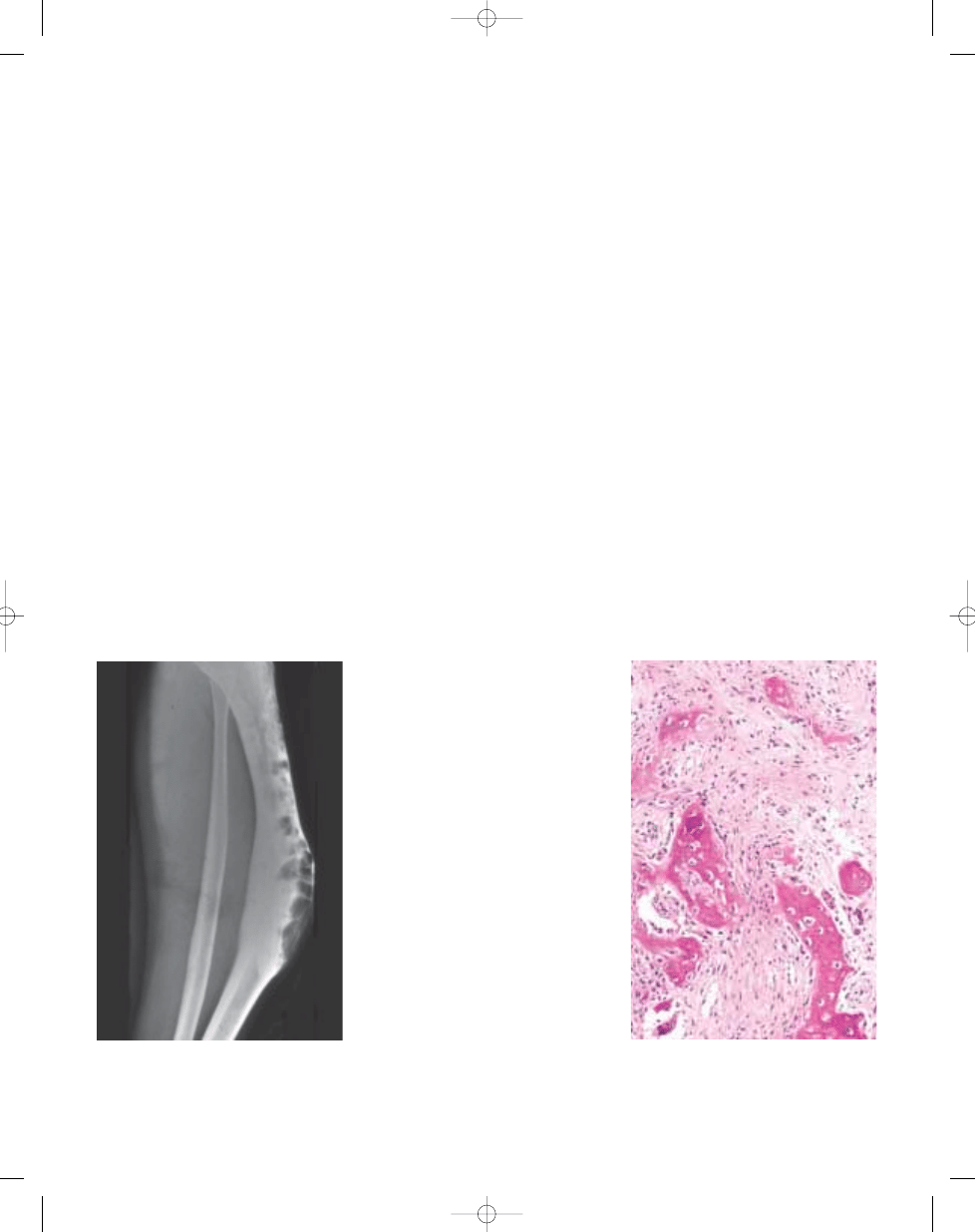
Fig. 20.12
Osteofibrous dysplasia. Expansile
lucent, longitudinally-oriented tibial lesion sur-
rounded by sclerosis and thinning of the anterior
cortex of the diaphysis of the tibia. Note the ante-
rior bowing of the tibia.
Fig. 20.13 Osteofibrous dysplasia. Low power
magnification of the lesion featuring hypocellular
spindle cell proliferation and spicules of bone. The
bony spicules display prominent osteoblastic
rimming.
343
Osteofibrous dysplasia
bb5_28.qxd 13.9.2006 14:16 Page 343

into the surrounding host bone {298}.
Secondary changes of hyalinization,
haemorrhage, xanthomatous change,
cyst formation and foci of giant cells are
rare. Cartilage or clusters of epithelial
cells are absent.
Immunophenotype
Osteofibrous dysplasia is positive for
vimentin and occasionally so for S100
and Leu7. Isolated cytokeratin positive
mast cells have been mentioned.
A tumour should be defined as OFD-
like adamantinoma when keratin-posi-
tive epithelial cells are found
{918,1534}.
Genetics
Numerical chromosomal abberations,
especially trisomy 7 and 8 have been
demonstrated {256, 267}, as well as
FOS and JUN proto-oncogene prod-
ucts.
Mutations of the alpha-subunit of sig-
nal transducing G-proteins with an
increase in cyclic AMP formation are
specifically absent {1845}.
Prognostic factors
The natural history of osteofibrous
dysplasia is that of gradual growth dur-
ing the first decade of life with stabi-
lization at about 15 years of age
followed by healing or spontaneous
resolution. The progression of OFD-like
adamantinoma (or ‘OFD with keratin
positive cells’) to classic adamantino-
ma has been shown in a few patients
{562,918, 1041,2016}. In many others,
there is at least strong suggestion of a
progression {381,2157, 2235}.
OFD-like adam- antinoma seldom
progresses to classic adamantinoma.
Table 20.01
Chromosomal abnormalities in osteofibrous dysplasia.
No./Author
Age/sex
Tumour (type)
Karyotype abnormality
1 Bridge {256}
11,M
OFD (R)
47,XY,+12 (FISH: also +8,+20)
2 Bridge {256}
19,M
OFD (R)
49,XY,+7,+8,+22
3 Bridge {257}
18,F
OFD (P/R)
52,XX,+5,+7,+7,+8,+21,+21
P, primary tumour; R, recurrence, FISH: fluorescence in situ hybridization. Cases 1/2:keratin-negative OFDs.
344
Tumours of undefined neoplastic nature
bb5_28.qxd 13.9.2006 14:16 Page 344
Definition
Langerhans cell histiocytosis is a neo-
plastic proliferation of Langerhans cells.
ICD-O codes
Langerhans cell histiocytosis,
NOS
9751/1
Langerhans cell histiocytosis,
unifocal
9752/1
Langerhans cell histiocytosis,
multifocal
9753/1
Langerhans cell histiocytosis,
disseminated
9754/3
Synonyms
Eosinophilic granuloma, Langerhans cell
granulomatosis, histiocytosis X. Clinical
variants have been referred to as Hand-
Schuller-Christian disease and Letterer-
Siwe disease.
Incidence
Langerhans cell histiocytosis (LCH) is a
relatively rare disorder, accounting for
less than 1% of all osseous lesions. LCH
involving bone has been reported in a
wide age distribution ranging from the
first months to the 8th decade of life with
80-85% of cases seen in patients under
the age of 30, and 60% under the age of
10. Males are affected twice as often as
females {1026,1253,1259,2253}.
Sites of involvement
Although any bone may be involved,
there is a predilection for LCH to involve
the bones of the skull, notably the calvar-
ium. Other frequently involved sites
include the femur, the bones of the
pelvis, and the mandible {1259,2253}. In
adults, the rib is the most frequent site of
involvement {2253}. Monostotic disease
is much more common than polyostotic.
Clinical features / Imaging
Pain and swelling of the affected area
occur most commonly. Other findings are
related to the bone involved. In cases of
temporal bone involvement, the present-
ing features can show significant clinical
overlap with otitis media or mastoiditis.
With mandibular involvement, loosening
or loss of teeth can be encountered.
Vertebral body disease may result in
compression fracture and possible neu-
rological impairment. In adults, the lesion
can present as an incidental finding on
imaging studies.
Early lesions may appear very aggres-
sive radiographically. Roentgenograms
generally show a purely lytic, well demar-
cated lesion, usually associated with
thick periosteal new bone formation.
Skull lesions are sometimes described
as representing a "hole in a hole" due to
uneven involvement of the two osseous
tables. In the vertebrae, the body is
involved producing collapse giving rise
to vertebra-plana.
Macroscopy
The involved tissue is soft and is red in
colour.
Histopathology
The diagnosis depends on the recogni-
tion of Langerhans cells, which are inter-
mediate size with indistinct cytoplasmic
B.R. De Young
K.K. Unni
Langerhans cell histiocytosis
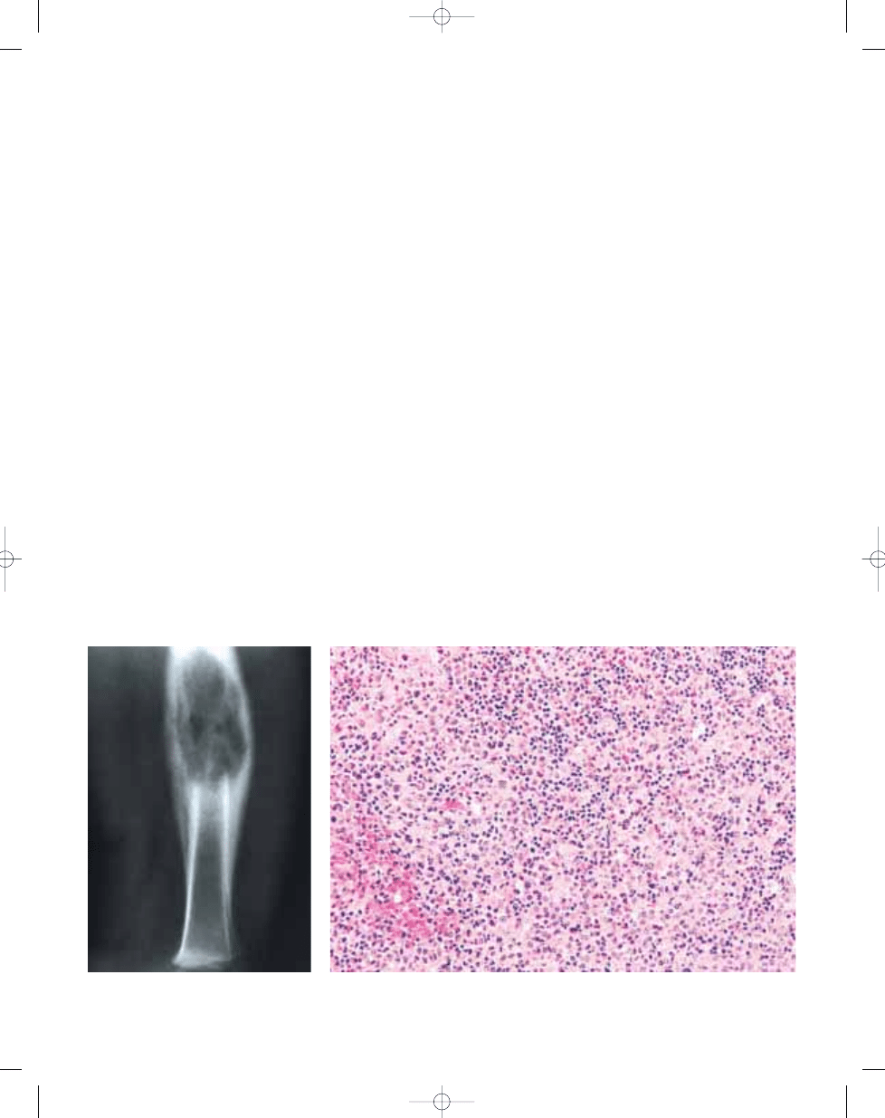
Fig. 20.14 Langerhans cell histiocytosis. Plain X-
ray showing lucency in the shaft of the femur asso-
ciated with thick periosteal new bone formation.
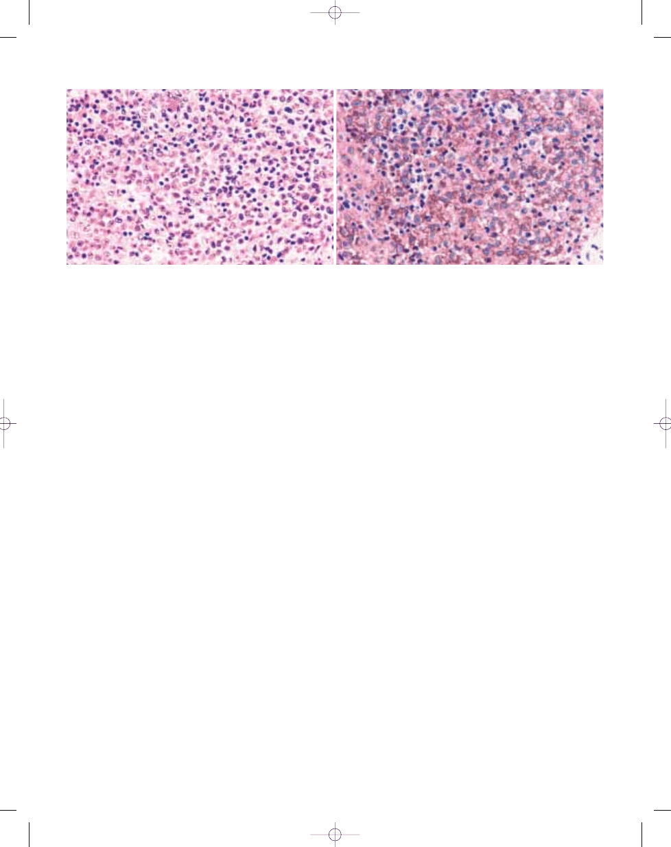
Fig. 20.15 Langerhans cell histiocytosis. Low power magnification shows loose aggregates of histiocytic
appearing cells in a mixed inflammatory background with prominent eosinophilia and evidence of recent
haemorrhage.
345
Langerhans cell histiocytosis
bb5_28.qxd 13.9.2006 14:16 Page 345
borders, eosinophilic to clear cytoplasm
with oval nuclei which frequently are
indented, irregular in outline, and typical-
ly possess nuclear grooves. Chromatin is
either diffusely dispersed or condensed
along the nuclear membrane. In osseous
LCH, the Langerhans cells are found in
nests or clusters. Diffuse sheet-like archi-
tecture is rare, and, if present, should
raise the suspicion of haematolymphoid
malignancy. The Langerhans cells are
frequently admixed with inflammatory
cells including large numbers of
eosinophils, as well as lymphocytes,
neutrophils and plasma cells. Necrosis is
common and does not portend an
aggressive clinical course. Multinucleat-
ed osteoclast-like giant cells and occas-
sionally lipid laden histiocytes may be
present. The cells of LCH can exhibit a
relatively brisk mitotic rate, with up to 5-6
mitoses per 10 high power fields.
Immunohistochemistry
Langerhans cells have a characteristic
immunophenotype which includes
expression of membrane based CD1a
{584} and S100 protein in both a nuclear
and cytoplasmic pattern {1530}. These
cells typically fail to express CD45.
Ultrastructure
Langerhans cells contain unique intracy-
toplasmic "tennis racket" shaped inclu-
sions known as Birbeck granules which
are thought to arise from the cell mem-
brane.
Genetics
Studies of X-chromosome inactivation
demonstrated that LCH is clonal {2275}.
Prognostic factors
The prognosis for patients with either
monostotic or limited polyostotic disease
is good. Death can result from LCH, but
this is a rare event and is associated only
with the disseminated forms of the dis-
ease and usually occurs in younger indi-
viduals less than three years at diagnosis
and with visceral involvement.
346
Tumours of undefined neoplastic nature
B
A
Fig. 20.16 Langerhans cell histiocytosis. A High power photomicrograph depicting Langerhans cells with ovoid to reniform nuclei with irregular notches and grooves.
B Langerhans cells show distinct membrane based immunoreactivity for CD1a.
bb5_28.qxd 13.9.2006 14:16 Page 346
Definition
Erdheim-Chester disease (ECD) is a rare
histiocytosis characterized by infiltration
of skeleton and viscera by lipid laden his-
tiocytes leading to fibrosis and osteo-
sclerosis.
Synonyms
Lipogranulomatosis, lipoidgranulomato-
sis, lipid (cholesterol) granulomatosis,
polyostotic sclerosing histiocytosis.
Epidemiology
The disease demonstrates a slight male
predominance with a peak incidence in
the 5th through the 7th decades (age
range is 7 to 84 years; mean age 53)
{2203}.
Sites of involvement
ECD predominantly affects the major
long bones of the extremities; but flat
bones can also be involved {306,664,
1138}. Extraskeletal manifestations occur
in more than 50% of cases, e.g.
kidney/retroperitoneum, heart/pericardi-
um, and lung.
Clinical features / Imaging
General symptoms consist of mild bone
pain, occasionally associated with soft
tissue swelling, fever, weight loss, and
weakness. Other manifestations include
exophthalmos, diabetes insipidus, kid-
ney failure, cardiac, pulmonary, or neuro-
logical symptoms, eyelid xanthomas,
and hepatosplenomegaly {627,1091,
1218,2045,2203}. Despite the impressive
lipid laden histiocytic infiltration, the
serum lipid profile is relatively normal.
The radiographic picture of ECD is
unique and includes bilateral, symmetric,
patchy or diffuse sclerosis of the
medullary cavity of major long bones,
with relative epiphyseal sparing {1785}.
One third of cases have a mixed oste-
olytic and sclerotic pattern {276,1463,
2045}. The sclerotic lesions show
increased uptake on bone scan. CT scan
serves to detect orbital, dural, and
retroperitoneal lesions. On MRI the lesion
is of low signal intensity on T1-weighted
sequences, enhances intensely after
gadolinium injection {2299}, and gives
mixed signal intensity on T2-weighted
sequences {118,2045}.
Macroscopy
On gross examination, the lesions
appear as sulphur-yellow and variably
firm.
Histopathology
The histology consists of a diffuse infiltra-
tion of marrow by foamy histiocytes asso-
ciated with dense fibrosis, lymphocytes,
plasma cells and Touton giant cells.
There is massive reactive sclerosis of
cortical and cancellous bone with irregu-
lar cement lines.
Immunophenotype
Immunohistochemistry confirms the
monocyte/macrophage lineage of the
lipid laden foamy histiocytes and giant
cells by their expression for lysozyme,
Mac387, CD68 (Kp-1), CD4 {2168},
alpha-1-antichymotrypsin, alpha-1-antit-
rypsin and S100 protein (variable)
{1615}. They are negative for CD1a.
Ultrastructure
Electron microscopy shows a predomi-
nance of histiocytes with indented nuclei,
abundant intracytoplasmic lipid vacuoles
and sparse mitochondria, lysosomes,
and endoplasmic reticulum. Birbeck
granules are absent {664}.
Prognostic factors
The majority of patients eventually die
within 3 years of renal, cardiovascular,
pulmonary, or CNS complications {2203}.
T.N. Vinh
D.E. Sweet
Erdheim-Chester disease
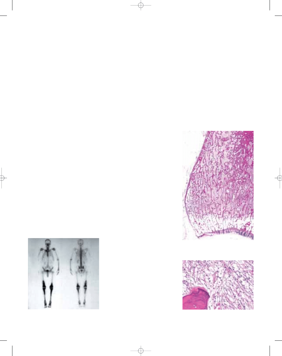
Fig. 20.17
Erdheim-Chester disease. Bone scan
highlights the increased uptake throughout the entire
length of the bones involved.
Fig. 20.18 Erdheim-Chester disease. Macrosection
of tibia showing medullary sclerosis, which abruptly
ends at the physis.
Fig. 20.19 Erdheim-Chester disease. Marrow infil-
tration by numerous foamy histiocytes associated
with dense fibrosis.
347
Erdheim-Chester disease
bb5_28.qxd 13.9.2006 14:16 Page 347
Definition
Chest wall hamartoma is a non-neoplas-
tic proliferation of mesenchymal tissue,
predominantly cartilage, admixed with
aneurysmal bone cyst elements. The
lesion develops during fetal life and pres-
ents at or shortly after birth with an
extrapleural mass arising from the rib
cage.
Synonyms
Vascular hamartoma of infancy, mes-
enchymal hamartoma of the chest wall,
mesenchymoma.
Epidemiology
The lesion is rare. To date only 59 cases
have been documented. In approximate-
ly 40% of cases, the mass is apparent at
birth. However, most cases present
between ages one month to one year
{97}. Less frequently, lesions may pres-
ent in children up to age eight. One adult
aged 26 was diagnosed with a chest wall
hamartoma {531}. The lesion has also
been diagnosed in utero with CT scans
or ultrasound {1351,1807}.
Sites of involvement
The lesion is an intrathoracic and
extraplural mass and arises from one or
more ribs. Almost always, the posterior or
lateral portions of the rib are affected.
Rarely, the lesion may be multifocal or
bilateral in the chest cavity {2132}.
Clinical features
Chest wall hamartoma presents as a
mass or fullness of the rib cage. Most
often, the bulk of the mass is intratho-
racic. As a result, infants frequently
develop respiratory distress.
Radiographically, chest wall hamartoma
is a partially mineralized mass arising
from the inside of the rib cage and
extending into the chest cavity. The
involved rib is partially destroyed, and
adjacent ribs are deformed. CT images
show an expansile mass and partial rib
destruction. Magnetic resonance images
shows alternating areas of high and low
signal on T1 and T2 sequences, reflect-
ing both solid and cystic components
{1886}.
Macroscopy
Lesions range from 3 to 7 cm in maxi-
mum dimension. Cut surface reveals
grey to white solid areas adjacent to cys-
tic cavities filled with blood.
Histopathology
Solid areas consist primarily of mature
hyaline cartilage, although areas resem-
bling chondroblastoma may be present.
The cartilage often shows enchondral
ossification. Areas with fibroblast-like
cells are also present. Cystic areas show
features typical of aneurysmal bone cyst:
blood-filled lakes are bounded by fibrous
septae which contain reactive bone and
osteoclast-like giant cells.
Prognostic factors
Complete surgical removal of the affect-
ed ribs results in cure. Scoliosis is an
occasional complication of surgery.
Rarely untreated patients may die of res-
piratory insufficiency {1379}. However,
most unoperated lesions remain stable.
Spontaneous regression has also been
reported {721}.
E.F. McCarthy
H. Dorfman
Chest wall hamartoma
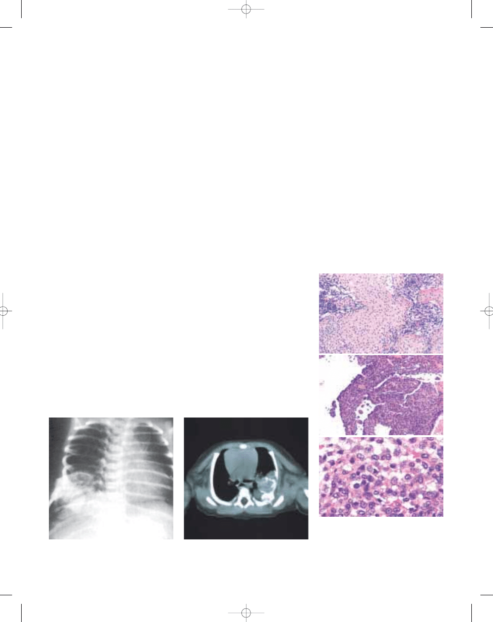
Fig. 20.20 Chest wall hamartoma. X-ray of a newborn
showing a lesion in the right lower rib cage, involving
several ribs and projecting into the chest cavity.
Fig. 20.21 CT scan of a chest wall hamartoma in a
three-day-old infant involving the inner aspect of a rib.
The lesion has a radiodense component.
348
Tumours of undefined neoplastic nature
Fig. 20.22 Chest wall hamartoma (A) showing the
typical chondroid matrix. B Histology similar to that
of a conventional aneurysmal bone cyst with blood-
filled lakes separated by septae composed of stromal
cells and multinucleated giant cells. C Immature
chondroblastoma-like cells.
B
A
C
bb5_28.qxd 13.9.2006 14:16 Page 348
Wyszukiwarka
Podobne podstrony:
bb5 chap3
bb5 chap8
bb5 chap1
BB5 BOX
bb5 chap16
bb5 chap15
bb5 contents
bb5 chap12
bb5 chap4
bb5 references
bb5 chap6
bb5 chap17
bb5 chap5
Lista wszystkich dostępnych polskich Product Code dla telefonów platformy BB5
bb5 chap21
bb5 source
CHAP20R
bb5 chap13
więcej podobnych podstron