CHAPTER 12
Fibrogenic Tumours
Tumours of fibrogenic origin do not have a mineralizing matrix
but generally produce collagen; high grade tumours may not
produce any matrix.
Desmoplastic fibroma is one of the most uncommon of bone
tumours. It is identical to the much more common soft tissue
desmoid and locally aggressive.
Fibrosarcomas range from the well differentiated tumours,
which are difficult to separate from desmoplastic fibroma, to
highly malignant tumours which are composed of small cells
and simulate Ewing sarcoma. Distinction from fibroblastic
osteosarcoma may be arbitrary and may depend on sampling.
bb5_20.qxd 13.9.2006 13:15 Page 287
Definition
Desmoplastic fibroma is a rare, benign
bone tumour composed of spindle cells
with minimal cytological atypia and
abundant collagen production.
ICD-O code
8823/0
Synonyms
Desmoid tumour of bone, intra-osseous
counterpart of soft tissue fibromatosis.
Epidemiology
The incidence is approximately 0.1% of
all primary bone tumours. It tends to
occur in adolescent and young adults
with near equal gender distribution.
Sites of involvement
Desmoplastic fibroma may involve any
bone but is most frequent in the
mandible.
Clinical features / Imaging
Patients present with a variety of symp-
toms. Some have pain, others present
because of deformity or loss of function.
Radiographically, desmoplastic fibroma
is usually a well defined, radiolucent
lesion that may expand the host bone.
Intralesional trabeculation is frequent.
Larger lesions may breach the
periostium and extend into soft tissue.
Such erosive, destructive pattern may
mimic other, more aggressive lesions.
Desmoplastic fibroma has low signal
intensity in both T1 and T2 weighted
MRI images. The extent of disease and
margins are best assessed with CT and
MRI.
Macroscopy
The tumour is firm and the cut surface is
creamy-white with a variegated whorled
pattern. The advancing surfaces of the
lesion tend to be scalloped and appar-
ently well defined. The tumour may
extend into soft tissue.
Histopathology
The lesion is composed of spindle cells
(fibroblasts/myofibroblasts) on a richly
collagenous, variably hyalinized back-
ground. The degree of cellularity is vari-
able but cellular atypia and pleomor-
phism are minimal or absent. Mitoses are
rare.
Genetics
FISH analyses of desmoplastic fibroma
suggest that trisomies 8 and 20 repre-
sent nonrandom aberrations in a subset
of these lesions, analogous to similar
findings in soft tissue desmoid tumours
{267}.
Prognostic factors
The tumour behaves in a locally
progressive/aggressive manner. Recur-
rence following curettage and resec-
tion are 72% and 17%, respectively
{832}. Local relapse has been reported
as late as eight years following primary
surgery. There is a single reported
case involving the spine that showed
little detectable change over a follow
up period of nine years without therapy
{1482}.
Desmoplastic fibroma of bone
V. Fornasier
K.P.H. Pritzker
J.A. Bridge
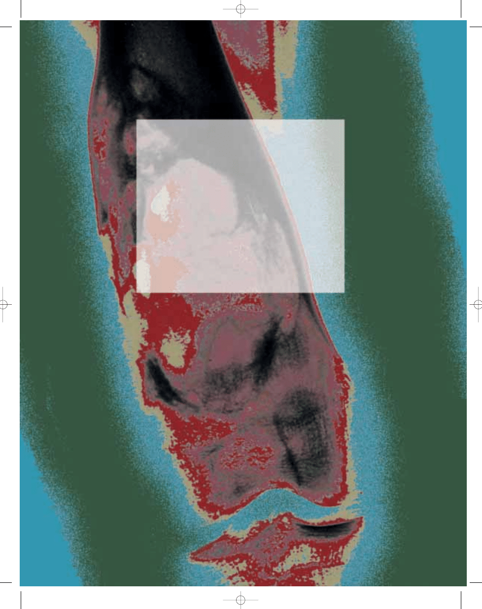
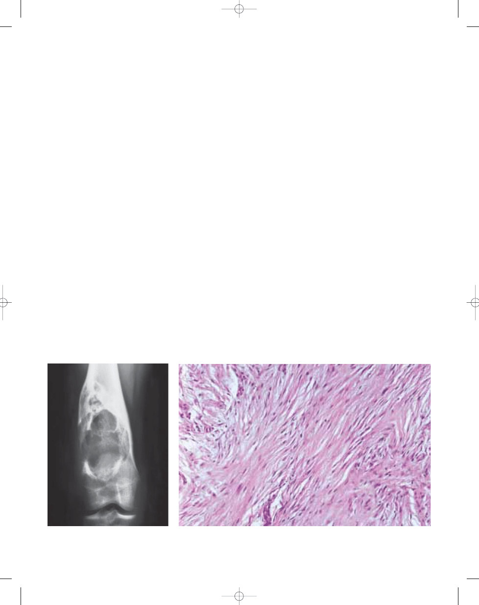
Fig. 12.01 Desmoplastic fibroma. Plain X-ray of a
tumour involving the distal femur. The lesion is
large, lobulated, and has a sclerotic rim.
288
Fibrogenic tumours
Fig. 12.02 Desmoplastic fibroma. High power magnification showing spindle cells without cytological atyp-
ia and large amounts of collagen.
bb5_20.qxd 13.9.2006 13:15 Page 288
Definition
A primary malignant spindle cell neo-
plasm of bone in which the tumour cells
are typically organized in a fascicular or
"herringbone" pattern.
ICD-O code
8810/3
Epidemiology
Precise epidemiological data pertaining
to fibrosarcoma of bone is difficult to
obtain due to inconsistent terminology
usage for fibrosarcoma versus malignant
fibrous histiocytoma.
Fibrosarcomas constitute up to 5% of all
primary malignant bone tumours, with
relatively uniform incidence over the sec-
ond to sixth decades and equal gender
distribution {991}. There have been
occasional reports of cases occurring
during infancy {167,425}.
Sites of involvement
Historical series indicate that fibrosarco-
mas most frequently involve the meta-
physes of long bones. In one large
series, the distal femur was involved in
48 of 102 of cases (47%) {2075}. Other
frequent sites of involvement were the
proximal femur (16%), distal humerus
(14%) and proximal tibia (11%). A series
of 130 cases also identified the distal
femur as the most common site (21%) of
involvement {991}.
Clinical features / Imaging
Pain and swelling are the usual symp-
toms. Up to one-third of patients have
pathological fracture {1221}.
Radiographically, fibrosarcoma usually
appears as a destructive geographic
lesion, but may have an ill defined per-
meative, "moth eaten" appearance with
cortical destruction and frequent soft tis-
sue extension. A periosteal reaction is
not infrequently present {2075}. The soft
tissue extension may be better visualised
by CT and MRI.
Aetiology
In most cases, the aetiology of fibrosar-
coma of bone is not known. However,
fibrosarcoma has been reported in asso-
ciation with a number of conditions
including prior radiation therapy, Paget
disease, giant cell tumour, osteochon-
droma, bone infarcts, chronic osteo-
myelitis, fibrous dysplasia, ameloblastic
fibroma and hereditary bone dysplasia
{85,644,886}.
Macroscopy
Well differentiated tumours produce
large amounts of collagen, resulting in a
firm consistency with a trabeculated,
white cut surface and circumscribed
margins. Poorly differentiated tumours
have a softer, fleshy consistency with foci
of necrosis; they vary in colour and are
poorly marginated.
Histopathology
Histologically, fibrosarcoma of bone is
composed of a uniformly cellular popula-
tion of spindle shaped cells arranged in
a fascicular or "herringbone" pattern with
a variable amount of collagen produc-
tion. Parts or all of the lesion may be
more myxoid and such lesions have
been labelled myxofibrosarcomas.
Higher grade lesions tend to be more
cellular with less collagen production,
exhibit greater nuclear atypia and a high-
er mitotic count including abnormal
mitoses than their better differentiated
counterparts. Areas of necrosis may be
seen.
Differential diagnosis
In cases with more severe cytological
atypia, including tumour giant cells,
fibrosarcoma may be difficult to distin-
guish from malignant fibrous histiocy-
toma. The presence of a storiform pat-
tern and epithelioid type cells with
"ground glass" cytoplasm would favour a
diagnosis of malignant fibrous histiocy-
toma. In view of the identical clinical,
L.B. Kahn
V. Vigorita
Fibrosarcoma of bone
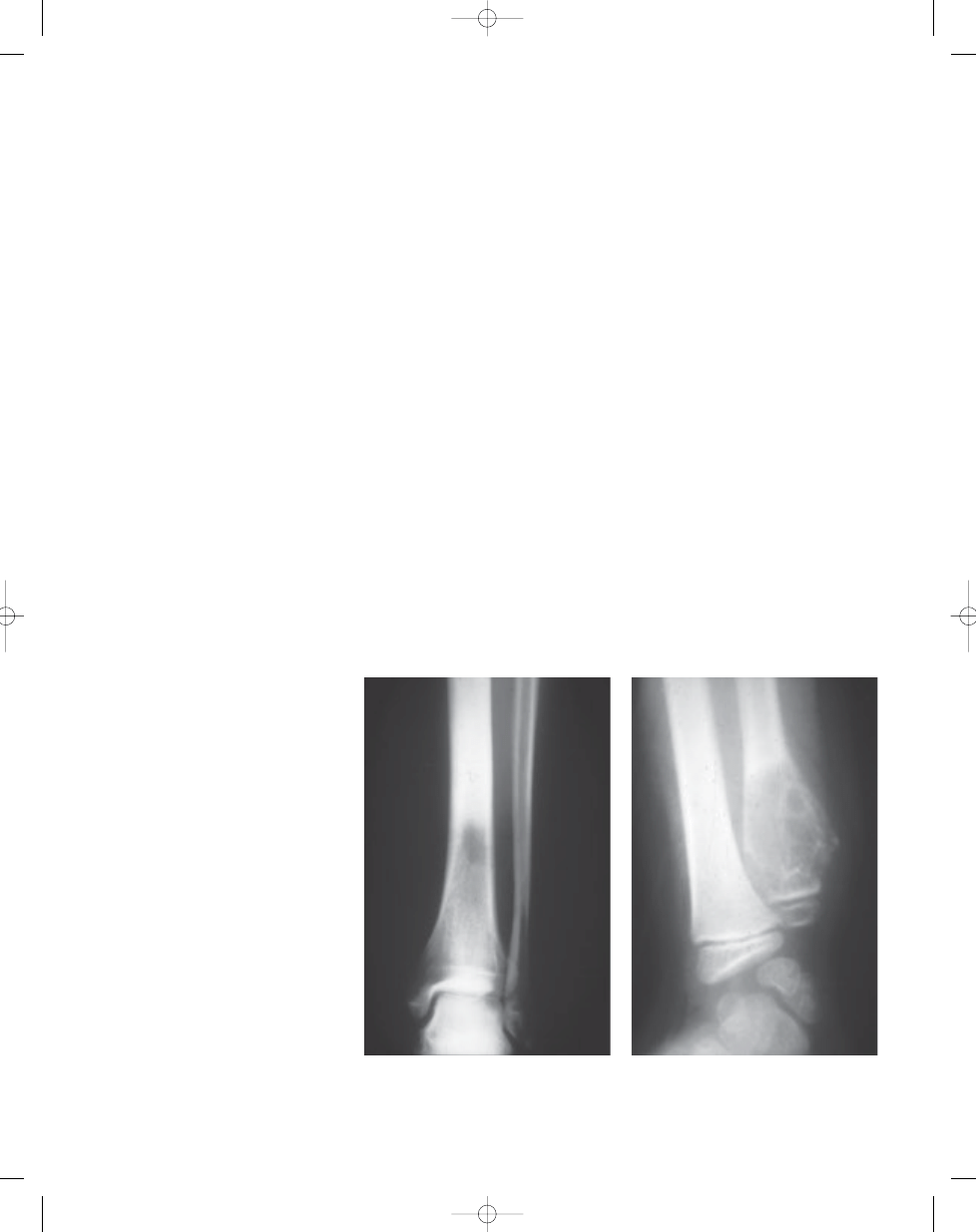
Fig. 12.03 Fibrosarcoma of tibia. Plain radiograph
demonstrating ill defined purely osteolytic lesion
involving distal third of tibia. The soft tissue exten-
sion of the tumour is not evident in this study.
Fig. 12.04 Fibrosarcoma of ulna. Plain radiograph
showing ill defined expansile osteolytic lesion of
the metaphysis with cortical destruction on the
medial aspect.
289
Fibrosarcoma of bone
bb5_20.qxd 13.9.2006 13:15 Page 289
radiological and even prognostic
features of these two lesions, some
investigators have chosen to include
them within the category of fibrosarco-
mas {2075}. Well differentiated fibrosar-
coma is distinguished from desmoplastic
fibroma by the presence of readily identi-
fiable mitoses and high cellularity in the
former and their extreme paucity or
absence in the latter.
Prognostic factors
Two series have reported an overall 5-
year survival approximating 34% {1647,
2075}. The most important prognostic
factor is histological tumour grade. In
one series, the 10 year survival was 83%
in low grade and 34% in high grade
fibrosarcoma {181}. Another series
reported an overall 10-year survival rate
of 28%, but there was a higher chance
of survival (48%) in primary tumours
originating from the cortical surface
{991}. In the latter series, metastases
occurred in 59/130 patients (45%),
most frequently involving lung and
other bones. In addition to poor histo-
logical differentiation, other ad- verse
prognostic factors included age over
40 years and axial skeletal location
{1647}.
290
Fibrogenic tumours
D
C
B
A
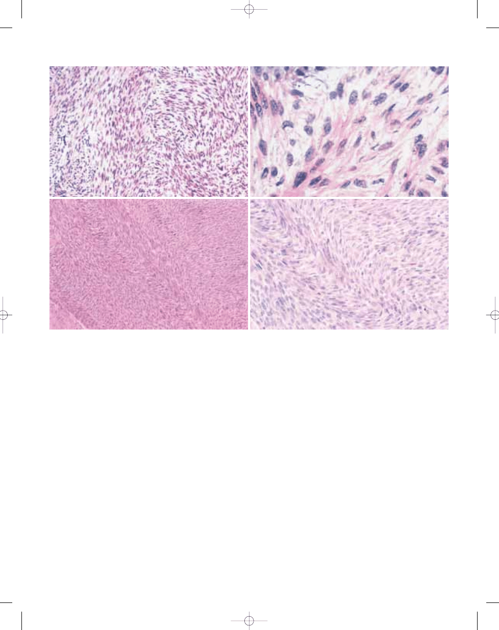
Fig. 12.05 Fibrosarcoma of tibia. A The fibrocytic cells are arranged in a haphazard fascicular rather than in the more typical "herring bone" pattern. B High power pho-
tomicrograph reveals a fairly uniform appearance of the neoplastic cells. The nuclei are ovoid, blunt-ended and have single small nucleoli and finely dispersed chromatin.
Collagen fibres appear to emanate from the nuclear poles. C Fibrosarcoma illustrating the characteristic "herringbone" pattern. D High power appearance of the pre-
vious photomicrograph.
bb5_20.qxd 13.9.2006 13:15 Page 290
Wyszukiwarka
Podobne podstrony:
bb5 chap3
bb5 chap8
chap12
bb5 chap1
BB5 BOX
bb5 chap16
bb5 chap15
bb5 contents
bb5 chap4
bb5 references
bb5 chap6
bb5 chap17
chap12 (2)
mcga shs capt guide chap12
bb5 chap20
bb5 chap5
Lista wszystkich dostępnych polskich Product Code dla telefonów platformy BB5
bb5 chap21
więcej podobnych podstron