CHAPTER 3
So-called Fibrohistiocytic Tumours
Over the past 10 years, the concept of fibrohistiocytic differenti-
ation has been challenged and is now regarded as a poorly
defined morphological descriptor of histiocytic differentiation.
Pleomorphic malignant fibrous histiocytoma (MFH) was previ-
ously regarded as a distinct tumour type representing the most
common adult soft tissue sarcoma. Today, this term is synony-
mous with undifferentiated pleomorphic sarcoma, which has
become a diagnosis of exclusion accounting for less than 5% of
adult sarcomas. Similarly, the morphological features formerly
regarded as characteristic of the giant cell and inflammatory
variants of MFH are shared by a variety of other, specific tumour
types. Myxofibrosarcoma (formerly known as myxoid MFH) and
so-called angiomatoid MFH remain as distinctive and discrete
entities (see Chapters 2 and 9).
Cutaneous fibrous histiocytomas, dermatofibrosarcoma protu-
berans (best classified as a fibroblastic neoplasm) and atypical
fibroxanthoma are described separately in the Skin volume.
Since the localized and diffuse forms of giant cell tumour of ten-
don sheath have more in common with the descriptive category
of fibrohistiocytic lesions than with true synovium, they are for
now included in this chapter.
bb5_8.qxd 13.9.2006 10:40 Page 109
Giant cell tumour of tendon sheath
N. de St. Aubain Somerhausen
P. Dal Cin
The term giant cell tumour of tendon
sheath encompasses a family of lesions
most often arising from the synovium of
joints, bursae and tendon sheath {1027}.
These tumours are usually divided
according to their site (intra- or extra-
articular) and growth pattern (localized
or diffuse) into several subtypes, which
differ in their clinical features and biolo-
gical behaviour.
Definition
The localized type of giant cell tumour of
tendon sheath is a circumscribed prolif-
eration of synovial-like mononuclear
cells, accompanied by a variable num-
ber of multinucleate osteoclast-like cells,
foam cells, siderophages and inflamma-
tory cells, most commonly occurring in
the digits.
ICD-O code
9252/0
Synonyms
Tenosynovial giant cell tumour, localized
type, nodular tenosynovitis.
Epidemiology
The localized form is frequent and the
most common subset of giant cell tu-
mours. Tumours may occur at any age
but usually between 30 and 50 years,
with a 2:1 female predominance {2163}.
Sites of involvement
Localized giant cell tumours occur pre-
dominantly in the hand where they prob-
ably represent the most common neo-
plasm. Approximately 85% of the
tumours occur in the fingers, in close
proximity to the synovium of the tendon
sheath or interphalangeal joint. The
lesions may infrequently erode or infil-
trate the nearby bone {2160}, or rarely
involve the skin.
Other sites include the wrist, ankle / foot,
knee, and very rarely the elbow and the
hip {1492,2163}.
Clinical features
The most common presenting symptom
is that of a painless swelling. The
tumours develop gradually over a long
period and a preoperative duration of
several years is often mentioned .
Antecedent trauma is reported in a vari-
able number of cases (from 1 to 50%)
{1492,2163}.
Radiological studies usually demon-
strate a well circumscribed soft tissue
mass, with occasional degenerative
changes of the adjacent joint or erosion
of the adjacent bone {1046}.
Aetiology
Tenosynovial giant cell tumours initially
were regarded as an inflammatory
process based on animal models, the
common history of trauma, the predilec-
tion for the first three fingers of the right
hand {1492} and one X-inactivation
study suggesting polyclonality {2295}.
However, the finding of aneuploidy in
some cases {7}, the demonstration of
clonal chromosomal abnormalities
{1774}, and the fact that these lesions
are capable of autonomous growth
strongly support a neoplastic origin.
Macroscopy
Grossly, most localized giant cell
tumours are small (between 0.5 and 4
cm), although lesions of greater size may
be found in large joints. Tumours are well
circumscribed and typically lobulated,
white to grey with yellowish and brown
areas.
Histopathology
Tumours are lobulated, well circum-
scribed and at least partially covered by
a fibrous capsule. Their microscopic
appearance is variable, depending on
the proportion of mononuclear cells,
multinucleate giant cells, foamy
macrophages, siderophages and the
amount of stroma. Osteoclast-like cells,
which contain a variable number of
nuclei (from 3-4 to more than 50), are
usually readily apparent but may be
110
Fibrohistiocytic tumours
B
A
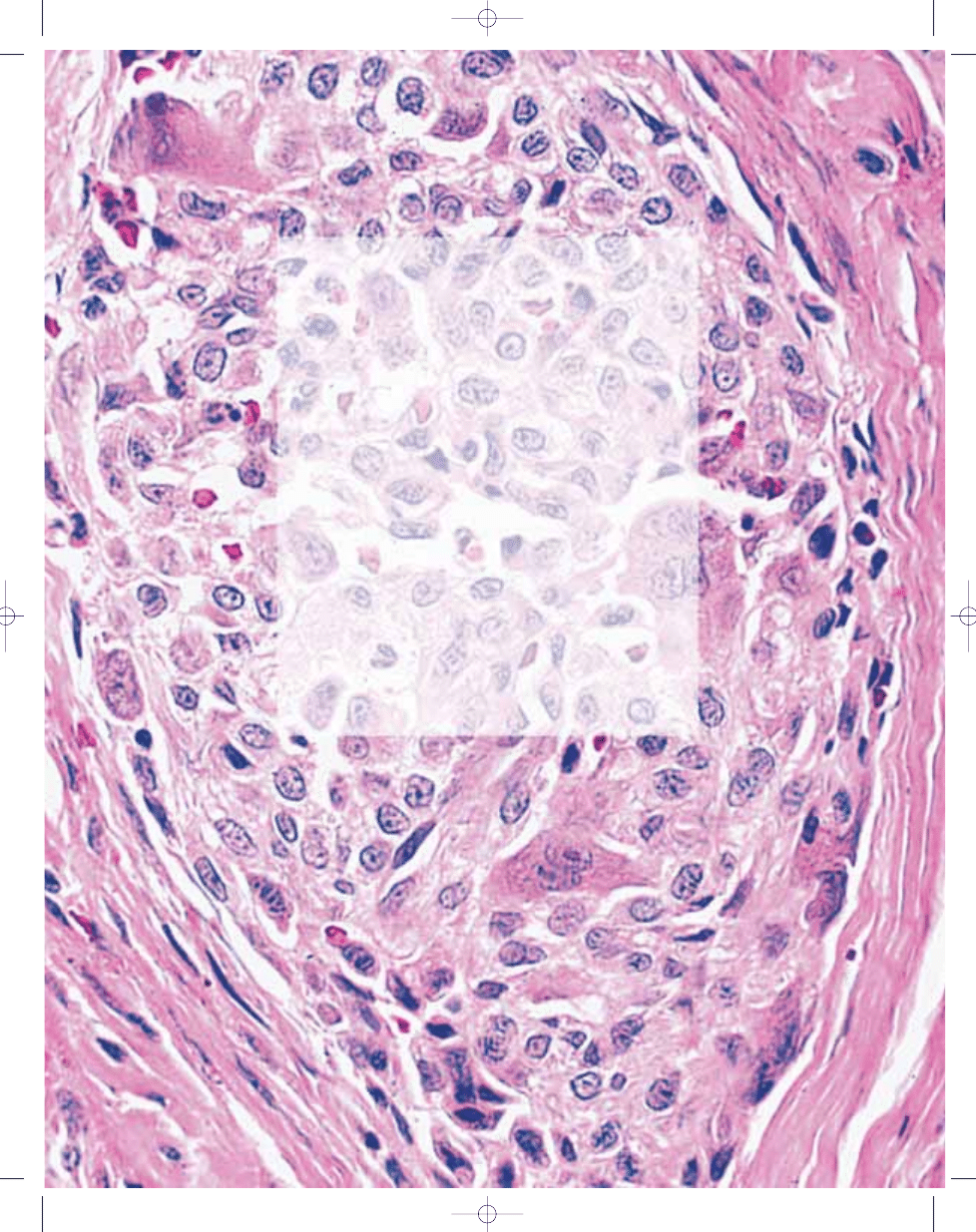
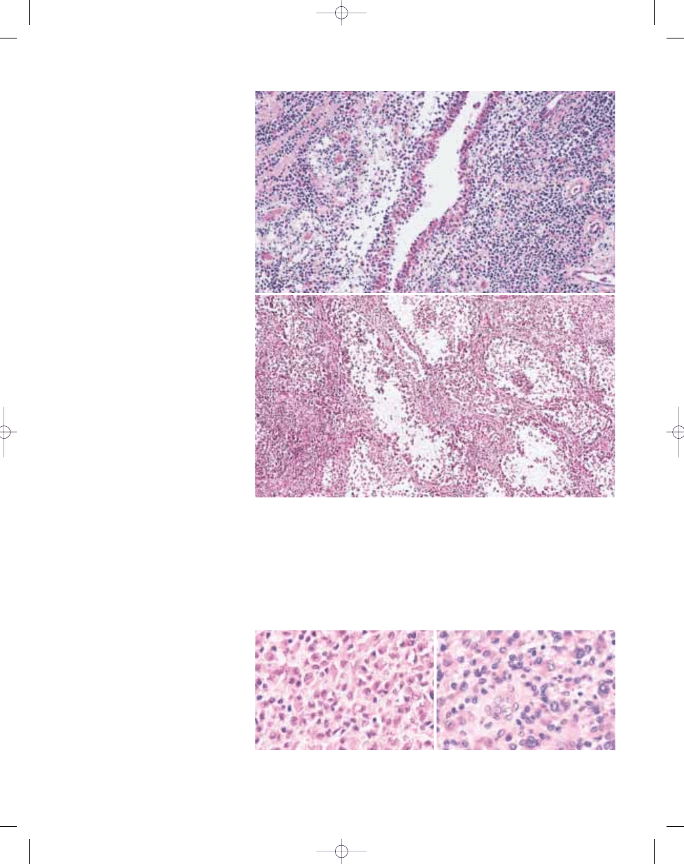
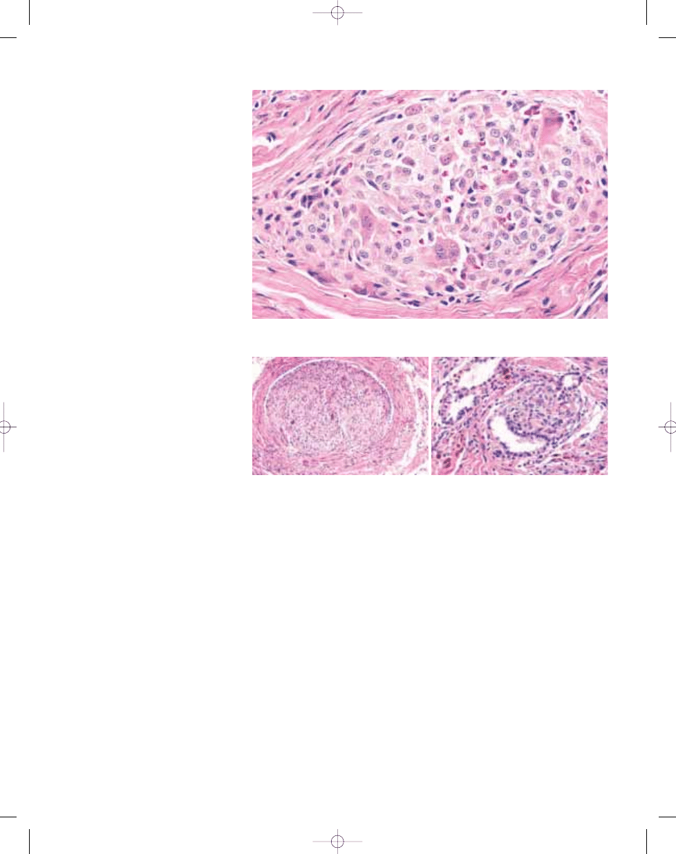
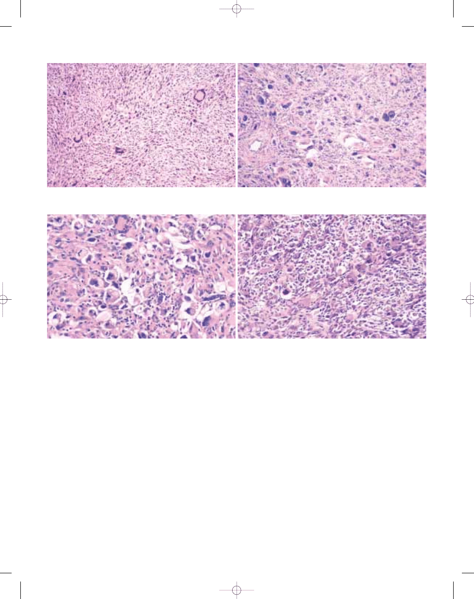
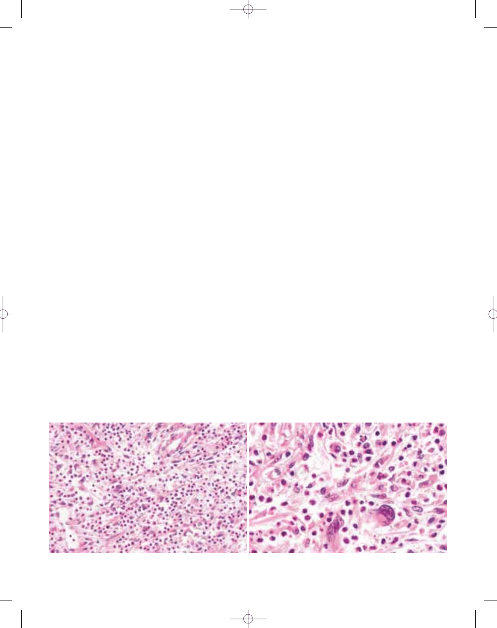
Fig. 3.01 Giant cell tumour of tendon sheath. A Typical admixture of histiocytoid cells, foamy cells and lymphocytes. In this case, giant cells are scanty. B Typical mononu-
clear histiocytoid cells with variably prominent eosinophilic cytoplasm and scattered osteoclastic giant cells.
bb5_8.qxd 13.9.2006 10:40 Page 110
inconspicuous in highly cellular tumours.
Most mononuclear cells are small, round
to spindle-shaped. They are character-
ized by pale cytoplasm and round or
reniform, often grooved nuclei. They are
accompanied by larger epithelioid cells
with glassy cytoplasm and rounded
vesicular nuclei. Xanthoma cells are fre-
quent, tend to aggregate locally near the
periphery of nodules and may be associ-
ated with cholesterol clefts.
Haemosiderin deposits are virtually
always identified. The stroma shows vari-
able degrees of hyalinization and may
occasionally have an osteoid-like
appearance. Cleft-like spaces are less
frequent than in the diffuse form {2163}.
Mitotic activity usually averages 3 to 5
mitoses per 10 HPF but may reach up to
20/10 HPF {2295}. Focal necrosis is
rarely seen.
Immunophenotype
Immunohistochemically, mononuclear
cells are positive for CD68. Some cells
may also express muscle-specific actin
(HHF35). A subset of desmin-positive
dendritic cells is reported in up to 50% of
cases {705}.
Multinucleate giant cells express CD68,
CD45 and markers such as tartrate
resistant acid phosphatase {449,1590}.
Ultrastructure
Ultrustructural studies have revealed an
heterogeneous cell population com-
posed of a majority of histiocyte-like
cells, accompanied by fibroblast-like
cells, intermediate cells, foam cells and
multinucleate giant cells {35,2163}.
Genetics
Cytogenetic aberrations have been
described in 11 giant cell tumours of ten-
don sheath. A near- or pseudodiploid
karyotype was seen in all cases, mostly
with simple structural changes {1910}.
The short arm of chromosome 1 is fre-
quently involved, with a clustering of
breakpoints to the region p11-p13 in 7/11
cases. A recurrent t(1;2)(p11;q35-36)
has been identified, but several other
translocation partners have been
described, including 3q21, 5q31, and
11q11. In addition, two cases without
1p11-13 rearrangement had transloca-
tions involving 16q24, thus possibly rep-
resenting an alternative primary cytoge-
netic change. Numerical changes seem
to be rare. In particular, it should be
noted that gain of chromosomes 5 and 7,
which is common in the diffuse type giant
cell tumour {1477}, has not been
described in the localized form {1910}.
Prognostic factors
Localized giant cell tumour is a benign
lesion with a capacity for local recur-
rence. Local excision is the treatment of
choice. 4 to 30 % of cases recur {1504,
1757,1774} but these recurrence are
usually non-destructive and are con-
trolled by surgical reexcision. It has been
suggested that recurrences develop
most often in highly cellular tumours or
lesions with a high mitotic count
{1757,2298}.
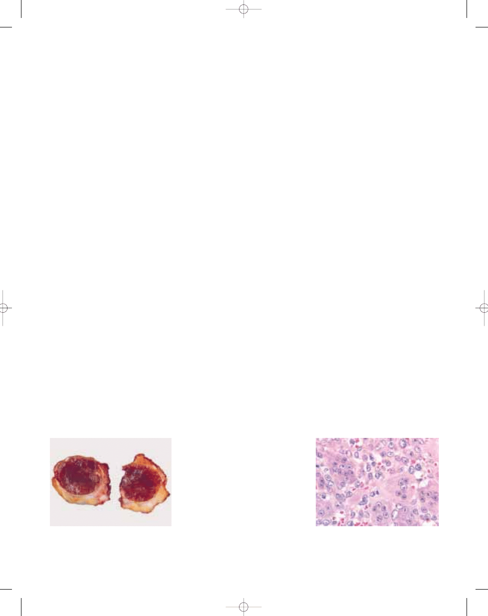
B
A
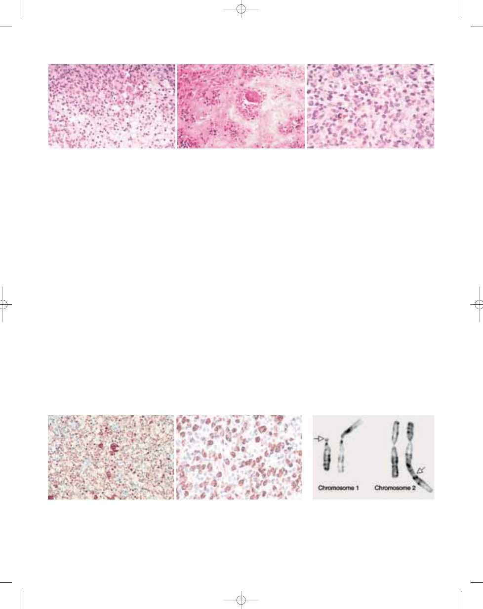
Fig. 3.03 Giant cell tumour of tendon sheath. A Localized giant cell tumours of tendons sheath are usually CD
68 positive. B Some cases of both localized and diffuse type contain numerous desmin-positive mononuclear
cells, sometimes with dendritic cytoplasmic porcesses.
Fig. 3.04 Giant cell tumour of tendon sheath. Partial
karyotype showing the characteristic t(1;2)(p13;q37)
translocation. Arrows indicate breakpoints.
B
C
A
Fig. 3.02 Giant cell tumour of tendon sheath. A Most cases show focal collections of xanthoma cells, while others (B) show extensive stromal hyalinization. C Small, his-
tiocyte-like cells with occasional nuclear grooves and larger cells with vesicular nuclei and abundant eosinophilic cytoplasm, frequently with a rim of haemosiderin.
Giant cell tumour of tendon sheath
111
bb5_8.qxd 13.9.2006 10:40 Page 111
112
Fibrohistiocytic tumours
Definition
Diffuse-type giant cell tumour is a
destructive proliferation of synovial-like
mononuclear cells, admixed with multi-
nucleate giant cells, foam cells,
siderophages and inflammatory cells.
The extraarticular form is defined by the
presence of an infiltrative soft tissue
mass, with or without involvement of the
adjacent joint.
The very uncommon malignant giant cell
tumour of tendon sheath is defined by
the coexistence of a benign giant cell
tumour with overtly malignant areas or by
the recurrence of a typical giant cell
tumour as a sarcoma.
ICD-O code
9251/0
Synonyms
Pigmented villonodular synovitis, pig-
mented villonodular tenosynovitis.
Epidemiology
Diffuse-type giant cell tumours tend to
affect younger patients than their local-
ized counterpart. The age of patients
varies widely but most lesions affect
young adults, under the age of 40. There
is a slight female predominance {1523,
1984,2164}.
Sites of involvement
Intraarticular lesions affect predominant-
ly the knee (75% of cases), followed by
the hip (15%), ankle, elbow and shoul-
der. Rare cases are reported in the tem-
poromandibular and spinal facet joints
{782,1899}. Extraarticular tumours most
commonly involve the knee region, thigh
and foot. Uncommon locations include
the finger, wrist, groin, elbow and toe {87,
1984,2164}.
Most extraarticular tumours are located
in periarticular soft tissues but these
lesions can be purely intramuscular or
predominantly subcutaneous {2164}.
Clinical features
Patients complain of pain, tenderness,
swelling or limitation of motion.
Haemorrhagic joint effusions are com-
mon. The symptoms are usually of rela-
tively long duration (often several years).
Radiographically, most tumours present
as ill defined peri-articular masses, fre-
quently associated with degenerative
joint disease and cystic lesions in the
adjacent bone {542}. On magnetic reso-
nance imaging, giant cell tumours show
decreased signal intensity in both T1-
and T2-weighted images {1036}.
Aetiology
Although these lesions have been
regarded as reactive, the presence of
clonal abnormalities {1910} and the
capacity for autonomous growth are now
widely regarded as evidence for a neo-
plastic origin.
Macroscopy
Diffuse-type giant cell tumours are usual-
ly large (often more than 5 cm), firm or
sponge-like. The typical villous pattern of
pigmented villonodular synovitis is usual-
ly lacking in extraarticular tumours. The
latter have a multinodular appearance
and a variegated colour, with alternation
of white, yellowish and brownish areas.
Histopathology
Most tumours are infiltrative and grow as
diffuse, expansile sheets. Their cellularity
is variable: compact areas alternate with
pale, loose, discohesive zones. Cleft-like
spaces are common and appear either
as artefactual tears or as synovial-lined
spaces. Blood-filled pseudoalveolar
spaces are seen in approximately 10% of
cases.
In comparison with the localized form,
osteoclastic giant cells are less common
and may be absent or extremely rare in
up to 20% of cases. They are irregularly
distributed throughout the lesions and
are more easily found around haemor-
rhagic foci.
The mononuclear component comprises
two types of cells: small histiocyte-like
cells, which represent the main cellular
component, and larger cells. Histiocyte-
like cells are ovoid or spindle-shaped,
with palely eosinophilic cytoplasm. Their
nuclei are small, ovoid or angulated, con-
tain fine chromatin, small nucleoli and
frequently display longitudinal grooves.
Larger cells are rounded or sometimes
show dendritic cytoplasmic processes.
Their cytoplasm is abundant, pale to
deeply eosinophilic, often contains a
N. de St. Aubain Somerhausen
P. Dal Cin
Diffuse-type giant cell tumour
B
A
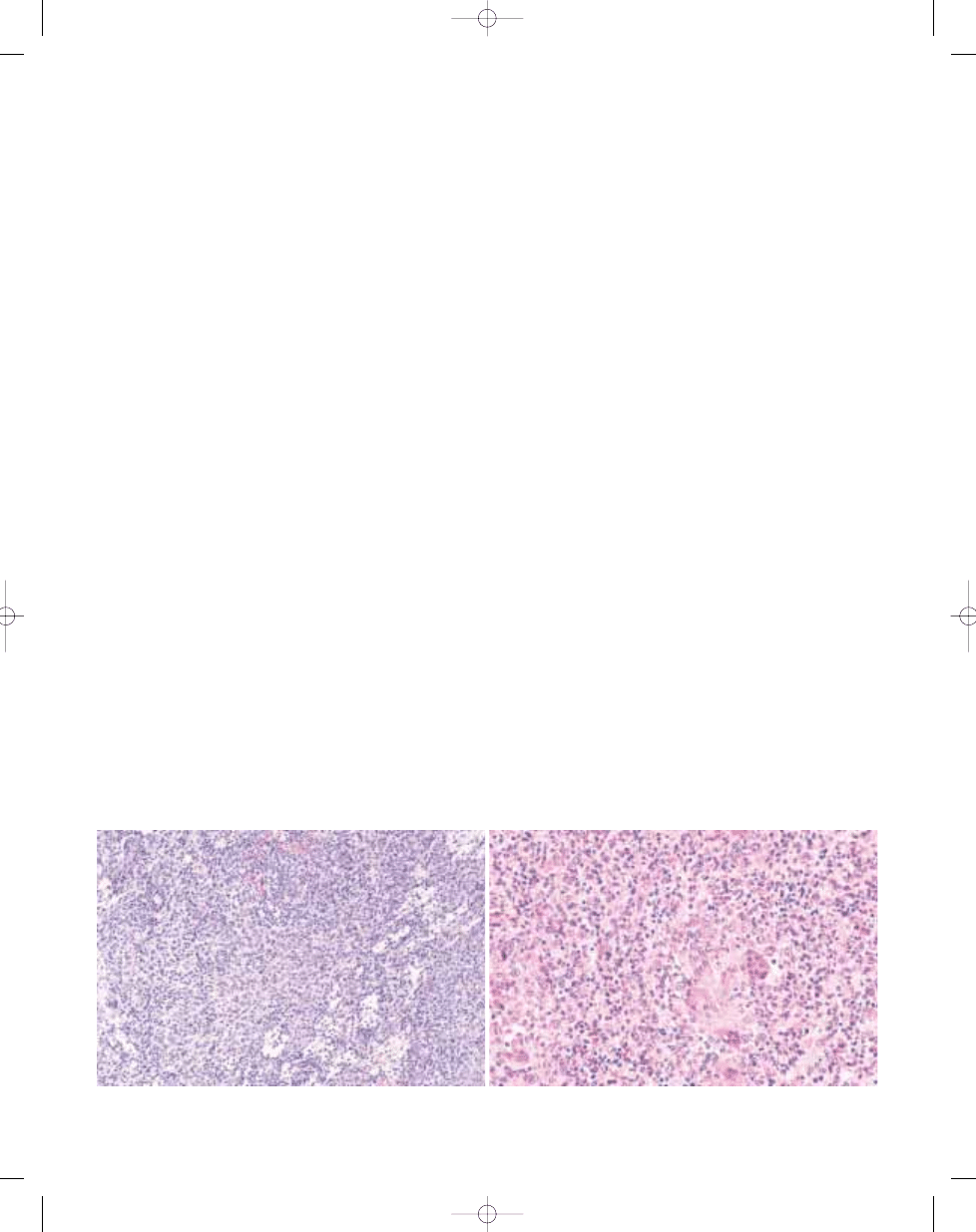
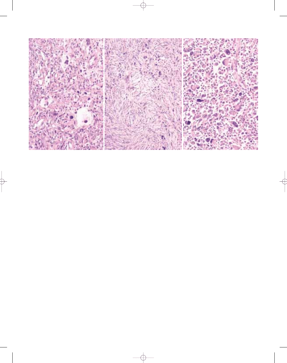
Fig. 3.05 A Villous appearance of an intra-articular diffuse-type giant cell tumour. B Low magnification of a com-
pletely extra-articular tumour showing infiltration of the muscular and adipose tissue.
Fig. 3.06 Diffuse-type giant cell tumour with promi-
nent inflammatory component and numerous large
dendritic cells with abundant cytoplasm.
bb5_8.qxd 13.9.2006 10:40 Page 112
peripheral rim of hemosiderin granules
and occasionally shows a paranuclear
eosinophilic filamentous inclusion. Nuclei
are characterized by reniform or lobulat-
ed shape, thick nuclear membranes,
vesicular chromatin and eosinophilic
nuclei. The occasional predominance of
these larger cells may obscure the typi-
cal features of giant cell tumour and lead
to a diagnosis of sarcoma. Sheets of
foam cells are frequently observed, usu-
ally in the periphery of lesions and vari-
able amounts of haemosiderin are identi-
fied in most cases. Giant cell tumours
may also contain a significant lymphocyt-
ic infiltrate. The stroma shows variable
degrees of fibrosis and may appear
hyalinized, although this is usually less
marked than in the localized form.
Mitoses are usually identifiable and
mitotic activity of more than 5 per 10 HPF
is not uncommon {1984,2164,2239}.
There have been several reports of typi-
cal giant cell tumours recurring as a his-
tologically malignant neoplasms and a
few series included primary histological-
ly malignant tumours of the tendon
sheath resembling giant cell tumours
{187,637,1555,1941,1984}. These neo-
plasms tended to show significantly
increased mitotic rate (more than 20
mitoses / 10 HPF), necrosis, enlarged
nuclei with nucleoli, spindling of mononu-
cleated cells, the presence of abundant
eosinophilic cytoplasm in histiocyte-like
cells, and stromal myxoid change,
although none of these features could be
used in isolation as a criterion for malig-
nancy {187,637,1984}.
In addition, two cases with banal histol-
ogy which developed metastatic disease
(in the lungs or lymph nodes) have been
reported to date {1984,2239}.
Immunophenotype
The immunohistochemical and ultra-
structural features of diffuse-type giant
cell tumour are similar to those of the
localized form. Mononuclear cells are
positive for CD68 and other macrophage
markers. Desmin stain highlights a popu-
lation of cells with dendritic features in 35
to 40% of cases; these frequently corre-
spond to the larger eosinophilic cells.
Giant cells are positive for CD68 and
CD45 {705,1590,1984}.
Genetics
Chromosomal aberrations have been
described in 17 cases, all with a near- or
pseudodiploid karyotype. Rearrange-
ments of the 1p11-13 region have been
detected in eight of them, one had a
t(1;2)(p22;q35-37), and one had involve-
ment of band 16q24, suggesting a close
cytogenetic relationship with the local-
ized form of giant cell tumour {1910}.
One difference, however, between these
two entities, is that trisomies for chromo-
somes 5 and 7, usually as the sole anom-
alies, have been detected only in diffuse-
type giant cell tumours {1477}. The sig-
Fig. 3.07 Diffuse-type giant cell tumour. A Pseudosynovial or 'pseudoglandular' spaces, surrounded by clusters
of xanthoma cells. B Pseudoalveolar spaces are commonly seen in diffuse-type giant cell tumours.
A
B
B
A
Fig. 3.08 Diffuse-type giant cell tumour. A Typical mononuclear histiocytoid cells, some of which have promi-
nent eosinophilic cytoplasm. B Note frequent nuclear grooves in the histiocytoid cells. Some tumour cells have
more prominent eosinophilic cytoplasm with haemosiderin granules.
113
Diffuse-type giant cell tumour
bb5_8.qxd 13.9.2006 10:40 Page 113
Deep benign fibrous histiocytoma
J.M. Coindre
Definition
A benign fibrous histiocytoma, which
develops entirely within subcutaneous
tissue, deep soft tissues or in parenchy-
mal organs.
ICD-O code
8830/0
Epidemiology
Deeply located fibrous histiocytomas are
rare. Based on the only published series,
they represent less than 1% of fibrohisti-
ocytic tumours {673}. Their exact fre-
quency is difficult to determine because
some cases published as deep fibrous
histiocytomas may represent solitary
fibrous tumours {673,706}. They may
develop at any age, but most affect
adults over 25 years old, with a predom-
inance in males.
Sites of involvement
The lower limb and the head and neck
region are the most common sites. Most
cases develop in subcutaneous tissue,
but a few cases have been reported in
muscle, mesentery, trachea and kidney
{673,869,1147,1843}.
nificance of trisomy 5 and 7 for tumour
development in this context is question-
able because the same aneuploidies are
frequent also in synovial samples from
patients with various forms of reactive
synovial lesion {1429}.
Prognostic factors
Recurrences are common, often multiple
and may severely compromise joint func-
tion. The recurrence rate has been esti-
mated between 18 and 46 % for intraar-
ticular lesions and between 33 and 50%
of cases for extraarticular tumours {1899,
1984,2164,2239}. The risk of recurrence
does not seem to be correlated with any
histological parameter other than posi-
tive excision margins. Therefore, diffuse-
type giant cell tumours should be regard-
ed as locally aggressive but nonmetasta-
sizing neoplasms and wide excision is
the treatment of choice.
Although the number of cases is limited,
malignant giant cell tumours of tendon
sheath showing obvious sarcomatous
areas are potentially aggressive and may
give rise to pulmonary metastasis {187,
1555,1941,1984}.
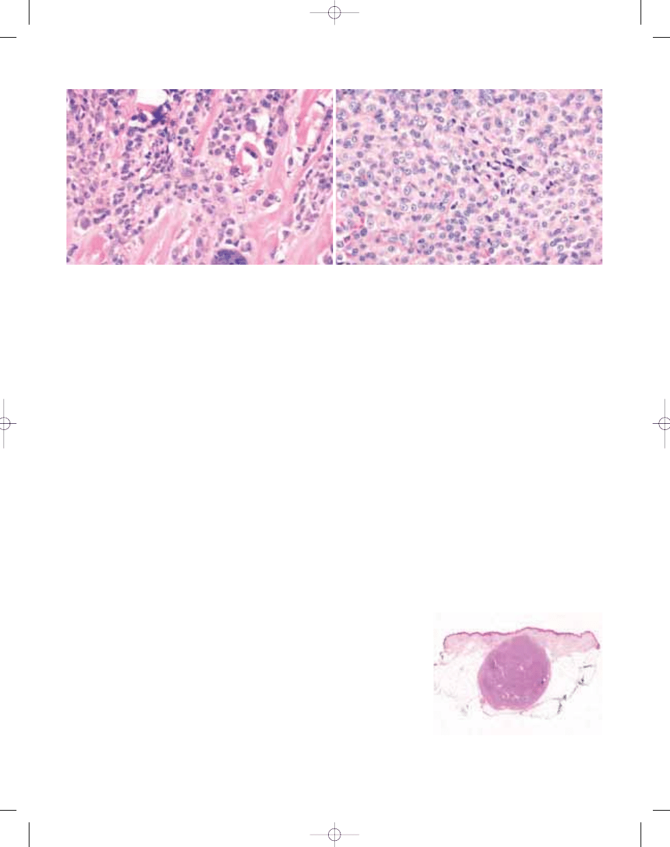
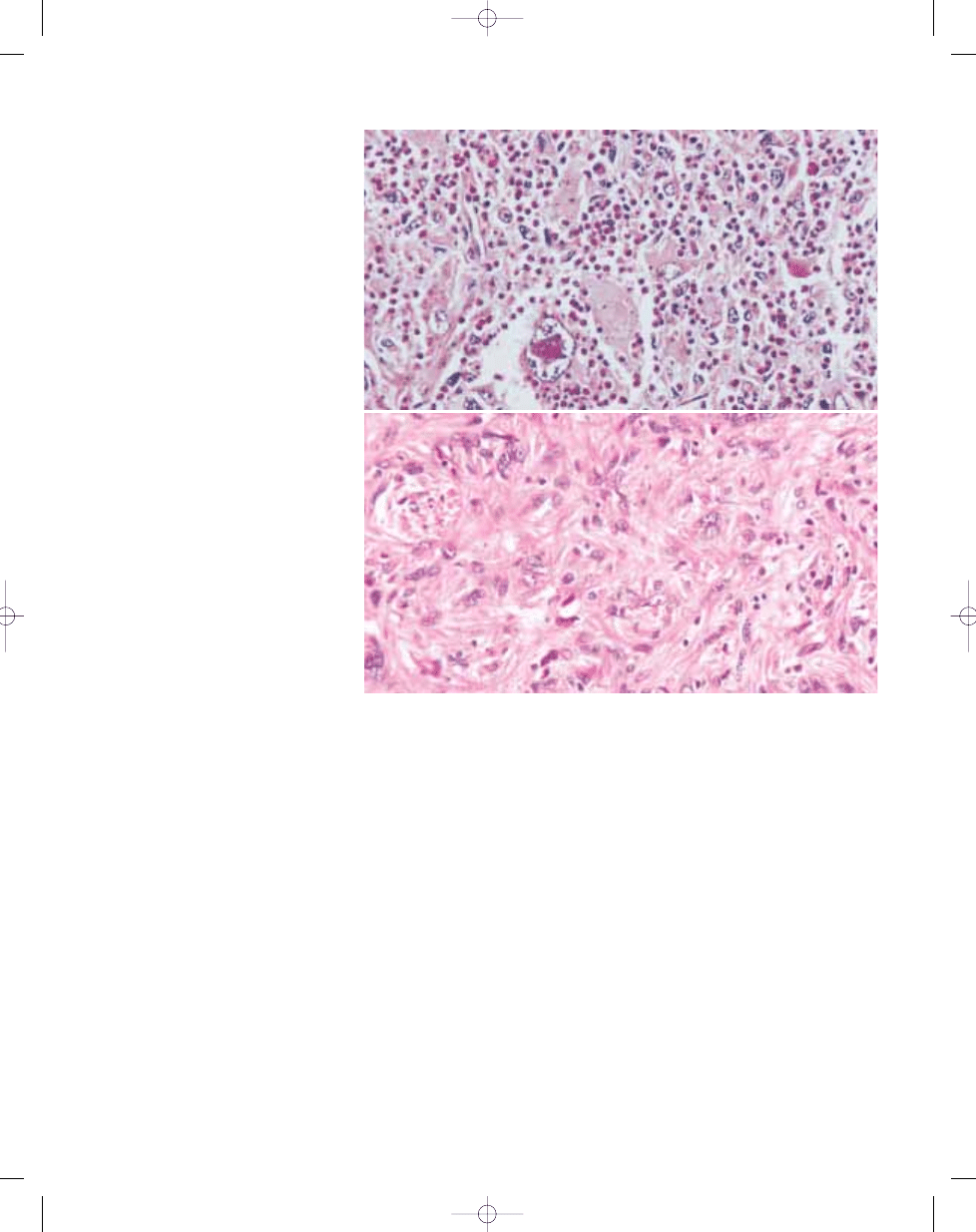
Fig. 3.10 Deep benign fibrous histiocytoma tends to
be more circumscribed than the cutaneous form and
pseudo-encapsulated.
114
Fibrohistiocytic tumours
B
A
Fig. 3.09 Malignant diffuse-type giant cell tumour. Although there is usually at least focal morphological overlap with usual giant cell tumour (A), closer examination
reveals increased cellularity and predominance of atypical large cells with prominent nucleoli (B).
bb5_8.qxd 13.9.2006 10:40 Page 114
Clinical features
Most lesions present as a painless and
slowly enlarging mass.
Macroscopy
Contrary to the cutaneous form, deep
lesions tend to be well circumscribed
and pseudo-encapsulated with occa-
sional areas of haemorrhage. Most
lesions are 4 cm or more when resected.
Histopathology
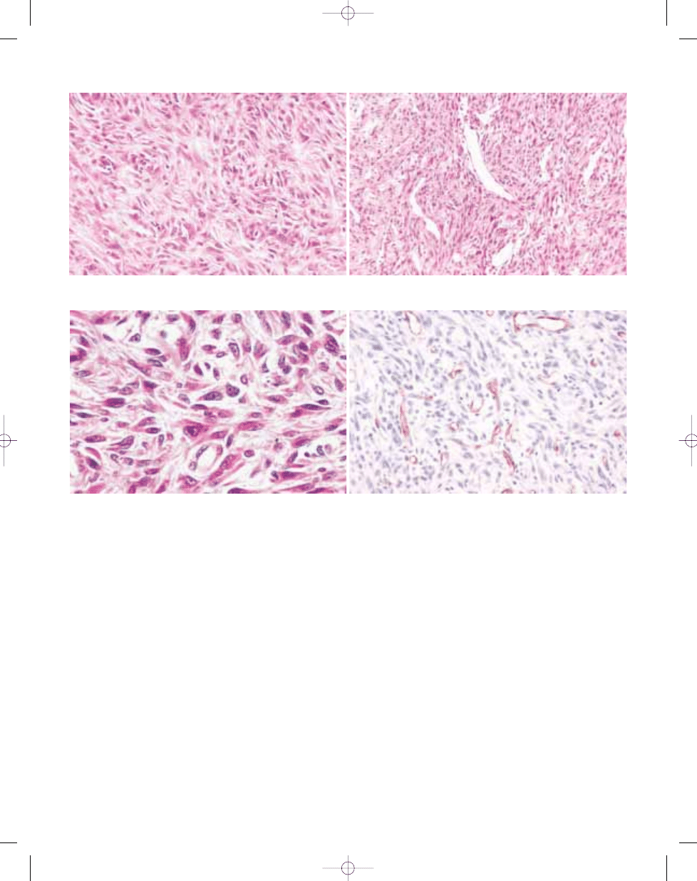
Deep fibrous histiocytomas usually show
a prominent storiform pattern, sometimes
combined with haemangiopericytoma-
like areas.
Contrary to conventional cutaneous
lesions, most lesions show monomor-
phism and usually lack secondary ele-
ments such as foamy cells and giant
cells but usually show scattered lympho-
cytes. Thus, they more closely resmble
the cellular variant of cutaneous fibrous
histiocytoma. The tumour cells are cyto-
logically bland and generally spindle-
shaped with elongated or plump vesicu-
lar nuclei and eosinophilic, ill defined
cytoplasm. There is no nuclear pleomor-
phism or hyperchromasia, and mitoses,
although commonly present, are usually
less than 5 per 10 high power fields. The
stroma may show myxoid change or
hyalinization and rarely osteoclast-like
giant cells or metaplastic ossification
{673,1973}. Small foci of necrosis may
be present.
Immunophenotype
Immunohistochemistry shows similar
results as in cutaneous lesions with neg-
ativity for epithelial markers, desmin and
S100 protein. Alpha smooth muscle actin
may be positive in some parts of the
lesion. CD34 is usually (but not always)
negative, but, if positive, solitary fibrous
tumour should be considered.
Prognostic factors
Deep fibrous histiocytoma may recur
locally {673}, particularly if incompletely
excised. No metastasis has been report-
ed so far.
B
A
Fig. 3.11 Deep benign fibrous histiocytoma. A A monomorphic storiform pattern is usually seen. B Branching pericytoma-like vessels are common.
115
Deep benign fibrous histiocytoma
B
A
Fig. 3.12 Deep benign fibrous histiocytoma. A These lesions show less cytologic polymorphism than their dermal counterparts. B Staining for CD34 is most often nega-
tive.
bb5_8.qxd 13.9.2006 10:40 Page 115
116
Fibrohistiocytic tumours
Definition
Plexiform fibrohistiocytic tumour (PFT) is
a mesenchymal neoplasm of children,
adolescents, and young adults, charac-
terized by fibrohistiocytic cytomorpholo-
gy, and a multinodular growth pattern. It
rarely metastasizes.
ICD-O code
8835/1
Epidemiology
PFT preferentially affects young individuals;
mean age at presentation is approximately
14.5 years {603,1782}. The tumour occurs
more often in female than in male patients,
with reported female-to-male ratios ranging
from 2.5:1 {603} to 6:1 {1782}. PFT has not
been reported to occur with greater fre-
quency in any particular race.
Sites of involvement
PFT involves the upper extremities in
approximately 65% of cases {603,1782},
with the hands and wrists being affected
in about 45% of cases {1782}. The lower
extremities are involved in approximately
27% of cases {1782}. PFT rarely occurs
in the head and neck region.
Clinical features
PFT usually presents as a small, poorly
demarcated, painless dermal or subcu-
taneous mass that slowly enlarges for
months to years {603,1782}. It is clinical-
ly characterized by slow growth, frequent
local recurrence, and rare regional lym-
phatic and systemic metastasis {603,
1782}.
Macroscopy
PFT is usually a multinodular, firm, poorly
circumscribed dermal or subcutaneous
mass that rarely exceeds 3 cm.
Histopathology
PFT is composed of small nodules or
elongated cellular clusters that are inter-
connected in a characteristic plexiform
arrangement. Three distinct cell types
are present in variable amounts:
mononuclear histiocyte-like cells, spindle
fibroblast-like cells, and multinucleate
giant cells. The nodules and clusters are
interconnected by spindle cells situated
at the periphery of the nodules. Three
histologic subtypes are recognized: a
fibrohistiocytic subtype composed main-
ly of nodules of mononuclear histiocyte-
like cells and multinucleated giant cells,
a fibroblastic subtype composed mainly
of elongated clusters and short fascicles
of fibroblast-like cells, and a mixed sub-
type composed of both patterns in equal
proportion. Cellular atypia and pleomor-
phism are minimal, mitotic count fre-
quently is low, and necrosis is absent.
Vascular invasion is observed in 10-20%
of cases. The nodules and clusters are
situated in subcutaneous tissue and
deep dermis, but extension into skeletal
muscle can occur. In pulmonary metas-
tases, PFT presents as small fibrohistio-
cytic nodules in subpleural and peribron-
chiolar locations.
Immunophenotype
PFT displays immunoreactivity for
vimentin, CD68 (KP1), and smooth mus-
cle actin {62,783,962,1782,2340}. CD68
immunoreactivity is mainly displayed by
multinucleated giant cells and mononu-
A.G. Nascimento
P. Dal Cin
Plexiform fibrohistiocytic tumour
Fig. 3.13 A Plexiform fibrohistiocytic tumour is composed of a mixture of small nodules and elongated fas-
cicles that interconnect with each other, forming a characteristic plexiform arrangement.
B The fibroblastic
subtype is composed mainly of elongated clusters and short fascicles of fibroblastlike cells, creating a picture
resembling fibromatosis. Scattered multinucleated giant cells are present.
A
B
bb5_8.qxd 13.9.2006 10:40 Page 116
117
Plexiform fibrohistiocytic tumour
clear histiocyte-like cells {1782,2340};
the fibroblast-like cells stain only rarely
with CD68. However, the fibroblast-like
cells and occasional histiocytelike cells
stain for smooth muscle actin {62,783,
962,2340}.
Ultrastructure
PFT cells have features of myofibroblasts
and histiocyte-like cells {62,783,962},
such as abundance of lysosomes, promi-
nent filopodia, and bundles of thin cytofil-
aments along the cytoplasmic mem-
brane {62}.
Genetics
Only two plexiform fibrohistiocytic
tumours with clonal chromosome aberra-
tions have been reported, and no shared
chromosome abnormalities were found
{1767,1974}.
Prognostic factors
PFT has been associated with a local
recurrence rate ranging from 12.5%
{1782} to 37.5% {603}, a regional lymph
node metastatic rate of 3/61 cases with
follow-up {603,1782} and a systemic
(lungs only, to date) metastatic rate of
3/61 cases {603}. Such significant
metastatic rates likely reflect the bias of
consultation practice. No clinicopatho-
logic or genetic factors seem to influence
the prognosis of patients with PFT {603,
1782}.
Fig. 3.14 The fibrohistiocytic subtype of plexiform fibrohistiocytic tumour is characterized by nodules of
mononuclear histiocyte-like cells and multinucleated giant cells.
B
A
Fig. 3.15 Plexiform fibrohistiocytic tumour. A Vascular invasion is occasionally present in 10-20% of cases. B
Small, peribronchiolar tumoural nodule in pulmonary metastasis of plexiform fibrohistiocytic tumour.
bb5_8.qxd 13.9.2006 10:40 Page 117
Definition
Giant cell tumour of soft tissue (GCT-ST)
is a primary soft tissue neoplasm that is
clinically and histologically similar to
giant cell tumour of bone; it very rarely
metastasizes.
ICD-O code
9251/1
Synonyms
Osteoclastoma of soft tissue, giant cell
tumour of low malignant potential.
Epidemiology
GCT-ST occurs predominantly in the fifth
decade of life but can affect patients
ranging in age from 5 to 89 years. GCT-
ST affects both sexes in equal numbers.
GCT-ST does not occur with greater fre-
quency in any particular race {702,1591,
1608}.
Sites of involvement
GCT-ST usually occurs in superficial soft
tissues of the upper and lower extremi-
ties (70% of tumours). Less frequently
affected are the trunk (20%) and head
and neck region (7%) {702,1591,1608}.
Clinical features
The tumours present as painless growing
masses {1591,1608}, with an average
duration of symptoms of 6 months
{1608}. As in giant cell tumour of bone
with soft tissue implants {397}, peripher-
al mineralization is exceedingly frequent
in GCT-ST, yielding a characteristic radi-
ographic appearance.
Aetiology
No aetiologic factors have been identi-
fied, but GCT-ST has occurred rarely in
patients with Paget disease of bone
{758} or after trauma {1608}.
Macroscopy
In the 3 major series of patients with
GCT-ST reported to date {702,1591,
1608}, tumours ranged in size from 0.7 to
10 cm (mean, 3 cm). Seventy percent of
the tumours involved subcutaneous adi-
pose tissue or dermis; only 30% were sit-
uated below the superficial fascia. GCT-
ST presents as a well circumscribed,
mostly solid, nodular mass with a fleshy,
red-brown or gray cut surface. Gritty
regions of mineralized bone frequently
are present at the periphery of the
tumours {1591}.
Histopathology
At low magnification, approximately 85%
of GCT-STs display a multinodular archi-
tecture, with the nodules ranging in size
from microscopic dimensions to 15 mm
{1608}. The cellular nodules are separat-
ed by fibroconnective tissue septa of
varying thickness and containing
haemosiderin-laden macrophages
{1591}. The nodules are composed of a
mixture of round to oval cells that are
mononuclear and osteoclastlike giant
cells that are multinucleated, with both
cell types immersed in a richly vascu-
larised stroma. The nuclei in the multinu-
cleate cells are similar to the nuclei in the
mononuclear cells.
Mitotic activity generally is present in
every GCT-ST; typical mitoses range from
1 to 30 figures per 10 high-power fields
{702,1591,1608}. Atypia, pleomorphism,
and tumoural giant cells are absent, and
necrosis is found rarely {702,1591,
1608}. Metaplastic bone formation is
present in approximately 50% of the
tumours; frequently it is in the form of a
peripheral shell of woven bone.
Secondary cystic changes and the for-
mation of blood-filled lakes, changes that
are similar to aneurysmal bone cystic
changes, are present in approximately
30% of tumours. Unquestionable foci of
vascular invasion are part of the histolog-
ical picture in about 30% of tumours
{702,1608}. Additional histological fea-
tures include stromal haemorrhage
(50%) and regressive changes in the
form of marked stromal fibrosis and clus-
ters of foamy macrophages (70%).
Immunophenotype
GCT-STs display immunoreactivity for
vimentin, CD68, and smooth muscle
actin {702,1591,1608}. CD68 strongly
marks the multinucleated giant cells; the
mononuclear cells show focal staining
only. Smooth muscle actin stains a few
mononuclear cells and does not mark
the multinucleated giant cells. Rarely,
tumours react focally with antibodies
against keratin and S100 protein {1608}.
Prognostic factors
In patients with clinical follow-up ranging
from 34 to 45 months, GCT-ST was asso-
ciated with a local recurrence rate of
12% and very rare metastasis and death
{702,1591,1608}. Incomplete surgical
excision is apparently followed by local
recurrence {702}. No clinicopathologic
factors are currently predictive of
metastatic behaviour associated with
GCT-ST {702,1591,1608}.
A.G. Nascimento
Giant cell tumour of soft tissue
118
Fibrohistiocytic tumours
Fig. 3.17 Cellular nodules in giant cell tumour of soft
tissue contain a mixture of round / oval mononuclear
and multinucleate osteoclast-like giant cells.
Fig. 3.16 Giant cell tumour of soft tissue, presenting
as well circumscribed, mostly solid nodule with a
fleshy, red-brown or grey cut surface.
bb5_8.qxd 13.9.2006 10:40 Page 118
B
A
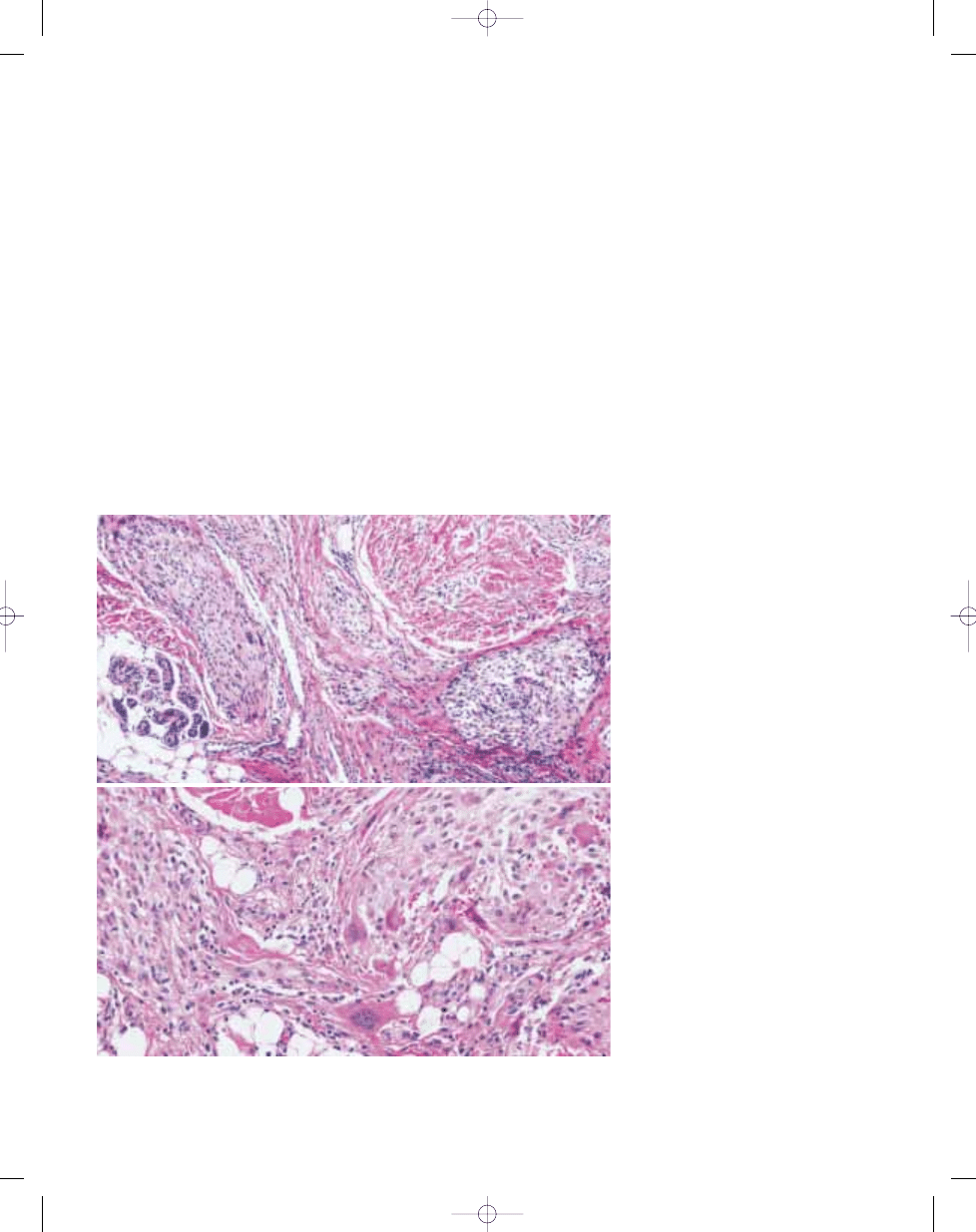
Fig. 3.18 A A multinodular growth pattern is present in approximately 85% of giant cell tumours of soft tissues. B Typical nodule with peripheral accumulation of osteo-
clast-like giant cells.
B
A
Fig. 3.19 A Secondary cystic changes, similar to aneurysmal bone cystic changes, occur in approximately 30% of giant cell tumours of soft tissue. B Metaplastic bone,
frequently in the form of a peripheral shell of woven bone, is present in approximately 50% of giant cell tumours of soft tissue.
119
Giant cell tumour of soft tissues
B
A
Fig. 3.20 A Clusters of foam macrophages reflecting regressive change in a giant cell tumour of soft tissue. B CD68 marks the multinucleate, osteoclastlike giant cells and
a few of the mononuclear cells in giant cell tumours of soft tissue.
bb5_8.qxd 13.9.2006 10:40 Page 119
Definition
The term pleomorphic malignant fibrous
histiocytoma is now reserved for a small
group of undifferentiated pleomorphic
sarcomas. Both terms may be used syn-
onymously. Current technology does not
show a definable line of differentiation.
ICD-O code
8830/3
Synonyms
Fibroxanthosarcoma {1088}; malignant
fibrous histiocytoma, storiform or fibrob-
lastic type; malignant fibrous xanthoma.
Historical annotation
For many years, pleomorphic malignant
fibrous histiocytoma (MFH) has been
regarded as the prototypical form of MFH
and the most common soft tissue sarco-
ma in adults {599,2233,2237}. Originally
defined, based on morphology and tis-
sue culture analysis, as a pleomorphic
spindle cell malignant neoplasm show-
ing fibroblastic and facultative histiocytic
differentiation, it is now widely accepted
that the morphologic pattern known as
so-called pleomorphic MFH may be
shared by a wide variety of poorly differ-
entiated malignant neoplasms {675}. It is
also now agreed that these tumours
show no evidence of true histiocytic dif-
ferentiation. This diagnostic term is now
reserved (by those who still use it) for the
much smaller group of pleomorphic sar-
comas which, by current technology,
show no definable line of differentiation
{2243}. As a consequence, the apparent
incidence of pleomorphic MFH has fallen
sharply over the past 10 years and it is
possible that this term may disappear
altogether at such time as criteria for the
diagnosis of pleomorphic sarcomas
showing fibroblastic or myofibroblastic
differentiation can be reproducibly
defined.
Epidemiology
The group of pleomorphic (MFH-like)
sarcomas collectively represent the most
common types of sarcoma in patients
over age 40. The overall incidence
among adults approximates to 1-2 cases
per 100,000 patients annually and the
incidence increases with age {861}.
Most undifferentiated high grade sarco-
mas occur in patients over age 40 with
peak incidence in the 6th and 7th
decades. Rare examples may be
encountered in adolescents and young
adults. There is a male predominance of
approximately 1.2:1.
Sites of involvement
Most undifferentiated high grade pleo-
morphic sarcomas occur in the extremi-
ties (especially the lower limb) and less
often the trunk. The majority of cases
arise in deep (subfascial) soft tissue,
while less than 10% are primarily subcu-
taneous. A notable exception among
pleomorphic sarcomas is dedifferentiat-
ed liposarcoma (see p. 38) which is most
common in the retroperitoneum.
Clinical features
Undifferentiated high grade pleomorphic
sarcomas are typically large deep-seat-
ed tumours which show progressive,
often rapid enlargement. Only those
which grow very rapidly tend to be
painful. Around 5% of patients have
metastases at presentation, most often to
lung. Although little is known about aeti-
ology of these lesions, a subset of pleo-
morphic sarcomas (<2-3%) arise at the
site of prior radiation therapy {1224} and
very rare cases arise at the site of chron-
ic ulceration or scarring.
Macroscopy
Most undifferentiated high grade pleo-
morphic sarcomas are well circum-
scribed, expansile masses which may
appear pseudoencapsulated. Tumour
size varies and, to some extent, depends
on location with subcutaneous lesions
often measuring <5 cm, while retroperi-
toneal tumours often exceed 20 cm. Most
tumours measure between 5 and 15 cm
in maximum diameter. Cut surface is vari-
able and may include pale fibrous or
fleshy areas, admixed with zones of
necrosis, haemorrhage or myxoid
change. Aside from an adjacent well-dif-
ferentiated component in dedifferentiat-
ed liposarcoma, there are no distinctive
macroscopic features which correlate
reliably with line of differentiation.
Histopathology
Undifferentiated high grade sarcoma is a
diagnosis of exclusion following thorough
sampling and judicious use of ancillary
diagnostic techniques. Tumours in the
general category of high grade pleomor-
phic (MFH-like) sarcomas are very het-
erogeneous in appearance and also in
cellularity, since some cases have an
extensive fibrous stroma. These tumours
have in common marked cytological and
nuclear pleomorphism, often with bizarre
tumour giant cells, admixed with spindle
cells and often rounded histiocyte-like
cells (which may have foamy cytoplasm)
in varying proportion {675}. A storiform
growth pattern and stromal chronic
inflammatory cells are common. The
spindle cell component most often
appears fibroblastic, myofibroblastic or
smooth muscle-like. Tumours showing
myogenic differentiation (pleomorphic
leiomyosarcoma or rhabdomyosarco-
ma), as well as carcinoma and
melanoma with MFH-like morphology,
often have more copious eosinophilic
cytoplasm and prominent large polygo-
nal cells. The presence of fascicular
spindle cell areas may suggest smooth
muscle or nerve sheath differentiation
(which needs to be proved immunohisto-
chemically or ultrastructurally). Thorough
C.D.M. Fletcher
E. van den Berg
W.M. Molenaar
Pleomorphic malignant fibrous
histiocytoma / Undifferentiated high
grade pleomorphic sarcoma
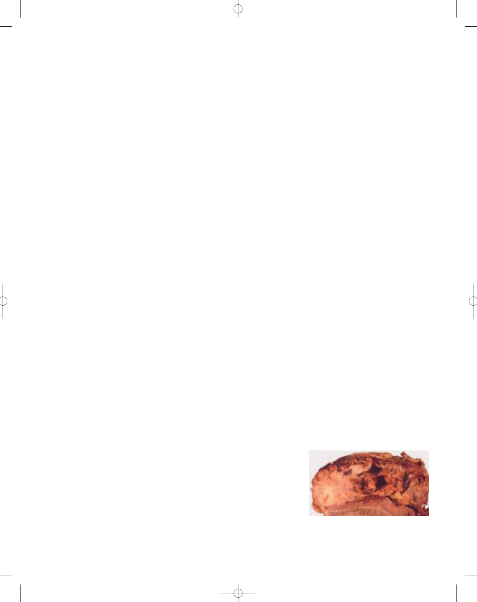
Fig. 3.21 Undifferentiated high grade pleomorphic
sarcomas are typically deep-seated and large, with
a variable cut surface; this case shows fleshy solid
areas, necrosis and cystic change.
120
Fibrohistiocytic tumours
bb5_8.qxd 13.9.2006 10:40 Page 120
sampling is critical in all cases to check
for the presence of lipoblasts or ‘malig-
nant’ osteoid.
Immunohistochemistry
The widespread introduction of immuno-
histochemistry has been one of the major
factors in demolition of the MFH concept.
Most high grade pleomorphic sarcomas
show a definable line of differentiation,
foremost among which are the pleomor-
phic variants of leiomyosarcoma, liposar-
coma, rhabdomyosarcoma and myxofi-
brosarcoma, after carcinomas,
melanomas and lymphomas have been
excluded {675}. Immunohistochemistry
was critical in helping to separate the lat-
ter non-mesenchymal malignancies.
Controversy exists as to the extent of
immunopositivity required for a given
antigen to define a specific line of differ-
entiation but diagnostic criteria have
been proposed for the different pleomor-
phic sarcomas and these appear to be
reproducible {683,1425}. The presence
of just rare cells showing positivity for
epithelial or myogenic antigens most
often has little significance and does not,
of itself, exclude this diagnosis. It is now
accepted that histiocytic antigens (such
as alpha-1-antitrypsin, alpha-1-antichy-
motrypsin, lysozyme and CD68) play no
useful role in the diagnosis of pleomor-
phic sarcomas.
Ultrastructure
Electron microscopic findings depend
upon the specific type of tumour giving
rise to the pleomorphic MFH pattern.
Inevitably almost all tumours in this cate-
gory are poorly differentiated so only a
minority of tumour cells may show ultra-
structural features of a specific lineage.
Many tumour cells show relatively undif-
ferentiated, non-specific fibroblast-like or
histiocyte-like features.
Genetics
The genetic aspects of malignant fibrous
histiocytomas (MFH) are difficult to eval-
uate because of the shifting diagnostic
criteria used throughout the years.
Bearing these shortcomings in mind,
cytogenetic aberrations have been
detected in more than 50 cases pub-
lished as storiform or pleomorphic MFH
or MFH NOS {1477}. Only a few cases of
giant cell or inflammatory MFH have
been investigated. In general, the kary-
otypes tend to be highly complex, with
extensive intratumoral heterogenity and
chromosome numbers in the triploid or
tetraploid range in the majority of cases
{1317,1477,1486,1635,1957}. Also near-
haploid karyotypes have been reported
in a few cases {92}. No specific structur-
B
A
Fig. 3.22 Undifferentiated high grade pleomorphic sarcoma. A Note the variable cellularity and striking cytological pleomorphism. This tumour proved to be a malignant
peripheral nerve sheath tumour. B In other areas this lesion turned out to be pleomorphic liposarcoma with prominent lipoblasts.
121
Pleomorphic malignant fibrous histiocytoma / Undifferentiated high grade pleomorphic sarcoma
B
A
Fig. 3.23 Undifferentiated high grade pleomorphic sarcoma. A Note the anaplastic cytomorphology in this unclassified sarcoma. B Many tumour cells show a promi-
nent eosinophilic cytoplasm and this case proved to be pleomorphic leiomyosarcoma.
bb5_8.qxd 13.9.2006 10:40 Page 121
al or numerical aberrations have
emerged, but telomeric associations,
ring chromosomes, and/or dicentric
chromosomes are frequent. Such chro-
mosomal abnormalities are, however,
common also in other fibrohistiocytic
lesions {1854}. Due to the presence of
numerous marker chromosomes in most
cases, the distribution of genomic imbal-
ances is impossible to asses reliably
from cytogenetic data.
Genomic imbalances, as detected by
comparative genomic hybridization
(CGH), frequently include loss of 2p24-
pter and 2q32-qter, and chromosomes
11, 13 and 16 {1219,1311,1651,1957,
2094}, as well as gain of 7p15-pter, 7q32,
and 1p31.
Several proto-oncogenes mapping to
chromosome region 12q13-15 appear to
participate in the development of MFH-
like pleomorphic sarcomas:
SAS, MDM2,
CDK4, DDIT3 (a.k.a. CHOP), and HMGIC
(a.k.a
HMGA2) have all been reported to
be amplified in MFH {172,1772,1842}. In
an amplicon at 8p23.1 a candidate gene
designated
MASL1 has been found
{1842}.
Alterations (mutations and/or deletions)
of
TP53, RB1 and CDKN2A have been
suggested to play a critical role in pleo-
morphic sarcoma development {341,
1772,1957,2097,2326}, but no clear rela-
tionship with clinical outcome has yet
been found. The significance of
HRAS
mutations and their relationship with
other genetic changes, such as
TP53
and
MDM2 gene status, remain to be
clarified {221,1790,2269}.
Prognostic factors
High grade pleomorphic sarcomas are
aggressive with an overall 5-year survival
probability of only 50-60% {861,2233}.
However, it has become clear that there
are prognostic subgroups among the
lesions formerly categorised as pleomor-
phic MFH {683}. For example, dediffer-
entiated liposarcoma has a metastatic
rate of only 15-20%, high grade myxofi-
brosarcoma has a metastatic rate of
around 30-35%, while pleomorphic myo-
genic sarcomas (leiomyosarcoma or
rhabdomyosarcoma) are especially
aggressive with much more frequent
metastasis and shorter relapse-free sur-
vival {1679}. The clinical and therapeutic
benefits of subclassifying pleomorphic
sarcomas are only just beginning to be
appreciated, hence the approach to sub-
classification and grading of pleomor-
phic sarcomas is likely to evolve.
122
Fibrohistiocytic tumours
Fig. 3.24 A Many pleomorphic sarcomas contain large bizarre cells with foamy cytoplasm, which in the past were mistakenly regarded as histiocytic in nature. B
A storiform growth pattern is a common feature shared by many of these undifferentiated high grade pleomorphic sarcomas, irrespective of lineage. C The pres-
ence of polygonal cells with prominent eosinophilic cytoplasm usually suggests myogenic, epithelial or less often melanocytic differentiation. This case proved to
be a pleomorphic rhabdomyosarcoma.
B
A
C
bb5_8.qxd 13.9.2006 10:40 Page 122
Definition
Formerly defined as a variant of malig-
nant fibrous histiocytoma (MFH) with
prominent osteoclastic giant cells, it is
now appreciated that this morphologic
pattern may be shared by a variety of
tumour types. The term giant cell MFH is
currently reserved for undifferentiated
pleomorphic sarcomas with prominent
osteoclastic giant cells.
ICD-O code
8830/3
Synonyms
Malignant giant cell tumour of soft parts,
malignant osteoclastoma, giant cell
sarcoma.
Historical annotation
Although formerly defined as a variant of
malignant fibrous histiocytoma (MFH)
with prominent osteoclastic giant cells
{599} (and frequently known as malig-
nant giant cell tumour of soft parts/tis-
sues {61,848}) it is now appreciated that
this morphologic pattern may be shared
by a variety of tumour types (most
notably giant cell tumour of soft tissues,
extraskeletal osteosarcoma, leiomyosar-
coma and osteoclast-rich carcinoma)
{961}. It is difficult to define giant cell
MFH as a discrete entity and this diagno-
sis is gradually disappearing from com-
mon usage in soft tissue pathology.
Epidemiology
All of the lesions previously subsumed
under this heading are very uncommon.
Arguably giant cell tumour of soft tissues
(see page 118) is the most frequent.
Almost all of the tumours which adopt the
pattern known as so-called giant cell
MFH occur in older adults with no sex
predilection. Rare examples of giant cell
tumour of soft tissue occur in children
and adolescents.
Sites of involvement
With the exception of giant cell tumour of
soft tissues (which shows a predilection
for subcutaneous tissue) {702,1591,
1608}, most tumours in this general cate-
gory occur in deep soft tissue of the
limbs or trunk. Organs in which giant cell-
rich or osteoclastoma-like carcinomas
are most common include pancreas, thy-
roid, breast and kidney.
Clinical features
Most tumours in this general category
present as an enlarging, painless, deep-
seated mass without distinctive features.
Macroscopy
With the exception of giant cell tumour of
soft tissues, most tumours in this general
category are high grade and thus tend to
be large tumours with haemorrhage and
necrosis. Tumour size is variable but
superficially located examples are small-
er than those in deep soft tissue.
Histopathology
The features shared by tumours previ-
C.D.M. Fletcher
Giant cell malignant fibrous
histiocytoma / Undifferentiated
pleomorphic sarcoma with giant cells
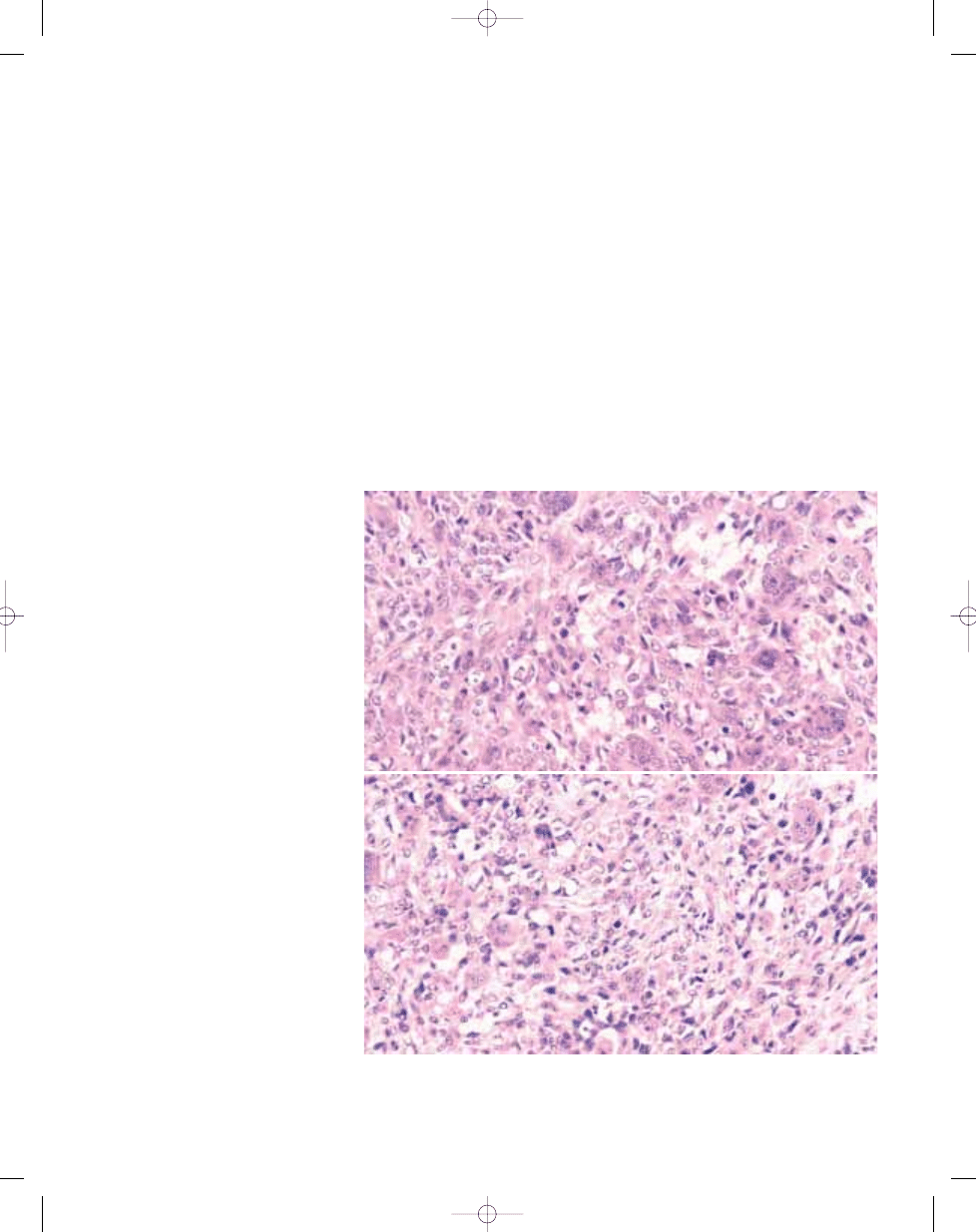
Fig. 3.25 Giant cell MFH. Two tumours showing the pattern often labelled as giant cell MFH, being character-
ized by atypical spindle-shaped and more epithelioid cells admixed with prominent osteoclastic giant cells. The
example on top (A) proved to be anaplastic carcinoma of thyroid, while the lower one (B) was a soft tissue
osteosarcoma.
A
B
123
Pleomorphic malignant fibrous histiocytoma
bb5_8.qxd 13.9.2006 10:40 Page 123
ously labelled as giant cell MFH include
variably pleomorphic ovoid-to-spindle-
shaped cells and a prominent stromal
osteoclastic giant cell reaction. In most
(but not all) lesions the giant cell compo-
nent lacks cytological features of malig-
nancy, but some tumours diagnosed as
giant cell MFH were notable for the pres-
ence of numerous bizarre multinucleate
tumour giant cells.
Aside from these similar (shared) fea-
tures, morphology is largely determined
by the specific tumour type. Giant cell-
rich soft tissue osteosarcoma (see page
182) definitionally shows variably promi-
nent ‘malignant’ osteoid being laid down
by cytologically atypical cells {355}.
Giant cell tumour of soft tissues (see
page 118) usually has a multinodular
growth pattern and cytologically resem-
bles giant cell tumour of bone {702,
1591,1608}. Leiomyosarcoma with
prominent osteoclastic giant cells has at
least small areas with conventional
smooth muscle cytomorphology and a
fascicular growth pattern {1411}. Other
sarcoma types may occasionally show
prominent osteoclastic giant cells {1415}.
Immunohistochemistry
Leiomyosarcoma with prominent osteo-
clastic giant cells usually shows positivi-
ty for smooth muscle actin and desmin in
the fascicular spindle cell component.
Unequivocal positivity for keratin is a
diagnostic requirement for osteoclas-
toma-like or giant cell-rich carcinoma,
with the exception of those cases show-
ing obvious morphologic transition to
usual carcinoma.
Prognostic factors
Undifferentiated high grade sarcomas
with prominent osteoclastic giant cells
behave similarly to other pleomorphic
sarcomas. Among neoplasms simulating
giant cell MFH, extraskeletal osteosarco-
ma and leiomyosarcoma are much more
aggressive than giant cell tumour of soft
tissues.
Fig. 3.28 Leiomyosarcoma mimicking so-called giant
cell MFH. Note with prominent osteoclastic giant
cells and the eosinophilic fascicular spindle cell
component..
Fig. 3.26 Giant cell MFH may resemble giant cell tumour of soft tissue, which has a multinodular growth pat-
tern, was often formerly labelled as giant cell MFH.
124
Fibrohistiocytic tumours
Fig. 3.27 Giant cell-rich soft tissue osteosarcoma.
This osteoclast-rich spindle cell malignant neoplasm
contains seams of osteoid produced by cytologically
malignant cells.
bb5_8.qxd 13.9.2006 10:40 Page 124
Definition
A malignant neoplasm characterized by
numerous xanthomatous cells, morpho-
logically both benign and malignant,
admixed with atypical spindle cells and
acute and chronic inflammatory cells.
Originally regarded as a variant of so-
called malignant fibrous histiocytoma
(MFH), differentiation in these tumours is
poorly understood and their morphology
may be shared by both mesenchymal
and epithelial neoplasms. The term
inflammatory MFH is now reserved for
undifferentiated pleomorphic sarcomas
with a prominent histiocytic and inflam-
matory infiltrate.
ICD-O code
8830/3
Synonyms
Xanthomatous MFH, malignant fibrous
xanthoma, xanthosarcoma.
Epidemiology
This is the rarest and the least document-
ed type of MFH, with only two published
series of 7 and 8 cases {1096,1198} and
a few case reports. There is no apparent
gender predominance, and patients are
usually more than 40 years old.
Sites of involvement
The most common site is the retroperi-
toneum but intra-abdominal and deep
soft tissue locations have also been
observed.
Clinical features
In addition to symptoms and imaging
features of a large retroperitoneal tumour,
inflammatory MFH may be associated
with fever, weight loss, leukocytosis,
eosinophilia, and leukaemoid reaction.
Analysis of tumour extracts and immuno-
histochemistry suggested that produc-
tion of specific cytokines by tumour cells
is responsible for the systemic symptoms
{1401,2076}.
Aetiology
There is no aetiology known for inflam-
matory MFH, but one post-radiation case
has been reported {735}.
Macroscopy
This tumour is usually large and often
displays a yellow colour due to large col-
lections of xanthoma cells.
Histopathology
Inflammatory MFH is characterized by
sheets of benign xanthoma cells with
numerous inflammatory cells including
neutrophils, eosinophils and a minor
component of lymphocytes and plasma
cells. Some cases show only a few or no
xanthoma cells but are predominantly
composed of neutrophils and eosino-
phils. There are scattered atypical large
cells, with one or more irregular, hyper-
chromatic nuclei with prominent nucleoli.
These cells may be rare and difficult to
find and occasionally resemble Reed-
Sternberg cells. Occasionally atypical
cells are xanthomatized and typically
display phagocytosis of neutrophils.
These cells may be set in a hyalinized
collagenous background. In most cases,
there are typical areas of pleomorphic
MFH-like sarcoma with spindle and pleo-
morphic cells arranged in a haphazard
growth pattern. Like pleomorphic MFH,
inflammatory MFH is a diagnosis of
exclusion and could represent an inflam-
matory dedifferentiated component
shared by different neoplasms such as
carcinomas, lymphomas, leiomyosarco-
mas, inflammatory myofibroblastic
tumours and liposarcomas {956,961}.
Among these, dedifferentiated liposarco-
ma is the most common simulant.
J.M. Coindre
Inflammatory malignant fibrous
histiocytoma / Undifferentiated
pleomorphic sarcoma with prominent
inflammation
B
A
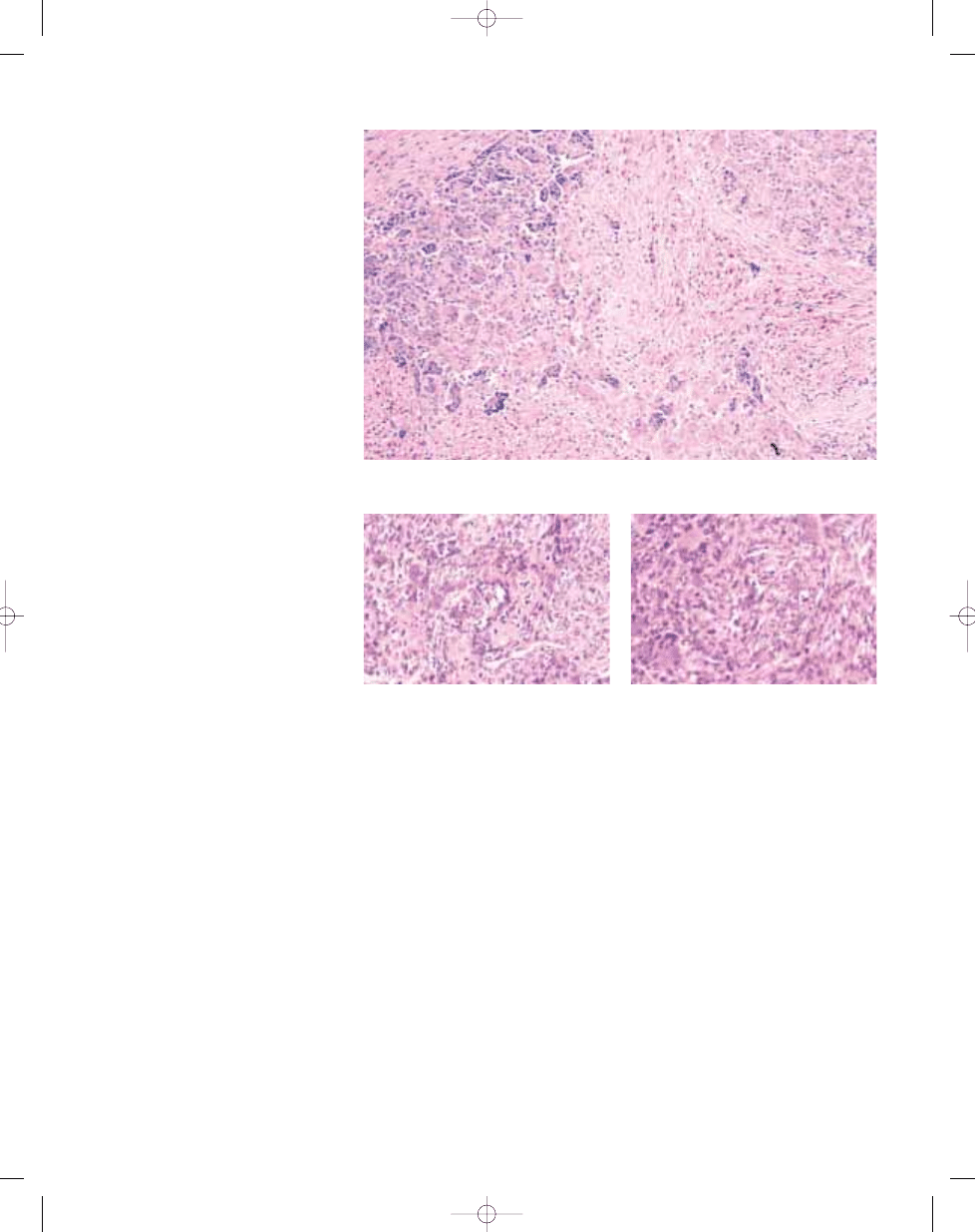
Fig. 3.29 Inflammatory malignant fibrous histiocytoma. A Pleomorphic spindle cells are associated with numerous inflammatory cells. B The atypical cells may be sug-
gestive of a lymphoid neoplasm.
125
Inflammatory malignant fibrous histiocytoma
bb5_8.qxd 13.9.2006 10:41 Page 125
Therefore inflammatory MFH areas may
often be associated with areas of more
specific tumours which should be care-
fully looked for.
Immunophenotype
Immunohistochemistry is useful for show-
ing a specific line of differentiation such
as epithelial, lymphoid or smooth muscu-
lar. In the other cases, the neoplastic
cells express vimentin, occasionally
CD68, but are negative for CD15, CD20,
CD30, CD43 and CD45 {1096}.
Ultrastructure
The tumour cells do not differ ultrastruc-
turally from tumour cells of pleomorphic
MFH.
Genetics
Genetic analysis may be particularly use-
ful for identifying a possible dedifferenti-
ated liposarcoma or other simulants such
as anaplastic large cell lymphoma.
Prognostic factors
From a review of the literature {961} and
a small series {1198}, it appears that two-
thirds of patients died of their tumour with
persistent or recurrent disease. About
one fourth of patients developed distant
metastasis. As in other retroperitoneal
sarcomas, this poor prognosis is proba-
bly related to the extent of the tumour
and its inaccessibility to proper surgery
at the time of the diagnosis.
126
Fibrohistiocytic tumours
Fig. 3.30 Inflammatory malignant fibrous histiocytoma. A Note the striking cytophagocytosis. B Pleomorphic
MFH-like areas with collagenous stroma are common.
A
B
bb5_8.qxd 22.9.2006 10:39 Page 126
Wyszukiwarka
Podobne podstrony:
bb5 chap8
bb5 chap1
BB5 BOX
bb5 chap16
bb5 chap15
bb5 contents
bb5 chap12
bb5 chap4
bb5 references
bb5 chap6
bb5 chap17
chap3 forward kinematics
bb5 chap20
chap3
CHAP3
bb5 chap5
Lista wszystkich dostępnych polskich Product Code dla telefonów platformy BB5
więcej podobnych podstron