CHAPTER 16
Giant Cell Tumours
Almost any kind of lesion in bone can contain giant cells, some-
times numerous. In order to qualify as a giant cell tumour, the
neoplasm has to have a combination of round to oval mono-
nuclear cells and more or less uniformly distributed giant cells.
Moreover, the nuclei of the giant cells should be very similar to
those of the mononuclear cells.
Giant cell tumours occur in skeletally mature individuals and
there is a slight female predominance. The ends of long bones
and the body of the vertebrae are typical sites. The tumour is
locally aggressive, but distant metastases are uncommon.
When metastases do occur, they rarely prove fatal and hence
the term benign metastases is appropriate.
Malignant change in giant cell tumour is uncommon. A sarcoma
may co-exist with a giant cell tumour (primary) or may arise at
the site of a previously diagnosed giant cell tumour (secondary).
bb5_24.qxd 13.9.2006 13:42 Page 309
Giant cell tumour
R. Reid
S.S. Banerjee
R. Sciot
Definition
Giant cell tumour is a benign, locally
aggressive neoplasm which is com-
posed of sheets of neoplastic ovoid
mononuclear cells interspersed with
uniformly distributed large, osteoclast-
like giant cells.
ICD-O code
9250/1
Synonym
Osteoclastoma.
Epidemiology
Giant cell tumour represents around 4-
5% of all primary bone tumours, and
approximately 20% of benign primary
bone tumours. The peak incidence is
between the ages of 20 and 45.
Although 10-15% of cases occur in the
second decade, giant cell tumour is sel-
dom seen in skeletally immature individ-
uals and very rarely in children below 10
years {299,538,1875,2155}. There is a
slight female predominance in some
large series. There is no striking racial
variation, but there may be some geo-
graphic variation.
Sites of involvement
Giant cell tumours typically affect the
ends of long bones, especially the dis-
tal femur, proximal tibia, distal radius
and proximal humerus. Around 5%
affect flat bones, especially those of the
pelvis. The sacrum is the commonest
site in the axial skeleton, while other ver-
tebral bodies are less often involved.
Fewer than 5% of cases affect the tubu-
lar bones of the hands and feet {200}.
Multicentric giant cell tumors are very
rare and tend to involve the small bones
of the distal extremities.
Rarely, tumours with the morphology of
giant cell tumour arise primarily within
soft tissue {702}.
Clinical features / Imaging
Patients with giant cell tumour typically
present with pain, swelling and often
limitation of joint movement; pathologi-
cal fracture is seen in 5-10% of patients.
Plain X-rays of lesions in long bones
usually show an expanding and eccen-
tric area of lysis. The lesion normally
involves the epiphysis and adjacent
metaphysis; frequently, there is exten-
sion up to the subchondral plate, some-
times with joint involvement. Rarely, the
tumour is confined to the metaphysis,
usually in adolescents where the tumour
lies in relation to an open growth plate,
but occasionally also in older adults.
Diaphyseal lesions are exceptional.
The margins of the lesion vary; this is
the basis of a radiological grading/stag-
ing system {299}. Type 1, ‘quiescent’,
lesions have a well-defined margin with
surrounding sclerosis and show little, if
any, cortical involvement. Type 2,
‘active’ tumours have well-defined mar-
gins, but lack sclerosis; the cortex is
thinned and expanded. Type 3, ‘aggres-
sive’ tumours have ill-defined margins
often with cortical destruction and soft
tissue extension. This grading system
does not correlate well with histological
appearances. On occasion, a giant cell
tumour has a trabeculated ‘soap-bub-
ble’ appearance. In the tubular bones of
the hands and feet, the x-ray appear-
ances are similar to those seen in long
bones. Tumours of sacrum and pelvic
bones are also lytic, commonly involve
adjacent soft tissues and may affect
sacro-iliac and hip joints.
There is seldom much reactive
periosteal new bone formation. Only
occasionally is radiologically evident
matrix produced within the tumour, usu-
ally in long standing lesions.
CT scanning gives a more accurate
assessment of cortical thinning and
penetration than plain radiographs. MR
imaging is most useful in assessing the
extent of intra-osseous spread and
defining soft tissue and joint involve-
ment. Giant cell tumour typically shows
low to intermediate signal intensity on
T1 weighted images and intermediate to
high intensity on T2 images. Large
amounts of haemosiderin are often
present giving areas of low signal in
both modalities.
Macroscopy
The appearance of an intact specimen
mirrors the radiological appearances in
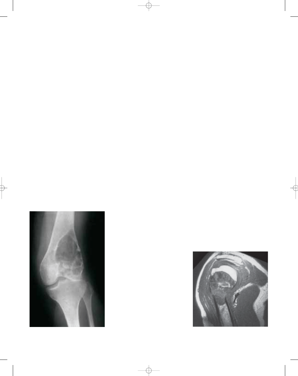
Fig. 16.01 Giant cell tumour. Large, expansile area
of lysis with a sclerotic border, cortical thinning,
and extension to the subchondral plate.
Fig. 16.02 Giant cell tumour of the proximal humerus.
MRI shows a well demarcated lesion with focal
destruction of cortex and extension into the epiphysis.
310
Giant cell tumours
bb5_24.qxd 13.9.2006 13:42 Page 310
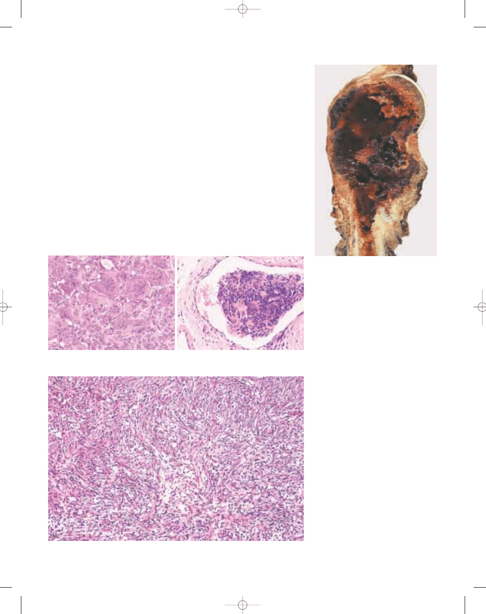
its eccentric location and fairly well
defined area of bone destruction. This is
often bounded by a thin and often
incomplete shell of reactive bone.
Although the tumour frequently erodes
the subchondral bone to reach the deep
surface of the articular cartilage, it sel-
dom penetrates it. The tissue is usually
soft and reddish brown, but there may
be yellowish areas corresponding to
xanthomatous change, and firmer whiter
areas where there is fibrosis. Blood-
filled cystic spaces are sometimes seen
and, when extensive, this may cause
confusion with an aneurysmal bone
cyst.
Histopathology
The characteristic histopathological
appearance is of round to oval polygo-
nal or elongated mononuclear cells
evenly mixed with numerous osteoclast-
like giant cells which may be very large
and contain 50 to 100 nuclei. The nuclei
of the stromal cells are very similar to
those of the osteoclasts, having an open
chromatin pattern and one or two small
nucleoli. The cytoplasm is ill-defined,
and there is little intercellular collagen.
Mitotic figures are invariably present;
they vary from 2 to 20 per ten high
power fields. Atypical mitoses are not,
however, seen and their presence
should point to a diagnosis of a giant
cell rich sarcoma. Occasional binucle-
ate and trinucleate cells are seen.
It is now generally accepted that the
characteristic large osteoclastic giant
cells are not neoplastic. The mononu-
clear cells, which represent the neo-
plastic component, are thought to arise
from primitive mesenchymal stromal
cells. They express RANKL, which stim-
ulates formation and maturation of
osteoclasts from osteoclast precursors
{1814,2342}; these cells of monocyte
lineage represent a second, minor, com-
ponent of the mononuclear cells.
There are variations from these standard
appearances. In some cases, the mono-
nuclear cells are more spindle shaped,
and they may be arranged in a storiform
growth pattern. Commonly, small num-
bers of foam cells are present, and in
rare cases this is the predominant pat-
tern thus simulating a fibrous histiocy-
toma. There may be areas of fibrosis,
while secondary aneurysmal bone cyst
change occurs in 10% or so. Small foci
of bone formation within the tumour are
found, especially after pathological frac-
ture or biopsy. When the tumour extends
into soft tissue or is present in lung, the
histological features are identical to the
primary lesion, and there is often a
peripheral shell of reactive bone. A strik-
ing feature, in one third of cases, is the
presence of intravascular plugs, partic-
ularly at the periphery of the tumour; this
does not appear to be of prognostic sig-
nificance. Areas of necrosis are com-
mon, especially in large lesions. These
may be accompanied by focal nuclear
atypia which may suggest malignancy.
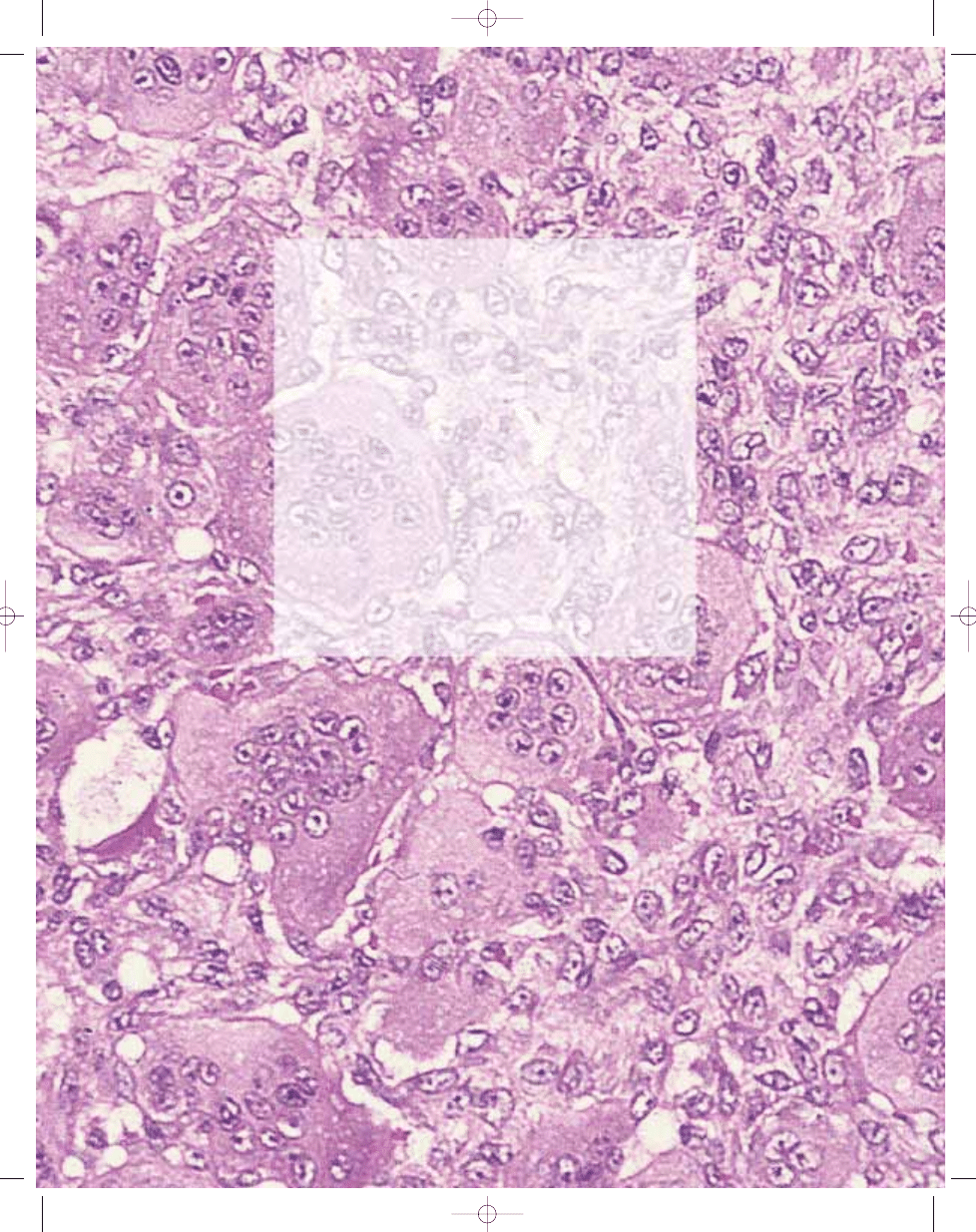
Fig. 16.05 Giant cell tumour. In some cases like this one, a storiform arrangement of fibroblasts and
macrophages resembles a benign fibrous histiocytoma.
B
A
Fig. 16.04 Giant cell tumour. A Typical appearance with large osteoclasts and uniform ovoid mononuclear
cells. B The vascular lumen contains a mixture of spindle and giant cells.
Fig. 16.03 Giant cell tumour. Large haemorrhagic
tumour of the proximal humerus with extensive corti-
cal destruction and soft tissue extension.
311
Giant cell tumour
bb5_24.qxd 13.9.2006 13:42 Page 311
Immunophenotype
The giant cells have the typical
immunophenotype of normal osteo-
clasts, expressing markers of histiocytic
lineage.
Genetics
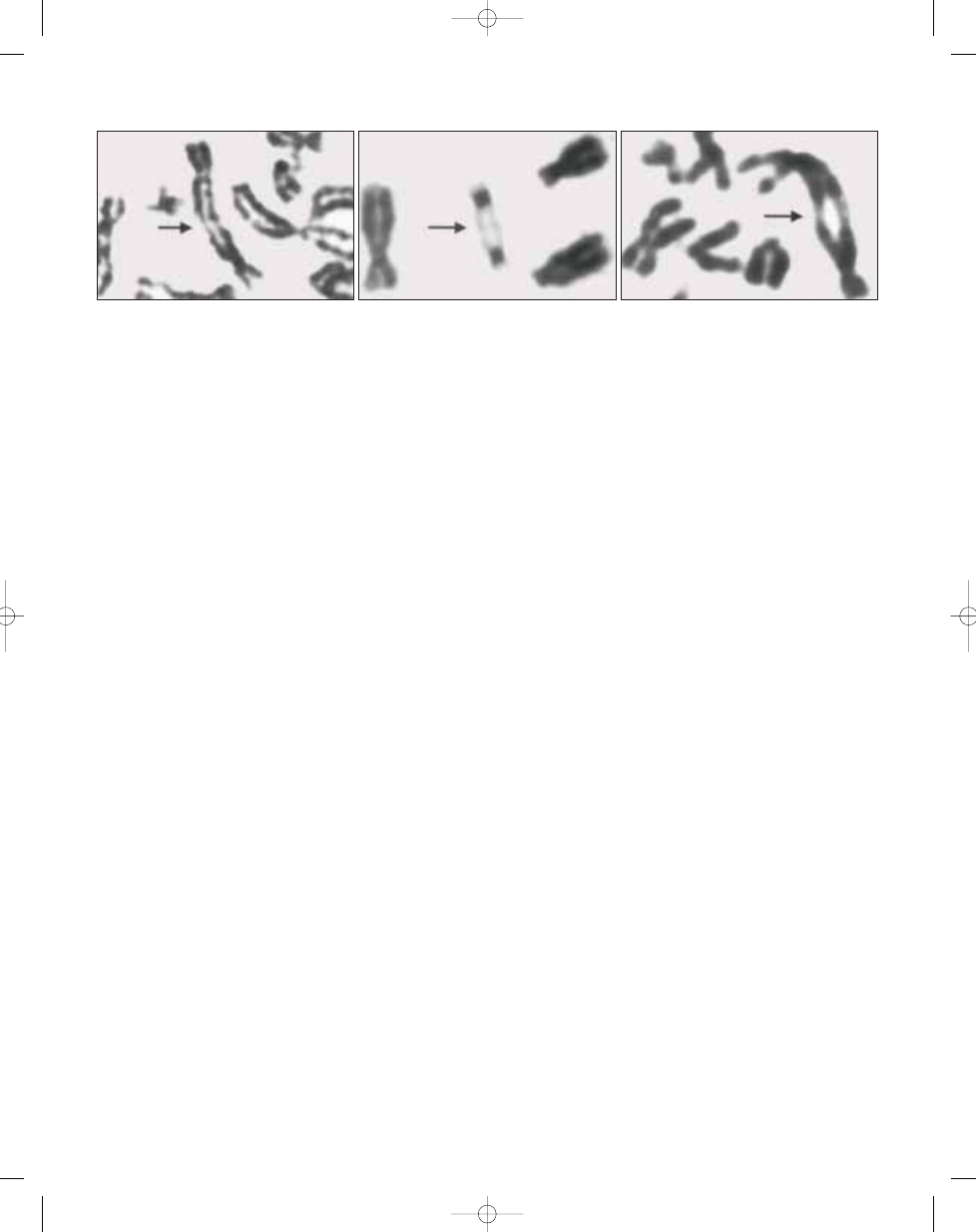
Telomeric association is the most fre-
quent chromosomal aberration. A re-
duction in telomere length (average loss
of 500 base pairs) has been demon-
strated in giant cell tumour cells when
compared to leukocytes from the same
patients {1898}. The telomeres most
commonly affected are 11p, 13p, 14p,
15p, 19q, 20q and 21p {262,1644,
1909,2090,2343}. Giant cell tumours
with a fibrohistiocytic reaction do not dif-
fer karyotypically from the others {1909}.
This observation supports the hypo-
thesis that these lesions are true giant
cell tumours rather than a different en-
tity like a fibroxanthoma. It is of interest
that four cases of giant cell tumour also
showed rearrangements in 16q22 or
17p13 {262,1488,1909}. These findings
might indicate the possible presence of
an associated aneurysmal bone cyst. It
has been suggested that there is an
association between the the presence
or absence of chromosomal aberrations
and clinical behaviour of giant cell
tumours {262}.
Prognostic factors
Giant cell tumour is capable of locally
aggressive behaviour and occasionally
of distant metastasis. Histology does not
predict the extent of local aggression.
Following treatment by curettage, sup-
plemented with bone grafting, cementa-
tion, cryotherapy, or instillation of phe-
nol, local recurrence occurs in approxi-
mately 25% of patients. Recurrence is
usually seen within 2 years. Block exci-
sion for lesions in small bones results in
fewer local recurrences. Pulmonary
metastases are seen in 2% of patients
with giant cell tumours, on average 3-4
years after primary diagnosis {1947}.
These may be solitary or multiple. Some
of these metastases are very slow grow-
ing (benign pulmonary implants) and
some regress spontaneously. A small
proportion are progressive and may
lead to the death of the patient.
Local recurrence, surgical manipula-
tion and location in distal radius may
increase the risk of metastasis {1350}.
Histological grading does not appear to
be of value in predicting which giant cell
tumour will metastasise, providing that
giant cell rich sarcomas have been
excluded. True malignant transformation
is rare {1346}, and often follows radio-
therapy.
312
Giant cell tumours
B
C
A
Fig. 16.06 Giant cell tumour. G-banded partial metaphase spreads (A,B,C). Telomeric associations are indicated by arrows.
bb5_24.qxd 13.9.2006 13:42 Page 312
313
Malignancy in giant cell tumour
Definition
Malignancy in GCT is a high grade sar-
coma arising in a giant cell tumour (pri-
mary) or at the site of previously docu-
mented giant cell tumour (secondary).
ICD-O code
9250/3
Synonyms
Malignant giant cell tumour, dedifferenti-
ated giant cell tumour.
Epidemiology
Malignancy arising in a giant cell tumour
can occur after treatment usually includ-
ing radiation or de novo. Most sarcomas
arise following radiation therapy.
Primary malignant giant cell tumour is
the least common type. Overall, malig-
nant transformation can be expected in
less than 1% of giant cell tumours. There
is a slight female predominance and
patients are generally about a decade
older than patients with giant cell
tumour.
Clinical features / Imaging
The recurrence of pain and swelling
many years following treatment of a
giant cell tumour should suggest the
possibility of malignant transformation.
The symptomatology of primary malig-
nant giant cell tumour is non specific. In
secondary malignant giant cell tumour
plain roentgenograms show a destruc-
tive process with poor margination situ-
ated at the site of a previously diag-
nosed giant cell tumour, usually at the
end of a long bone. Mineralization may
be present. In primary malignancy in
giant cell tumour, the tumour presents
as a lytic process extending to the
end of a long bone. Rarely the roen-
tgenograms show typical features of
giant cell tumour and a sclerotic
destructive tumour juxtaposed to it.
Sites of involvement
Bones involved with giant cell tumour
are also affected by malignancy in giant
cell tumour. The distal femur and the
proximal tibia are the most common
sites. There have been no cases report-
ed in the small bones of the hands and
feet or the skull.
Macroscopy
The gross appearance of a secondary
malignant giant cell tumour is that of any
high grade sarcoma: a large fleshy
white tumour with soft tissue extension.
Primary malignant giant cell tumours
occur at the ends of bones and have
dark red or tan colour.
Histopathology
In secondary giant cell tumour the
neoplasm is a high grade spindle cell
sarcoma which may or may not pro-
duce osteoid. No residual giant cell
tumour is usually present. In primary
malignant giant cell tumour areas
of conventional giant cell tumour with
proliferations of round to oval mononu-
clear cells and multinucleated giant
cells are present. There is an abrupt
transition to a spindle cell tumour with
marked cytological atypia. Multinuc-
leated giant cell may or may not be
present.
Prognostic factors
The prognosis in secondary malignant
giant cell tumours is similar to that of a
high grade spindle cell sarcoma. The
prognosis in primary malignant giant
cell tumours has been reported to be
better {1536}. In this series of eight
patients only one died of disease.
P.G. Bullough
M. Bansal
Malignancy in giant cell tumour
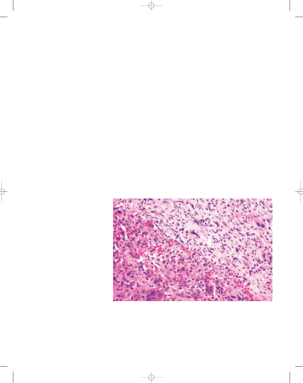
Fig. 16.07 Malignancy in giant cell tumour. Photomicrograph of conventional giant cell tumour (lower left) with
mononuclear cells uniformly interspersed with multinucleated giant cells and an adjacent area of malignant
anaplastic tumour cells (upper right).
bb5_24.qxd 13.9.2006 13:42 Page 313
Wyszukiwarka
Podobne podstrony:
bb5 chap3
bb5 chap8
bb5 chap1
BB5 BOX
bb5 chap15
bb5 contents
bb5 chap12
bb5 chap4
bb5 references
bb5 chap6
bb5 chap17
bb5 chap20
bb5 chap5
Lista wszystkich dostępnych polskich Product Code dla telefonów platformy BB5
bb5 chap21
bb5 source
bb5 chap13
bb5 chap19
więcej podobnych podstron