CHAPTER 4
Smooth Muscle Tumours
Smooth muscle tumours arising at non-cutaneous, non-uterine
locations have been the focus of a considerable conceptual shift
in recent years and this is ongoing. Specifically, it has been
uncertain whether or not there exist benign leiomyomas of deep
soft tissue, but these lesions are now becoming better
recognized and defined. The vast majority of so-called smooth
muscle tumours arising in the gastrointestinal tract, mesentery
and omentum are, in fact, gastrointestinal stromal tumours
defined by the presence of activating
KIT mutations and expres-
sion of KIT protein. These lesions, described in the Digestive
System volume, also account for most cases formerly classified
as epithelioid smooth muscle tumours, or smooth muscle
tumours of uncertain malignant potential.
During the past decade, it has been recognized, mainly through
immunohistochemistry, that soft tissue leiomyosarcoma is more
common than formerly believed and that a rare but histological-
ly distinct subset of these lesions is related to Epstein Barr virus
infection in immunocompromised patients.
Pilar leiomyoma and cutaneous leiomyosarcoma are described
in the Skin volume. Smooth muscle tumours of the external
genitalia (vulvovaginal region, scrotum and nipple), as well as
leiomyomatosis peritonealis disseminata, are described in the
respective WHO Blue Books.
bb5_9.qxd 13.9.2006 10:47 Page 127
Angioleiomyoma
H. Hashimoto
B. Quade
Definition
A frequently painful, benign subcuta-
neous or deep dermal tumour composed
of mature smooth muscle bundles which
surround and intersect between vascular
channels. These tumours form a mor-
phological continuum with myopericy-
toma and myofibroma.
ICD-O code
8894/0
Synonyms
Angiomyoma, vascular leiomyoma.
Epidemiology
Angioleiomyoma is a relatively common
neoplasm. In the largest series reported
by Hachisuga et al., 562 cases of angio-
leiomyoma accounted for approximately
4.4 % of a total of 12,663 cases of benign
soft tissue tumours {863}.
Sites of involvement
Most angioleiomyomas occur in the
extremities, especially the lower extre-
mity, and other sites include the head and
the trunk {1309}. The tumours are usually
located in the subcutis and less often
in the deep dermis. Most of the solid
histological subtype (see below) develop
in the lower extremity, and most of the
cavernous subtype in the upper extre-
mity {863}. Tumours of the venous type
develop more often in the head than do
the other subtypes. In contrast to pilar
leiomyoma (see volume on Skin Tumours),
almost all angioleiomyomas are solitary.
Clinical features
Angioleiomyomas occur more frequently
in women {555, 1500}, although tumours
located in the upper extremity and the
head appear more frequent in men than
in women {863}. The lesions usually
develop between the fourth and sixth
decades of life.
Most angioleiomyomas present as a
small, slowly enlarging mass usually of
several years’ duration. Pain is the most
characteristic subjective complaint in
about half of patients with angioleiomy-
oma {555}. In some patients the pain is
exacerbated by wind, cold, pressure,
pregnancy, or menses.
Macroscopy
Angioleiomyomas are sharply demar-
cated, spherical, grey-white or brown
nodules, and most are less than 2 cm in
diameter. Tumours of the solid type are
smaller than those of the other two types.
Histopathology
Angioleiomyomas may be separated into
three subtypes according to the domi-
nant histological pattern: solid, venous
and cavernous. Smooth muscle cells of
angioleiomyoma are mature and well dif-
ferentiated. Mitotic figures are usually
absent or very rare. In tumours of the
solid type smooth muscle bundles are
closely compacted, and intersect with
one another. Vascular channels in this
type of tumour are large in number but
usually small in size and slit-like. Tumours
of the venous type have vascular chan-
nels of venous type with thick muscular
walls, and lesional smooth muscle bun-
dles are not so compact. The outer layers
of the smooth muscle in the vascular
walls blend with intervascular smooth
muscle bundles. Tumours of the cav-
ernous type are composed of dilated
vascular channels with small amounts of
smooth muscle, and the muscular walls
of these vessels are difficult to distinguish
from intervening smooth muscle bundles.
Although two different histological pat-
terns are seen occasionally in the same
tumour, one of the above histological
subtypes is generally identified as the
dominant histology. According to this
subclassification, the angioleiomyomas
reported by Hachisuga et al. were sepa-
rated into 374 cases (66%) of the solid
type, 127 (23%) of the venous type, and
61 (11%) of the cavernous type {863}.
Rarely, the nuclei of smooth muscle cells
are enlarged and hyperchromatic, proba-
bly displaying degenerative nuclear atyp-
ia {307,1076,1344}. Areas of hyaliniza-
tion, calcification, myxoid change, haem-
orrhage, and small groups of mature fat
cells may be seen {863}. Because there
is no evidence of any relationship
between those fat-containing angi-
oleiomyomas and renal or retroperitoneal
angiomyolipomas, nor with tuberous
sclerosis, they should not be labelled
"subcutaneous angiomyolipoma".
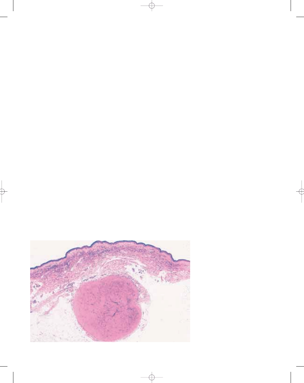
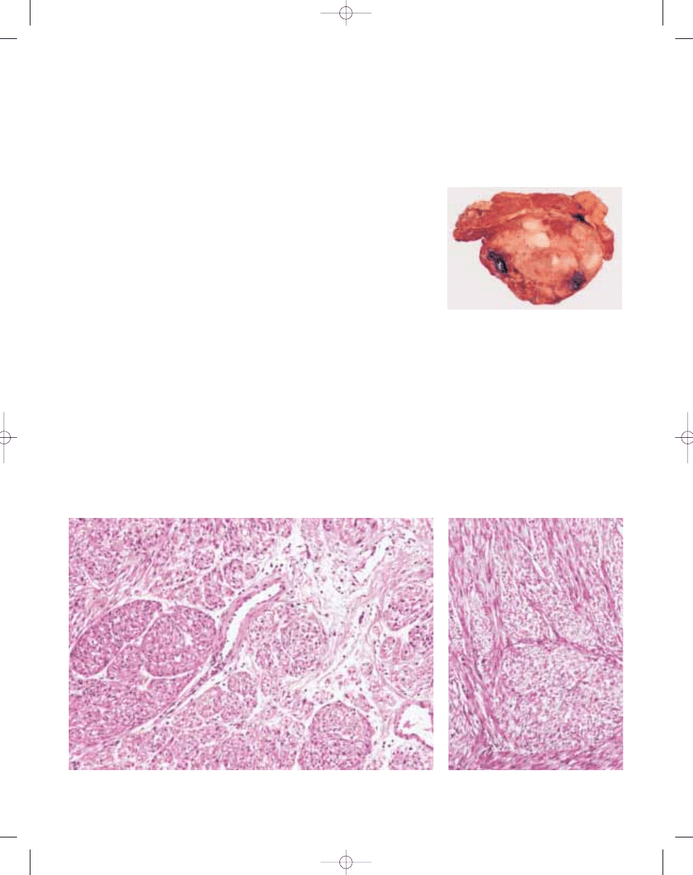
Fig. 4.01 Solid type angioleiomyoma located in the subcutis showing sharp demarcation.
128
Smooth muscle tumours
bb5_9.qxd 13.9.2006 10:47 Page 128
129
Angioleiomyoma
Immunohistochemistry
Most cells are positive for alpha-
smooth muscle actin, desmin, vimentin
and collagen type IV. According to a
study by Hasegawa et al., in more
than half of cases, small nerve fibres
positive for both S100 protein and
PGP9.5 are seen within the capsule of
tumours and tumour stroma {899}. The
peculiar pain of angioleiomyomas is
possibly mediated by these nerve
fibres. In contrast to renal and retroperi-
toneal angiomyolipoma, angioleiomyo-
mas (including the fat-containing
examples) are consistently negative for
HMB45.
Genetics
Cytogenetic data exist for only four
angioleiomyomas from different sites.
All had near-diploid karyotypes, but no
consistent abnormality has been
detected among them {926,936,1567,
1989}.
Prognostic factors
Angioleiomyoma is benign. Simple
local excision is adequate treatment,
and recurrence after excision is
exceptional.
D
C
B
A
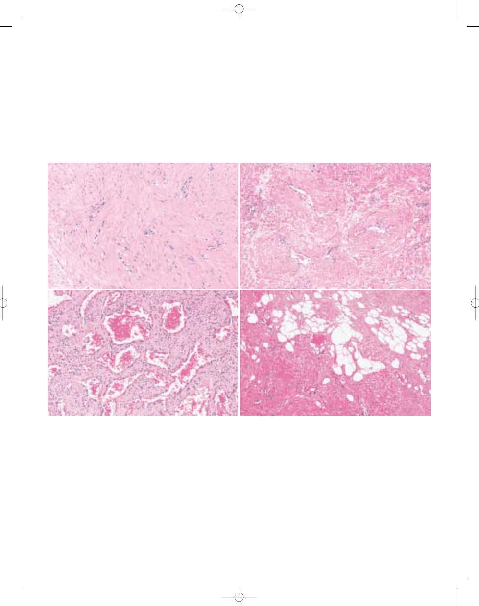
Fig. 4.02 A Angioleiomyomas are typically composed of monomorphic well differentiated smooth muscle cells. B Solid type angioleiomyoma composed of closely com-
pacted vascular and muscle elements. C Cavernous type angioleiomyoma showing dilated vascular channels with little muscular thickening of the walls.
D Angioleiomyoma with groups of mature fat cells.
bb5_9.qxd 13.9.2006 10:47 Page 129
Definition
A very rare type of leiomyoma that
occurs in the deep somatic soft tissue or
retroperitoneum/abdominal cavity.
ICD-O code
8890/0
Epidemiology
The existence and diagnostic criteria of
leiomyomas of deep soft tissue have
been controversial, and only sporadic
cases reports of leiomyomas arising in
the deep soft tissue have been reported,
except for the recent three large series
by Kilpatrick et al. {1106}, Billings et al.
{196}, and Paal and Miettinen {1636},
respectively.
Sites of involvement
The extremities are the most common
site in the deep somatic soft tissue. They
arise in the deep subcutis or skeletal
muscle. Pelvic retroperitoneum and
abdominal cavity, including the mesen-
tery and omentum, are other deep soft
tissues where leiomyomas may occur.
They are always distinct from the uterus
and independent soft tissue primaries
rather than parasitic leiomyomas of the
uterus.
Clinical features
Leiomyomas of the deep somatic soft tis-
sue affect both sexes equally, whereas
leiomyomas of the retroperitoneum or
abdominal cavity occur almost exclu-
sively in women {196,1636}. Most
patients in both groups are young adults
or middle-aged. Many lesions are calci-
fied, so they may be detected radi-
ographically.
Macroscopy
Leiomyomas of the deep soft tissue are
well circumscribed, grey-white tumours.
The greatest diameter of 11 leiomyomas
of the deep somatic soft tissue reported
by Kilpatrick et al. ranged 2.5 – 15 cm
(mean 7.7 cm), and most measured 5 cm
or more, exceeding the usual size of
angioleiomyomas {1106}. Twenty retro-
peritoneal and 3 abdominal leiomyomas
reported by Billings et al. ranged in size
3.2-37 cm (mean 14 cm) {196}. The
greatest diameter of 51 retroperitoneal
leiomyomas reported by Paal and
Miettinen ranged 2.5 - 31 cm (mean 16.2
cm), and the tumour weight ranged 28 -
5400 g (mean 1600 g) {1636}. Myxoid
change is common.
Histopathology
Leiomyomas of deep soft tissue are
composed of cells that closely resemble
normal smooth muscle cells because
they have eosinophilic cytoplasm with
haematoxylin and eosin, fuchsinophilic,
red-staining cytoplasm with Masson’s
trichrome technique and bland, uniform
blunt-ended, cigar-shaped nuclei. They
are arranged in orderly intersecting fas-
cicles. They are highly differentiated,
possess little or no atypia and, at most,
an extremely low level of mitotic activity.
In limb lesions and intra-abdominal
lesions in males, mitoses number less
than 1/50 HPF. In peritoneal / retroperi-
toneal lesions in females (showing posi-
tivity for hormonal receptors) mitoses
may number up to 5/50 HPF. Necrosis
should not been present in deep leio-
myoma. Most lesions are paucicellu-
lar, and degenerative or regressive
changes, such as fibrosis, hyaliniza-
tion, calcification and myxoid change,
are common in large leiomyomas.
Ossification, focal epithelioid change,
clear cell change and fatty differentia-
tion {1393} are also occasionally seen. If
the fatty change is prominent, such
tumours should be termed myolipoma
(see page 29). The significance of focal
degenerative nuclear atypia is as yet
not fully defined and should always
prompt a careful search for mitoses and
additional sampling.
Immunohistochemistry
Tumour cells are always positive for
actin, desmin and h-caldesmon at least
focally. S100 protein is negative. Billings
et al. reported that all six of the retroperi-
toneal leiomyomas tested were positive
for progesterone receptors and five
of six were positive for oestrogen recep-
tors, probably indicating that the tumours
arise from hormonally sensitive smooth
muscle {196}, whereas none of the
somatic leiomyomas {196} or retroperi-
toneal leiomyosarcomas {1636} ex-
pressed either hormone receptor protein.
Prognostic factors
Tumours categorized as leiomyomas of
the deep soft tissue should be cured by
complete excision. If they recur, the
recurrence should be nondestructive.
Long-term follow-up did not reveal
metastases, but one of 29 patients
reported by Billings et al {196} and two of
36 patients reported by Paal and
Miettinen {1636} had local recurrence;
however, none of the patients with recur-
rence demonstrated disease progres-
sion in follow-up.
H. Hashimoto
B. Quade
Leiomyoma of deep soft tissue
130
Smooth muscle tumours
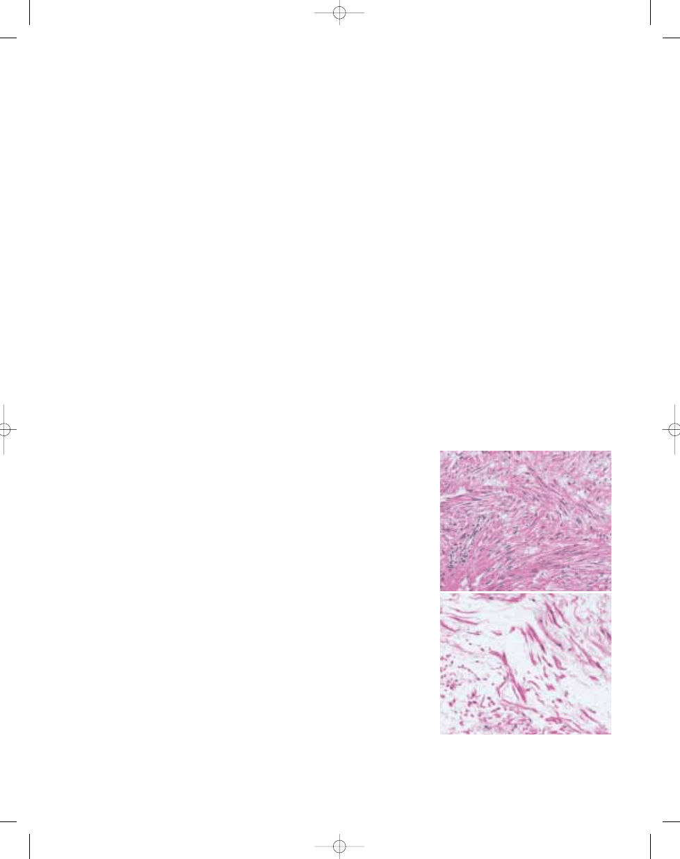
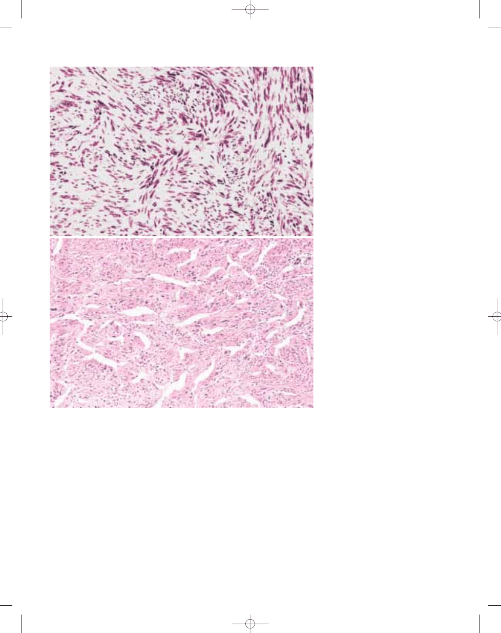
Fig. 4.03 A Leiomyoma of the retroperitoneum com-
posed of interlacing fascicles of bland smooth mus-
cle cells. B Leiomyoma of the retroperitoneum show-
ing myxoid change.
A
B
bb5_9.qxd 13.9.2006 10:47 Page 130
Definition
Leiomyosarcoma is a malignant tumour
composed of cells showing distinct
smooth muscle features.
ICD-O code
8890/3
Epidemiology
Soft-tissue leiomyosarcoma usually
occurs in middle-aged or older persons,
although it may develop in young adults
and even in children {1839, 2066}.
Leiomyosarcoma forms a significant
percentage of retroperitoneal (including
pelvic) sarcomas {906,1749,1754,1945,
2268} and is the predominant sarcoma
arising from larger blood vessels {166,
1095,1243,2192}. Aside from these loca-
tions, it is a comparatively less common
sarcoma, accounting for perhaps 10-15%
of limb sarcomas. The sex incidence
depends on tumour location, with women
forming a clear majority of patients with
retroperitoneal and inferior vena cava
leiomyosarcomas but not of those with
leiomyosarcomas in other soft tissue sites.
Sites of involvement
The most common location of soft tissue
leiomyosarcoma is the retroperitoneum,
including the pelvis. Another distinctive
subgroup consists of leiomyosarcomas
that arise in large blood vessels, most
commonly the inferior vena cava and the
large veins of the lower extremity. Arterial
origin occurs but is rare; sarcomas of the
pulmonary artery and other large arteries
generally do not have the features of
leiomyosarcoma and are better classified
as intimal sarcomas (see page 223).
Leiomyosarcomas involving nonretro-
peritoneal soft tissue sites constitute a
third group {423,642,903,2039}. These
are found most frequently in the lower
extremity but may develop elsewhere.
Intramuscular and subcutaneous local-
izations occur in approximately equal
proportion, and some of these tumours
show evidence of origin from a
small to medium sized (unnamed) vein.
Leiomyosarcomas also develop in the
dermis, but these are discussed in the
volume on tumours of the skin.
Clinical features
Soft tissue leiomyosarcoma generally
presents as a mass lesion. With retroperi-
toneal tumours, pain may also be present.
The symptoms produced by leiomyosar-
coma of the inferior vena cava depend on
the portion involved. When the tumour is
in the upper portion, it obstructs the
hepatic veins and produces the Budd-
Chiari syndrome, with haepatomegaly,
jaundice, and ascites.
H.L. Evans
J. Shipley
Leiomyosarcoma
Fig. 4.04 Leiomyosarcoma. This high grade lesion (19
cm) from the quadriceps muscle shows extensive
necrosis and haemorrhage.
Fig. 4.05 Leiomyosarcoma composed of nodules and bundles of eosinophilic spindle cells.
Fig. 4.06 Leiomyosarcoma with typical intersecting
groups of spindle cells.
131
Leiomyoma of deep soft tissue
bb5_9.qxd 13.9.2006 10:47 Page 131
Location in the middle portion may
result in blockage of the renal veins
and consequent renal dysfunction,
whereas involvement of the lower
portion may cause leg oedema. The
latter may also occur with leiomyosar-
comas of the large veins of the lower
extremity.
Imaging studies of leiomyosarcoma
demonstrate a nonspecific soft tissue
mass but are helpful in delineating the
relationship to adjacent structures, par-
ticularly in the retroperitoneum.
In the instance of leiomyosarcoma of
vein origin, venogram may demon-
strate an intraluminal component.
Aetiology
The cause of soft tissue leiomyosar-
coma is unknown. The predominant
occurrence of retroperitoneal and
inferior vena cava leiomyosarcomas in
women raises the question of hormonal
influence, but this is unclear.
Macroscopy
Leiomyosarcoma of soft tissue typically
forms a fleshy mass, with colours
varying from grey to white to tan. A
whorled character may be evident to
some degree. Larger examples often
display haemorrhage, necrosis, or
cystic change. The tumour border
frequently appears well circumscribed,
although obvious infiltrativeness may
also be found. In the retroperitoneum
there may be extension into adjacent
organs.
Histopathology
The typical histological pattern of
leiomyosarcoma is that of intersecting,
sharply marginated groups of spindle
cells. This pattern may be less well
defined in areas of some tumours, and
occasionally there is a focal storiform,
palisaded, or haemangiopericytoma-like
arrangement. The tumours are usually
compactly cellular, but fibrosis or myx-
oid change may be present; in the latter
instance, a retiform or microcystic pat-
tern may result. Hyalinized, hypocellular
zones and coagulative tumour necrosis
are frequent in larger leiomyosarcomas.
Rarely there is abundant chronic or
acute inflammation {1421}.
The tumour cell nuclei are characteristi-
cally elongated and blunt-ended and
may be indented or lobated. Nuclear
hyperchromatism and pleomorphism
are generally notable, although they
may be focal, mild, or occasionally
absent. Mitotic figures can usually be
found readily, although they may be few
or patchy; and atypical mitoses are
often seen. The cytoplasm varies from
typically eosinophilic to pale, and in the
former instance is often distinctly fibril-
lar. Cytoplasmic vacuolation is frequent-
ly apparent, particularly in cells cut
transversely. Epithelioid cytomorpholo-
gy, multinucleated osteoclastlike giant
cells {1411}, very prominent chronic
inflammatory cells {1421}, and granular
cytoplasmic change {1573} are unusual
findings that are normally present in only
part of a tumour when identified.
Occasional soft tissue leiomyosarcomas
contain areas with a nonspecific, poorly
differentiated, pleomorphic appearance
in addition to typical areas {1594}.
These could be regarded as "dediffer-
entiated leiomyosarcomas" although this
term is not in common use. Rarely, an
osteosarcomalike or rhabdomyosarco-
matous component is associated with
leiomyosarcoma (see "malignant mes-
enchymoma").
Immunophenotype
SMA, desmin and h-caldesmon are
positive in a great majority of soft tissue
leiomyosarcomas. However, none of
132
Smooth muscle tumours
Fig. 4.07 Leiomyosarcoma (A) showing fascicles which intersect at 90
o
and, in another case (B), showing a peri-
cytoma-like vascular pattern.
A
B
bb5_9.qxd 22.9.2006 9:20 Page 132
these is absolutely specific for smooth
muscle (or indeed muscle in general),
and positivity for two of these markers
is more supportive of leiomyosarco-
ma than positivity for one alone.
"Dedifferentiated" areas may be ne-
gative for SMA and desmin, but total
negativity for both in a tumour would
cast great doubt on the diagnosis of
leiomyosarcoma. Stains that may be
positive, at least focally, include
keratin, EMA, CD34, and S100 protein.
KIT (CD117) is normally negative, in
contrast to gastrointestinal stromal
tumours. In general, the diagnosis of
soft tissue leiomyosarcoma should not
be made on the basis of immunostains
in the absence of appropriate morpho-
logic features.
Ultrastructure
Soft tissue leiomyosarcomas usually
demonstrate at least some of the ultra-
structural features of normal smooth
muscle cells, namely cytoplasmic fila-
ments with densities, cell junctions,
pinocytotic vesicles, and basement
membrane. However, any, or occasio-
nally, all of these may be focal or absent,
and the findings may be nonspecific. It
is particularly important to note that
filaments with densities are present in
myofibroblasts and can occur in other
cells. Electron microscopy is not gene-
rally needed for the diagnosis of soft
tissue leiomyosarcoma, and ultrastruc-
tural observations should always be
correlated with the light microscopic
appearance.
Genetics
Cytogenetics
Karyotypes from around 100 leiomyo-
sarcomas have been reported {1477}.
Most karyotypes are complex and no
consistent aberrations have been noted
{2215}. Frequently lost chromosome
regions include 3p21-23, 8p21-pter,
13q12-13, 13q32-qter, whereas the
1q21-31 region is often gained {1314}.
No striking differences among different
subtypes have been identified {1314}.
Comparative genomic hybridization
(CGH) has confirmed frequent numerical
changes, including gain of material from
chromosomes 1, 15, 17, 19, 20, 22 and
X and loss from 1q, 2, 4q, 9p, 10, 11q,
13q and 16, and has identified regions
of amplification, e.g., 1q21, 5p14-pter,
12q13-15, 13q31, 17p11 and 20q13)
{2215}. Tumour size-related changes
have been observed, such as an
association of gain of 16p and 17p with
smaller tumours and gain of 6q and 8q
with larger tumours {577}.
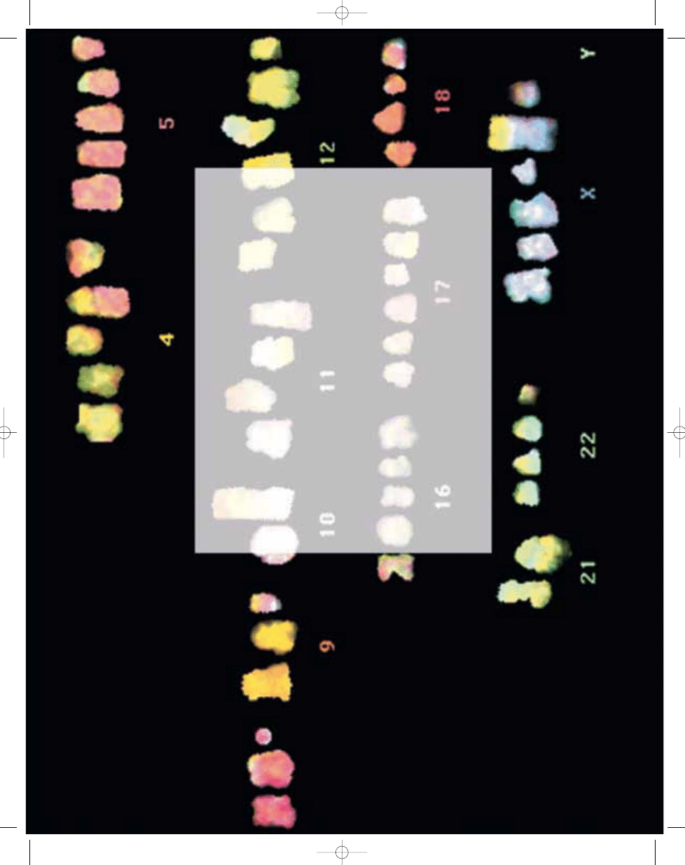
Fig. 4.10 24-colour karyotype and corresponding reverse DAPI-banded image from a soft tissue leiomyosar-
coma showing multiple copies of chromosomes and many rearranged chromosomes.
133
Leiomyoma of deep soft tissue
B
C
A
Fig. 4.08 Leiomyosarcoma showing (A) a myxoid, reticular appearance, (B) necrosis and hyalinization, and (C) abrupt transition to more pleomorphic tumour indicative
of dedifferentiation.
Fig. 4.09
Leiomyosarcoma. Tumour cells contain
prominent longitudinal filament bundles with focal
densities. Note also the external lamina.
bb5_9.qxd 13.9.2006 10:47 Page 133
134
Smooth muscle tumours
Molecular genetics
The
RB1 gene has been implicated,
which is consistent with loss of
chromosome 13 material {2042}. Ana-
lysis of the genes and proteins
in the Rb-cyclinD pathway (
RB1,
CDKN2A, CCND1, and CCND3) has
revealed frequent abnormalities in
leiomyosarcomas {488}. Involvement of
TP53 and MDM2 appears less frequent
than in other sarcoma types {488,692},
although such abnormalities have
been suggested to correlate with a
poorer prognosis in leiomyosarcomas
{1668}. Amplification at a number of
loci suggest candidate genes in these
regions including
MDM2, GLI, CDK4
and
SAS at 12q13-15, the FLF and
PRUNE genes at 1q21, and the critical
region involved in Smith-Magenis
syndrome at 17p11.2 {579,692,708,
709,712,1627}.
Prognostic factors
Soft tissue leiomyosarcomas are
capable of both local recurrence
and distant metastasis. Regional
(or other) lymph node metastasis is
rare. The most important prognostic
factors by far are tumour location and
size, which are strongly interrelated.
Retroperitoneal leiomyosarcomas are
fatal in the great majority of cases; they
are typically large (over 10 cm), often
difficult or impossible to excise with
clear margins, and prone to both
local recurrence and metastasis.
Leiomyosarcomas of large vessels
also tend to have a poor prognosis,
although local control rates are higher
except for those in the upper in-
ferior vena cava, and very small exam-
ples (1-2 cm) may be less prone to
metastasize. Nonretroperitoneal soft
tissue leiomyosarcomas are generally
smaller than those in the retroperi-
toneum, more amenable to local con-
trol, and more favourable in outlook
overall. In some studies, intramuscular
rather than subcutaneous location
{903} and larger tumour size {642,
1479} were related to increased me-
tastasis and poorer patient survival
within this group. Histological grading
as well as osseous and vascular
involvement are reliable prognostic
indicators.
Local recurrences and metastases of
soft tissue leiomyosarcoma usually
become manifest within the first few
years after diagnosis but may appear
as much as 10 years later. For
retroperitoneal leiomyosarcomas, the
most common sites of metastases are
the lungs and liver, whereas the lungs
are the dominant location when the pri-
mary tumour is nonretroperitoneal.
Metastases also occur with some fre-
quency in skin, soft tissue, and bone.
Smooth muscle tumours in
immunocompromised patients
Smooth muscle tumours in immunocom-
promised individuals, to this point
described only in single case reports and
small series, form a distinctive subgroup.
These usually involve parenchymal
organs rather than soft tissue, occur pre-
dominantly in children and young adults
who are HIV positive {323,1368,1811,
2179} or post-transplant, and are asso-
ciated with Epstein-Barr virus. The
tumours may be multifocal, and at least in
some instances this appears to represent
true multicentricity rather than metastasis
{1811,1985}. Histologically, they range
from bland to mitotically active, may have
a variable lymphocytic infiltrate of uncer-
tain significance and may show a perivas-
cular growth pattern.
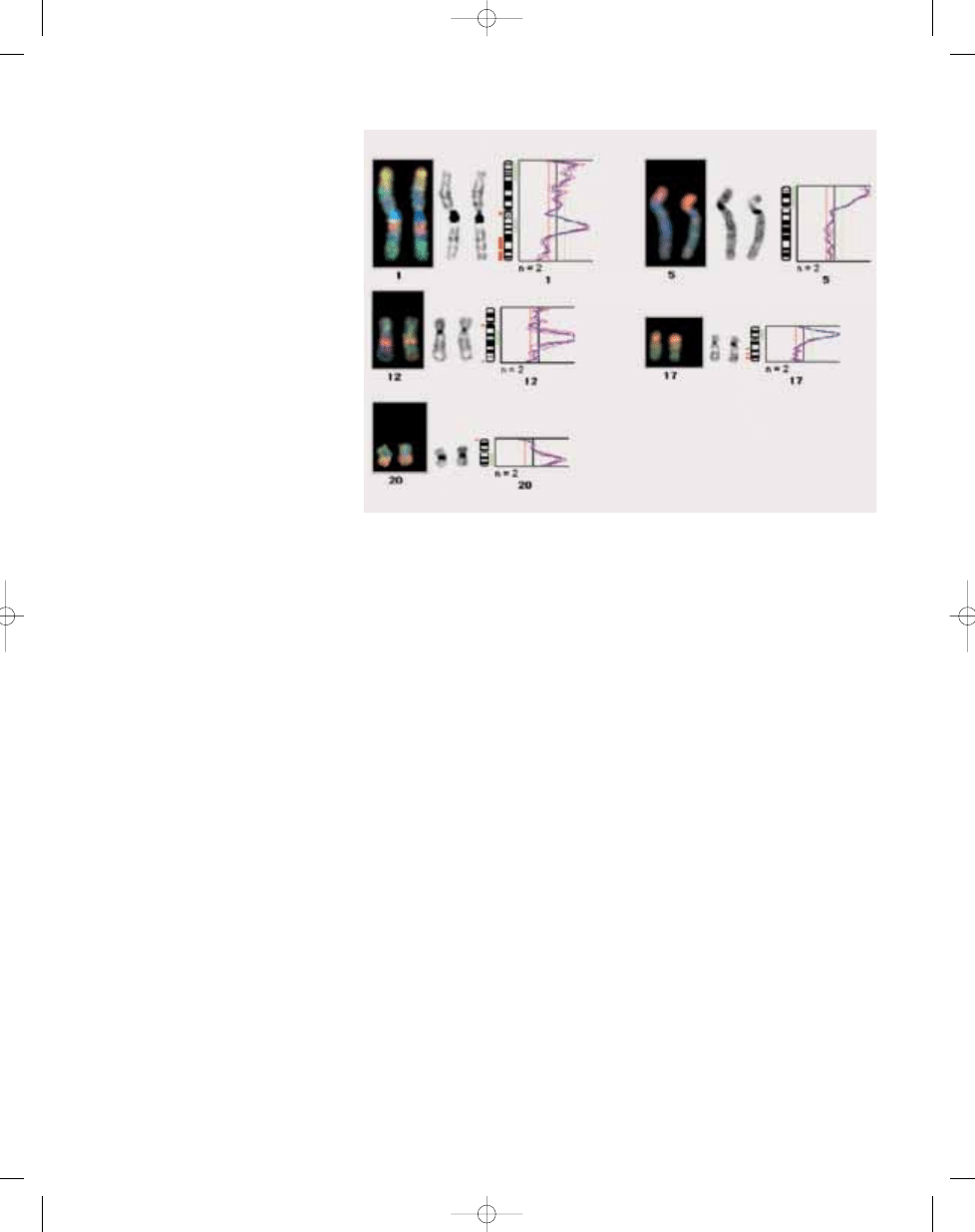
Fig. 4.11 Amplification in leiomyosarcomas identified by CGH analysis. CGH images, reverse DAPI-banded
chromosomes and corresponding profiles of the red to green fluorescence intensities are shown and indi-
cate amplification at 1q21-q25, 5p, 12q13-q21, 17q11.2-q12 and 20q, respectively.
bb5_9.qxd 13.9.2006 10:47 Page 134
Wyszukiwarka
Podobne podstrony:
bb5 chap3
bb5 chap8
bb5 chap1
BB5 BOX
bb5 chap16
bb5 chap15
bb5 contents
bb5 chap12
bb5 references
bb5 chap6
bb5 chap17
bb5 chap20
bb5 chap5
CHAP4
Lista wszystkich dostępnych polskich Product Code dla telefonów platformy BB5
bb5 chap21
bb5 source
bb5 chap13
więcej podobnych podstron