CHAPTER 21
Congenital and Inherited Syndromes Associated with
Bone and Soft Tissue Tumours
During the past decade, rapid progress has been made in our
understanding of how inherited genetic aberrations may influ-
ence cancer risk. A large number of neoplasia-associated syn-
dromes following Mendelian inheritance has been defined both
clinically and at the DNA level, providing a solid basis for genet-
ic counselling of patients and their families. The identification of
specific genes involved in inherited cancer predisposition pro-
vides, in addition, important insights into genetic pathways
involved in the development of sporadic neoplasia.
Although inherited susceptibility accounts for only a minority of
all bone and soft tissue tumours, several syndromes and disor-
ders have been identified, and for many of them the underlying
genetic cause has been identified. In the attached Table, well
characterized familial disorders associated with bone and soft
tissue tumours are listed, including congenital malformation syn-
dromes in which no clear pattern of inheritance has as yet been
noted.
On the following pages, a more detailed description of the clini-
cal, histopathological, and genetic data is provided for those
syndromes that are well characterized at the DNA level, or for
which the associated neoplasms display features that are dis-
tinct from those of their sporadic counterparts. Cowden disease,
Li-Fraumeni syndrome and neurofibromatosis type 1 and 2 have
been dealt with in the WHO Classification of Tumours of the
Nervous System.
bb5_29.qxd 13.9.2006 13:58 Page 349
350
Congenital and inherited syndromes associated with bone and soft tissue tumours
OMIM
a
Disorder
b
Inheritance
Locus
c
Gene
Bone and soft tissue tumours
103580
Albright hereditary osteodystrophy
AD
20q13
GNAS1
Soft tissue calcification and osteomas
153480
Bannayan-Riley-Ruvalcaba syndrome AD
10q23
PTEN
Lipomas, haemangiomas
130650
Beckwith-Wiedemann syndrome
Sporadic/AD
11p15
Complex,
Embryonal rhabdomyosarcomas,
incl.
CDKN1C
myxomas, fibromas, hamartomas
and
IGF2
210900
Bloom syndrome
AR
15q26
BLM
Osteosarcomas
160980,
Carney complex
AD
17q23-24
PRKAR1AK
Cardiac and other myxomas,
605244
2p16
-
melanocytic schwannomas
112250
Diaphyseal medullary stenosis
AD
9p21-22
-
Malignant fibrous histiocytomas of bone
with malignant fibrous histiocytoma
151623
Li-Fraumeni syndrome
AD
17p13
TP53
Osteosarcomas, rhabdomyosarcomas
22q11
CHEK2
and other soft tissue sarcomas
151800
Lipomatosis, symmetrical
Sporadic
-
-
Lipomas, lipomatosis of the head and neck
166000
Maffucci syndrome
Sporadic
-
-
Enchondromas, chondrosarcomas,
spindle cell haemangiomas,
haemangiomas, angiosarcomas
-
Mazabraud syndrome
Sporadic
20q13
GNAS1
Polyostotic fibrous dysplasia,
osteosarcomas, intramuscular myxomas
174800
McCune-Albright syndrome
Sporadic
20q13
GNAS1
Polyostotic fibrous dysplasia,
osteosarcomas
133700, Multiple osteochondromas,
AD
8q24,
EXT1
Osteochondromas, chondrosarcomas
133701
non-syndromic
11p11-12
EXT2
228550
Myofibromatosis
AR
-
-
Myofibromas
162200
Neurofibromatosis type 1
AD
17q11
NF1
Neurofibromas, malignant peripheral
nerve sheath tumours
101000
Neurofibromatosis type 2
AD
22q12
NF2
Schwannomas
166000
Ollier disease (enchondromatosis)
Sporadic
3p21-22
PTHR1
Enchondromas, chondrosarcomas
Table 21.01
Congenital syndromes associated with bone and soft tissue tumours.
bb5_29.qxd 13.9.2006 13:58 Page 350
351
Congenital syndromes associated with bone and soft tissue tumours
OMIM
a
Disorder
b
Inheritance
Locus
c
Gene
Bone and soft tissue tumours
167250;
Paget disease of bone, familial
AD
18q21
TNFRSF11A
Osteosarcomas
602080
5q31
-
5q35
-
176920
Proteus syndrome
Sporadic
-
-
Lipomas
180200
Retinoblastoma
AD
13q14
RB1
Osteosarcomas, soft tissue sarcomas
601607
Rhabdoid predisposition syndrome
AD
22q11
SMARCB1
Malignant rhabdoid tumours
268400
Rothmund-Thomson syndrome
AR
8q24
RECQL4
Osteosarcomas
180849
Rubinstein-Taybi syndrome
AD
16p13
CREBBP
Myogenic sarcomas
138000
Venous malformations
AD
1p21-22
-
Glomus tumors
with glomus cells
277700
Werner syndrome
AR
8p11-12
WRN
Various bone and soft tissue sarcomas
a OMIM = entry number in McKusick’s Online Mendelian Inheritance in Man {1376}.
b Syndromes associated with tumours affecting only the skin or parenchymatous organs are not included.
cAD = autosomal dominant; AR = autosomal recessive.
bb5_29.qxd 13.9.2006 13:58 Page 351
Familial adenomatous polyposis
M. Nilbert
C.M. Coffin
Definition
Familial adenomatous polyposis (FAP)
is characterized by the development of
multiple colorectal polyps, which are
premalignant lesions with a strong ten-
dency to progress into carcinomas.
Gardner syndrome, characterized by
colorectal polyps as well as extra-
colonic manifestations such as dental
abnormalities, osteomas, epidermoid
cysts and desmoid tumours, was initial-
ly considered a separate entity, but has
now been recognized as a variant of
FAP. FAP is caused by mutations in the
adenomatosis polyposis coli (
APC)
gene on chromosome 5.
OMIM number
175100
Synonyms
Bussey-Gardner polyposis, adenoma-
tous polyposis coli, familial polyposis
coli, familial multiple polyposis, etc.
Incidence
Estimates of the incidence of FAP vary
between 1/7,000 and 1/30,000 {1033}.
Whereas dental abnormalities and
osteomas occur in more than half of the
patients, desmoid tumours and epider-
moid cysts develop in a minority of the
patients. Overall, FAP accounts for less
than 1% of all colorectal cancers.
Diagnostic criteria
The diagnosis of FAP requires 1) at least
100 colorectal adenomas or 2) a
germline, disease-causing mutation of
the
APC gene or 3) a family history of
FAP and at least one of the following:
epidermoid cysts, osteomas or desmoid
tumour. Other types of extracolonic
manifestations are associated with FAP,
including adenomatous polyps of the
upper gastrointestinal tract, congenital
hypertrophy of the retinal pigment
epithelium (CHRPE), an increased risk
of hepatoblastoma and tumours of the
endocrine system, most commonly pap-
illary carcinoma of the thyroid. Further-
more, an association with brain
tumours, especially medulloblastomas,
occurs in the Turcot syndrome, which in
two-thirds of the cases is caused by
APC mutations. In familial infiltrative
fibromatosis (OMIM No. 135250), which
is also caused by germline mutations of
APC, there is an inherited predisposition
to desmoid tumours, but only few or no
colonic polyps.
Clinical features
Colorectal adenomas usually develop
into endoscopically detectable lesions
at 10-20 years of age and increase in
number and size over time. Untreated
FAP patients develop colorectal cancer
at a median age of about 40 years. FAP
patients should be screened with
endoscopy with 1-2 year intervals from
10-15 years of age up to 40 years of age
and prophylactic colectomy is per-
formed when adenomas are detected.
Extraintestinal manifestations, in partic-
ular epidermoid cysts, dental abnormal-
ities, osteomas and CHRPE often pre-
ceed the development of adenomas
and may serve as clinical markers of
FAP.
Bone and soft tissue tumours
The description of Gardner syndrome in
the 1950’s highlighted the association of
familial polyposis coli with a spectrum of
extracolonic manifestations, including
lesions of soft tissue and bone {766-
769}. The most commonly encountered
bone and soft tissue lesions are osteo-
mas, cortical thickening of bone, epi-
dermoid cysts, and desmoid-type fibro-
matoses {766,768,1544,1606,1705}. In
addition to these lesions, a variety of
other soft tissue masses have been clin-
ically described, with varying extents of
pathological analysis. These include ill
defined connective tissue masses, "lipo-
mas" {1705}, "fibrous dysplastic lesions"
{1544}, "familial infiltrative fibromatosis"
{1913}, fibromatous mesenteric plaques
{363}, juvenile nasopharyngeal angiofi-
broma {784}, Gardner fibroma {2227},
and rhabdomyosarcoma {84}.
The association of desmoids, including
those with childhood onset, with adeno-
matous polyposis of the coli is now well
recognized {175,312,361,362,566,768,
769,1032,1068,1913}. The incidence of
desmoid tumours in patients with poly-
posis has been estimated to be around
10%. Pathological features of desmoid-
type fibromatosis are described else-
where in this book (see page 83).
Particular
APC mutation types are asso-
ciated with a higher frequency of des-
moid tumours {175, 312, 859, 931, 957,
1015,1047,1137,1286,1685,1799,1993}.
The Gardner fibroma {2227}, described
elsewhere in this book (see page 76),
is similar to the fibromatous mesenteric
plaques reported in patients with ade-
nomatous polyposis coli {363}. These
lesions are associated with develop-
ment of desmoid-type fibromatosis in
the same site, either following surgery
or de novo {361,2227}. Recognition of
the Gardner fibroma in childhood can
serve as the sentinel event for diagnosis
of adenomatous polyposis of the colon
{2227}. Juvenile nasopharyngealangio-
fibroma has also been reported in asso-
ciation with adenomatous polyposis of
the colon {8,10,784}. However, some
have questioned whether this associa-
tion is coincidental or whether it is actu-
ally related to another alteration of the
APC gene {850}. Rhabdomyosar-coma
has been reported in rare instances in
individuals or families with adenoma-
tous polyposis of the colon {84,1299},
but it is unclear whether this is a spo-
radic occurrence or another syndromic
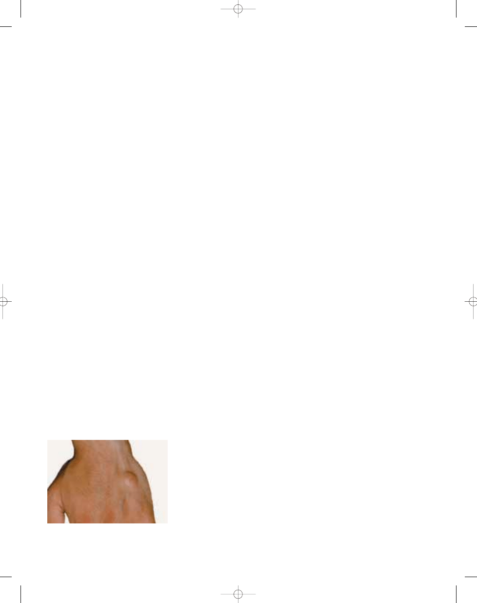
Fig. 21.01 Epidermoid cyst on the dorsal surface of
the hand of a patient with familial adenomatous
polyposis.
352
Congenital and inherited syndromes associated with bone and soft tissue tumours
bb5_29.qxd 13.9.2006 13:58 Page 352
manifestation.
Bone lesions associated with adenoma-
tous polyposis of the colon are entirely
benign and are viewed as dysplasias.
Multiple osteomas formed by membra-
nous ossification, especially of calvarial
and mandibular surfaces, characterize
the "ivory exostosis" of Gardner syn-
drome {285, 331, 1075, 1690}. Histologi-
cally, the Gardner osteoma is a nodular
excrescence of mature lamellar bone
involving the cortical surface, especially
the outer table of the skull, the mandibu-
lar cortex, or rarely other sites. Like
desmoid fibromatosis, particular
APC
gene mutations are associated with
more severe osseous manifestations
{451, 1180, 2080}. Diffuse craniofacial
sclerotic bone changes and dental mal-
formations are also encountered. The
bony lesions of adenomatous polyposis
of the colon do not evolve into other
benign neoplasms, such as osteoblas-
tomas, or into malignant lesions.
Genetics
Germline mutations of the
APC gene is
the only identified cause of FAP. FAP is
autosomally dominantly inherited with
an almost complete penetrance.
However, at least one-fifth of the
patients lack a family history and are
thus assumed to carry de novo muta-
tions of the
APC gene {204}.
Gene structure
The
APC gene was in 1986 localized to
5q21-22 through observation of a
patient with polyposis and a constitu-
tional interstitial deletion of 5q followed
by an establishment of linkage to this
locus in several FAP kindreds {940,
1241}. The
APC gene was isolated in
1991 and was found to be mutated in
the germline of patients with FAP {840,
1123}. The gene spans 120 kb, is com-
posed of 15 coding exons and contains
an open reading frame of 8,538 bp.
Several alternatively spliced forms of
APC with different 5´regions have been
identified.
Gene expression
The 2,843 amino-acid APC protein is
ubiquitously expressed in most normal
tissues with the highest expression
found in the central nervous system.
APC is a multifunctional protein with
several functional domains through
which APC exerts its main function as a
negative regulator of the Wnt signalling
pathway {312,693,921,1819}. Normal
Wnt signalling inhibits the function
of glycogen synthase 3ß (GSK3B),
dephosphorylates axin / conductin and
thereby targets ß-catenin for degrada-
tion. ß-catenin is involved in the
cytoskeletal organisation with micro-
tubule binding and in cell adhesion
through interaction with E-cadherin.
APC mutations, presumably trough loss
of binding sites and degradation sites
for ß-catenin lead to intracellular accu-
mulation of ß-catenin, which is trans-
ferred to the nucleus and through inter-
action with transcription factors of the
TCF/LEF family regulates expression of
downstream target genes such as
MYC
and
CCND1 {2011, 2104}. The C-termi-
nal mediates binding to microtubule-
associated proteins of the EB1/RP1
family. Truncated APC thereby promotes
chromosomal instability through disrupt-
ed interaction between the kinetochores
and the spindle microtubules {693}.
Mutations
Analyses of the
APC gene in patients
with FAP reveal mutations in about 80%
of the kindreds examined, and the
remaining patient are likely to carry
APC
gene mutations leading to large dele-
tions or impaired protein expression.
Over 95% of the mutations identified
result in protein truncation, which large-
ly result from nonsense point mutations
or deletions causing frameshifts.
Genotype-phenotype correlations exist;
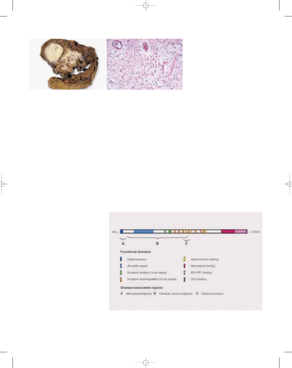
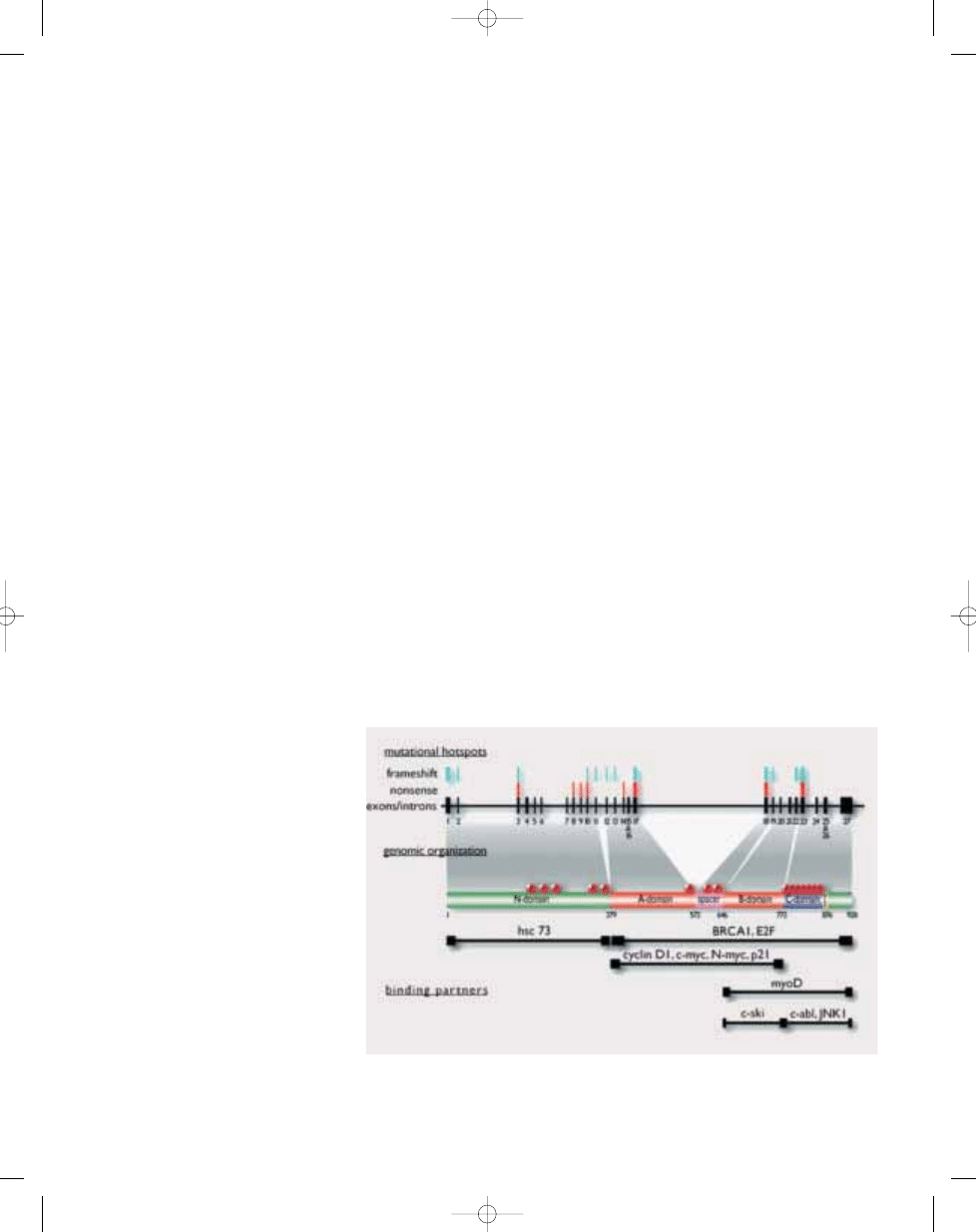
Fig. 21.03 Functional and disease-related domains of the APC gene. ß-catenin binding is achieved through
the 15-amino acid and 20-amino acid repeat-containing regions and the C-terminal of APC which interacts
with microtubule-associated proteins of the EB/RP family and with DLG, a human homologue of the
Drosophila discs large tumour suppressor protein. Mutations between codons 1403 and 1578 have been
associated with the extracolonic manifestations, e.g. desmoid tumours.
353
Familial adenomatous polyposis
B
A
Fig. 21.02 Mesenteric fibromatosis (desmoid tumour) in a patient with FAP. A The lesion entraps loops of
small intestine. B Histopathology is dominated by collagen bands and small vessels.
bb5_29.qxd 13.9.2006 13:59 Page 353

354
Congenital and inherited syndromes associated with bone and soft tissue tumours
truncating mutations in the 5´ end of
the gene have been associated with
attenuated FAP, mutations in the central
region of gene, including the mutational
hotspot at codon 1309, are associated
with multiple polyps at young age, and
mutations between codons 1444 and
1578 are associated with an increased
incidence of desmoid tumours {451,
1124, 2011}. However, patients with
identical mutations can develop dissi-
milar clinical features and the genotype
clinically serves as a risk determinant
rather than as an absolute predictor of
the extent of the disease.
Definition
The Beckwith-Wiedemann syndrome
(BWS) is a complex overgrowth disorder
caused by a number of genes that are
subject to genomic imprinting. A high
incidence of solid childhood tumours,
including rhabdomyosarcoma, is seen in
patients that present with BWS.
OMIM number
130650
Synonyms
EMG syndrome (Exomphalos-Macro-
glossa-Gigantism syndrome), WBS
(Wiedemann-Beckwith syndrome).
Incidence
The syndrome occurs with an estimated
incidence of 1:13,700 and most cases
(85%) are sporadic.
Diagnostic criteria
Patients can be classified as having BWS
according to the clinical criteria pro-
posed by Elliot or DeBaun {479, 580}
although cases of BWS are known that
do not comply with either set of criteria.
Elliot classifies patients as BWS when
they present with three major features or
two major features plus three or more
minor features (major features: anterior
abdominal wall defects, macroglossia
and pre- and/or postnatal growth > 90th
centile; Minor features: ear creases or
pits, naevus flammeus, hypoglycaemia,
nephromegaly and hemihypertrophy).
DeBaun is less strict in his classification
i.e. two or more of the five most common
features (macroglossia, birth weight >
90th percentile, hypoglaecemia in the
first month of life, ear creases/pits and
abdominal wall defects).
BWS can be diagnosed in the laboratory
by chromosome banding analysis (< 5%)
or DNA-diagnostics. The current major
test involves methylation assays or loss
of imprinting (LOI) studies at the RNA
level. The majority of cases (50-80%)
demonstrates aberrant methylation of
KCNQ1OT1, with or without aberrant
methylation of
IGF2/H19. These latter
cases often show uniparental disomy
(UPD), in a mosaic form, for 11p15,
which explains this aberrant methylation.
However, the majority of cases with
KCNQ1OT1 defects and some cases
with
H19/IGF2 defects have no UPD
11p15. Therefore, an imprinting switch
can be assumed involving an imprinting
centre, analogous to the Prader-Willi and
Angelman syndromes. The current data
are most compatible with two distinct
imprinting centres for either
KCNQ1OT1
or
IGF2/H19. CDKN1C mutation analy-
ses might be considered, especially in
familial cases of BWS. The increased
tumour risk for BWS patients seems to be
associated with UPD in general and
H19
methylation defects in particular.
KCNQ1OT1 methylation defects only
seem to be a favourable prognostic fac-
tor since tumours are not, or only very
rarely associated with this group of
patients. Recurrence risks for a second
pregnancy can be assessed with UPD
studies. In case of a UPD in a mosaic
form, there is no increased recurrence
risk for BWS in a second pregnancy
since the genetic defect occurred post-
fertilisation.
Clinical features
The BWS is a disorder first described by
Beckwith in 1963 at the 11th annual
meeting of the Western Society for
Pediatric Research. Later, Wiedemann
and Beckwith described the syndrome in
more detail {149, 2266}. BWS is charac-
terized by a great variety of clinical fea-
tures, among which are abdominal wall
defects, macroglossia, pre- and postna-
tal gigantism, earlobe pits or creases,
facial nevus flammeus, hypoglycemia,
renal abnormalities and hemihypertrophy.
Tumours
BWS patients have a risk of 7.5% for the
development of (mostly intra-abdominal)
childhood tumours. Tumours most fre-
quently found are Wilms’ tumour, adreno-
cortical carcinoma, embryonal rhab-
domyosarcoma, and hepatoblastoma.
Also myxomas, fibromas, and chest wall
hamartomas have been reported to
occur at increased frequencies.
M. Mannens
Beckwith-Wiedemann syndrome
bb5_29.qxd 13.9.2006 13:59 Page 354
355
Beckwith-Wiedemann Syndrome
Genetics
BWS is caused by genetic changes in
chromosome band 11q15, as shown by
linkage studies, and the detection of
chromosome abnormalities, LOI, and
gene mutations. The syndrome is sub-
ject to genomic imprinting since mater-
nal transmission seems to be predomi-
nant. In addition, chromosomal translo-
cations are of maternal origin, dupli-
cations and UPD of paternal origin. All
hitherto known causative genes are
imprinted. The translocation breakpoints
on chromosome 11 map to three distinct
regions within 11p15.3-pter. Beckwith-
Wiedemann syndrome chromosome
region 1 (BWSCR1) near
INS/IGF2,
BWSCR2 5 Mb proximal to BWSCR1,
and BWSCR3 2 Mb even more proximal
{967}. This already points to genetic
heterogeneity but also at the clinical
level there seems to be heterogeneity.
Chromosomal translocations in BWSCR1
and BWSCR3 are associated with the
classical BWS phenotype and BWSCR2
with minor BWS features but pronoun-
ced hemihypertrophy. BWSCR 1 and
BWSCR2 have been cloned and genes
isolated from these regions were shown
to be involved in the development of this
disorder. All genes involved are subject
to genomic imprinting {1326, 2023}.
BWSCR1
This region consists of a number of
imprinted genes. All known transloca-
tion breakpoints disrupt
KCNQ1, a
gene coding for a potassium channel
in-volved also in inherited cardiac
arrhythmia syndromes. This imprinted
gene, however, is most likely not direct-
ly involved in BWS. A gene transcribed
in the antisense orientation of
KCNQ1
clearly is. This gene,
KCNQ1OT1, shows
aberrant methylation in 50-80% of BWS
cases. It does not code for a protein
and may function through its RNA.
CDKN1C is an inhibitor of cyclin-
dependent kinases. Heterozygous
mutations have been identified in
about 20% of BWS patients in two stud-
ies. Others, however, have not been
able to confirm this mutation frequency.
Although not a major cause of BWS, it is
possible that in certain countries, e.g.,
in Asia, the mutation frequency is ele-
vated. In addition, it has been
reported that this gene is more fre-
quently involved in familial cases
of BWS.
CDKN1C
mouse models
revealed some of the clinical BWS
features such as omphalocele and renal
adrenal cortex anomalies. In humans,
CDKN1C also seems to be more fre-
quently associated with abdominal wall
defects. Another strong candidate for
involvement in the aetiology of BWS is
IGF2. Mouse models overexpressing
Igf2 displayed a phenotype overlapping
with the BWS phenotype. Loss of
IGF2
imprinting is often seen in BWS patients.
Down-stream from
IGF2 lies H19, again
a non-coding gene. The expression of
IGF2 and H19 seems to be linked. H19
is important for the maintenance of the
imprinting status of
IGF2. Mouse studies
underline the link between
IGF2 and
H19 expression and overgrowth phe-
notypes were found.
H19 loss of im-
printing is frequently seen in BWS
cases although not always in combi-
nation with
IGF2 LOI. Finally, a gene
called
ASCL2 is localized to the 11p15-
imprinted region. Although no direct
involvement in the BWS aetiology is
known, this gene might account for
the fact that most, if not all, BWS cases
with UPD present in a mosaic form.
The mouse homologue codes for a
transcription factor, which is expressed
during early mouse development and
is essential for the development of the
placenta. Therefore, also in humans,
complete lack of expression might be
lethal.
BWSCR2
Two patients define this second chromo-
somal region, one of whom developed a
Wilms tumour {34}. Both translocations
in 11p15.4 disrupt a paternally imprint-
ed zinc-binding finger gene
ZNF215.
Parts of the 3’ end of this gene are tran-
scribed from the antisense strand of a
second zinc-finger gene,
ZNF214.
Although putative mutations in these
genes in other sporadic BWS cases
were found, their involvement in BWS
needs to be further elucidated by func-
tional studies.
More detailed information on the struc-
ture and expression of genes involved in
BWS could be found at:
http://www.infobiogen.fr/services/
chromcancer/Kprones/Beckwith
WiedemannID10037.html
{1325}.
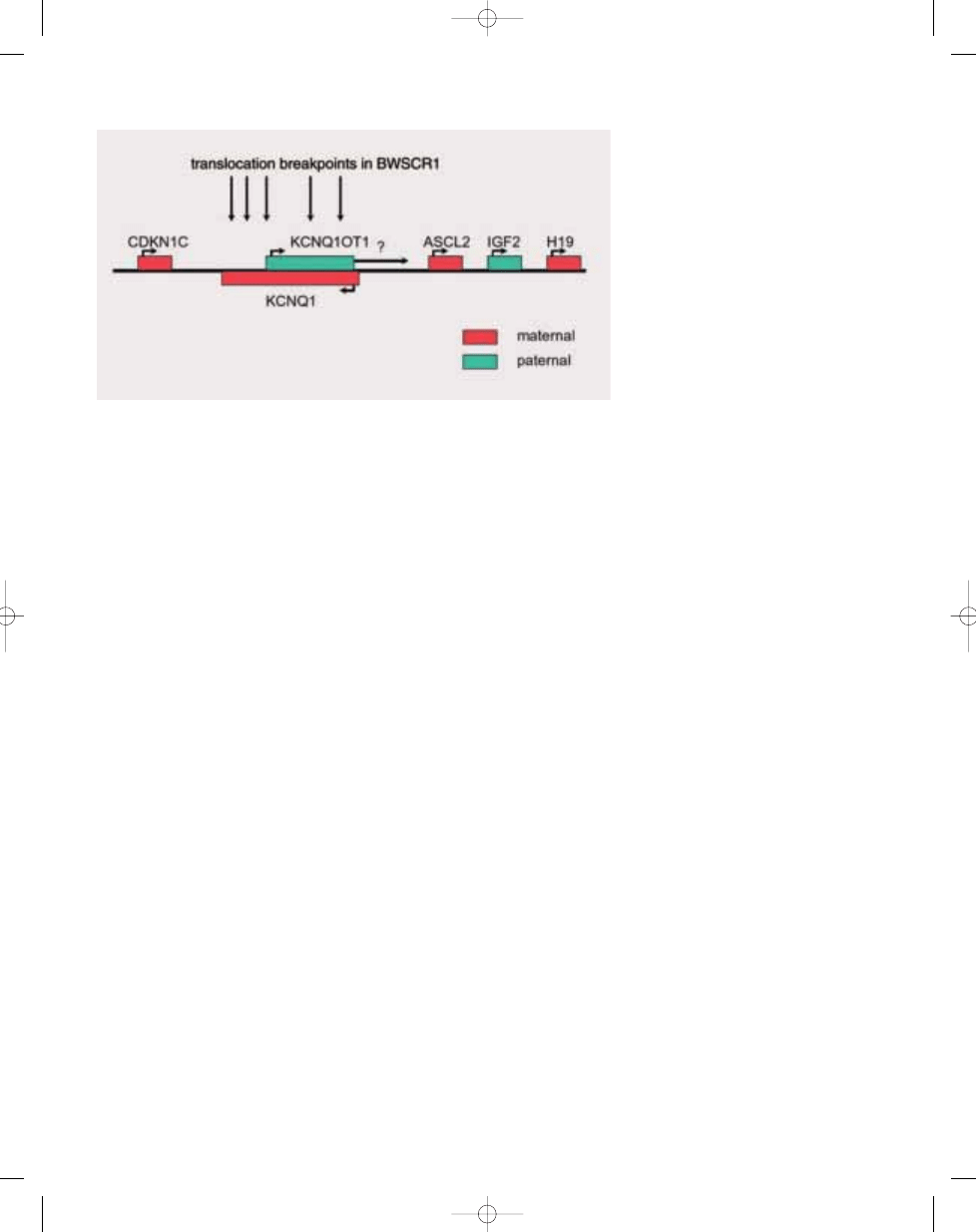
Fig. 21.04 Imprinted genes on 11p15 involved in BWS. The parental expression (imprinting) of these genes
is indicated.
bb5_29.qxd 13.9.2006 13:59 Page 355
Definition
Ollier disease is a developmental disor-
der characterized by the occurrence of
multiple cartilaginous masses, particular-
ly affecting the short and long tubular
bones of the limbs. When cutaneous, soft
tissue or visceral haemangiomas are
also present, the disorder is referred to
as Maffucci syndrome.
OMIM number
16600L {1376}
Synonyms
Ollier disease is also referred to as multi-
ple enchondromas or dyschondroplasia.
Incidence
Rare, but exact incidence is unknown.
Enchondromatosis has been described
in many different ethnic groups, and
there is no significant gender bias.
Diagnostic criteria
The diagnosis is based on the
roentgenographic appearance and clini-
cal features. No distinctive genetic or
biochemical marker for either Ollier dis-
ease or Maffucci syndrome has as yet
been identified.
Clinical features and tumours
Ollier disease usually manifests already
in early childhood, commonly presenting
as swelling of the fingers. Enchondromas
in the metaphyseal regions of long bones
may also result in deformity and limb
asymmetry, as well as pathological frac-
tures. Although careful examination will
reveal that the vast majority of patients
have bilateral enchondromatosis, there is
a tendency for one side of the body to be
more severely affected. The extent of a
patient’s orthopedic complications,
which is highly variable and difficult to
predict, is largely dependent on the num-
ber and skeletal distribution of enchon-
dromas.
The enchondromas primarily affect the
short and long tubular bones of the
extremities, but flat bones, such as the
pelvis and ribs, may be involved. The
craniofacial bones and vertebrae, how-
ever, are usually spared. With few excep-
tions, the enchondromatous lesions stop
growing at puberty. Continued or
renewed growth in adults should raise
the suspicion of malignancy. Whereas
sarcomatous transformation of solitary
enchondromas is rare, patients with
Ollier have a markedly increased risk,
ranging from 15 to 30%, of developing
malignant bone tumours, in particular
chondrosarcomas {1274,1901}. Some
patients even develop multiple sarcomas
{303}.
Most patients with Maffucci syndrome
present at birth or in early childhood with
cavernous haemangiomas, varying in size
from a few millimetres to several centime-
tres, that are typically located in the dermis
or subcutaneously on the distal parts of
the limbs. However, haemangiomas may
also be found in internal organs. In addi-
tion, spindle cell haemangioma, a vascu-
lar lesion with a high propensity for local
recurrence but no potential for metastasis,
is overrepresented among patients with
Maffucci syndrome {639,1688}.
The skeletal features in Maffucci syn-
drome are indistinguishable from those in
Ollier disease, but the risk of developing
chondrosarcoma is possibly even higher
among patients with Maffucci syndrome,
with incidence figures reaching 20-30%
in some series {1067,2055}. An
increased incidence has also been sug-
gested for other malignancies, including
angiosarcomas, brain tumours, and
tumours of the hepatobiliary system {538,
1901}, as well as certain benign tumours.
In both forms of enchondromatosis, care-
ful surgical and orthopedic intervention
may avoid or minimise deformities.
Furthermore, all patients should be
instructed to pay close attention to signs
or symptoms heralding malignant trans-
formation.
The more widespread the disease, the
greater is the likelihood for malignant
transformation {538}. The prognosis for
patients developing secondary chon-
drosarcoma is similar to that for patients
with sporadic chondrosarcomas, and
depends on tumour size and location,
and histological malignancy grade
{1230}.
Roentgenographic features
Roentgenographic features of Ollier dis-
ease and Maffucci syndrome are similar
except for the presence of phleboliths in the
soft tissue haemangiomas in the latter con-
dition. The cartilage present has expansile
masses at the metaphyseal region with cal-
cification in the form of longitudinal striation.
F. Mertens
K. Unni
Enchondromatosis:
Ollier disease and Maffucci syndrome
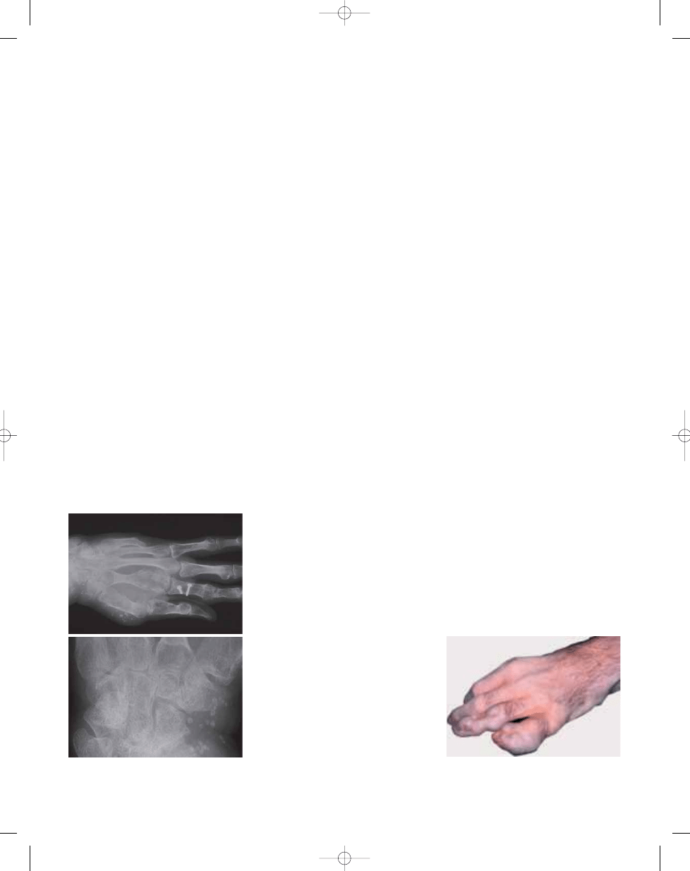
Fig. 21.05 Enchondromas and calcified thrombi in
soft tissue haemangiomas in the left hand of a
patient with Maffucci syndrome.
A
B
Fig. 21.06
Multiple enchondromas causing
swelling and angular deformity in the left hand of a
patient with Ollier disease.
356
Congenital and inherited syndromes associated with bone and soft tissue tumours
bb5_29.qxd 13.9.2006 13:59 Page 356
Definition
McCune-Albright syndrome (MAS) is a
sporadically occurring disorder consist-
ing of polyostotic fibrous dysplasia, café-
au-lait spots, and hyperfunctioning
endocrinopathies. The syndrome is
caused by mutations in the
GNAS1 gene.
OMIM number
174800L
Incidence
No accurate incidence has ever been
determined for MAS. Fibrous dysplasia
may occur without MAS and the over-
whelming majority of these cases are
monostotic. Polyostotic fibrous dyspla-
sia occurs much less frequently and
about 3% of the these cases represent
MAS {382,383}.
Diagnostic criteria
Polyostotic fibrous dysplasia, café-au-lait
spots, and hyperfunctioning endocrino-
pathies {31-33,1375}.
Clinical features
Cardinal features include café-au-lait
spots, polyostotic fibrous dysplasia, mul-
tiple endocrinopathies including sexual
precocity, pituitary adenoma, and hyper-
thyroidism. There is high expression of
the
FOS proto-oncogene in cells popu-
lating the bone marrow spaces. Many
other abnormalities are found with low
frequency: gastrointestinal polyps;
hyperplasia of the thymus, spleen, and
pancreatic islet cells; hepatobiliary dis-
ease; cardiac disease; failure to thrive;
metabolic acidosis; abnormalities in
serum electrolytes, glucose, or insulin
Microscopic features
The cartilage in enchondromas is pre-
sent as well circumscribed nodules in
the medullary cavity and occasionally on
the surface. The matrix does not show
myxoid change. The lesion is hypercellu-
lar and the chondrocyte nuclei are
enlarged and irregular.
Genetics
Most cases of enchondromatosis are
sporadic, but families with multiple
affected members have been reported,
possibly suggesting autosomal domi-
nant inheritance with reduced pene-
trance {1376}. Molecular genetic analy-
sis of a high grade chondrosarcoma
from a patient with Ollier disease re-
vealed loss of heterozygosity for the
chromosomal bands harbouring the
RB1
and
CDKN2A tumour suppressor genes
as well as TP53 overexpression, but
none of these changes were found in
tissue from an enchondroma {243}.
Recently, a study of patients with Ollier
disease revealed mutations of the
PTHR1 gene, encoding a receptor for
parathyroid hormone and parathyroid
hormone-related protein (PTH/PTHrP), in
two of six cases; in one as a germline
mutation, and in one as a somatic mu-
tation in enchondroma tissue {968}. The
detected mutation, resulting in an R150C
substitution in the extracellular domain
of PTHR1, was shown to cause in-
creased cAMP signalling, which is ana-
logous to the situation in Jansen meta-
physeal chondrodysplasia (OMIM
156400), an autosomal dominant disor-
der sharing some radiographic and his-
tological features with Ollier disease.
The hypothesis that a mutant PTH/PTHrP
receptor could delay the differentiation
of proliferating chondrocytes by consti-
tutively activating Hedgehog signalling
{1885} was further substantiated by
studies of transgenic mice carrying the
same R150C
PTHR1 mutation {968}. The
R150C substitution could not be detec-
ted in a series of 50 sporadic chon-
drosarcomas {968}.
M.M. Cohen, Jr.
G.P. Siegal
McCune-Albright syndrome
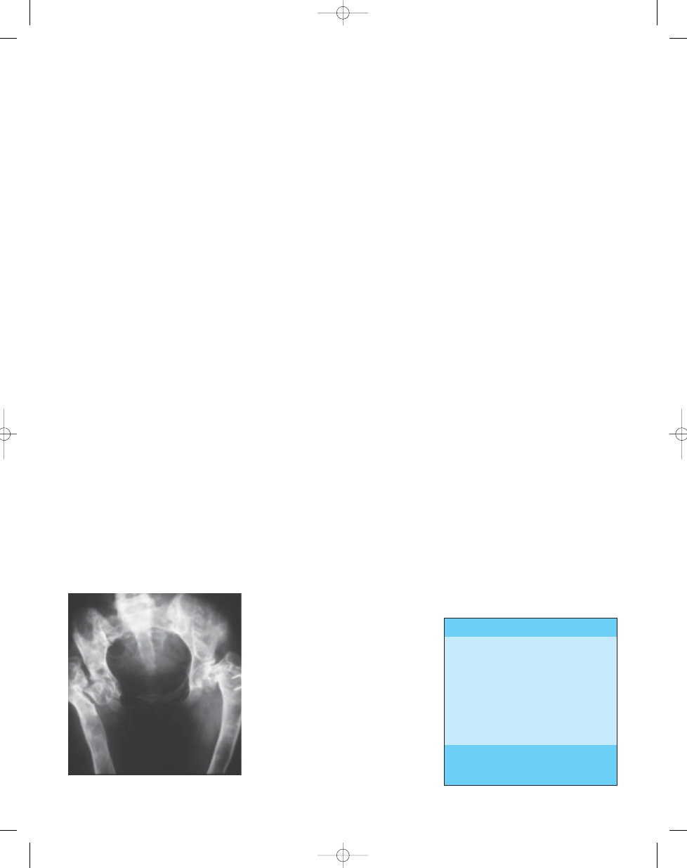
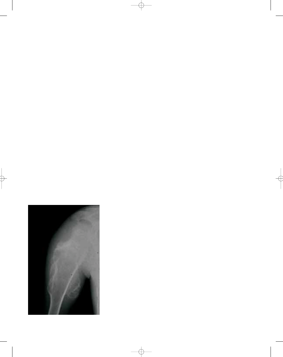
Fig. 21.07 Fibrous dysplasia in Albright syndrome.
Table 21.02
GNAS1 mutations in solitary, sporadic neoplasms.
Neoplasm
* Mazabraud syndrome, the combination of polyosto-
tic fibrous dysplasia and intramuscular myxomas, is
also caused by
GNAS1 mutations. From Cohen {382}
Osteosarcoma
Pituitary adenoma
Thyroid adenoma
Thyroid carcinoma
Parathyroid adenoma
Leydig cell tumour
Ovarian cyst
Intramuscular myxoma*
Breast carcinoma
357
Enchondromatosis: Ollier disease and Maffucci syndrome / McCune-Albright syndrome
bb5_29.qxd 13.9.2006 13:59 Page 357
levels; hyperphosphaturic hypophos-
phatemia; osteo-sarcoma (4%); devel-
opmental delay; microcephaly; and sud-
den or premature death {302,382,383,
392,1936}.
Bone and soft tissue tumours
As noted above, one of the primary
pathological conditions which defines
MAS is polyostotic fibrous dysplasia.
Other benign lesions associated with
this condition include mucoceles of
the head and neck {547,745}, simple
(unicameral) bone cysts {1001,1129}
and aneurysmal bone cysts {76,
1288,1759}. Perhaps the best known
concordance is with soft tissue, usu-
ally intramuscular, myxomas, known as
the Mazabraud syndrome {2108}.
Interestingly, activating mutation in the
GNAS1 gene have been detected in
myxoma cells {1605}, but not in leuko-
cytes or fibroblasts, from patients with
Mazabraud syndrome.
Malignant bone tumours have also
been associated with the fibrous
dysplasia seen in MAS. Osteosarco-
ma, and possibly also conventional
and dedifferentiated chondrosarco-
ma, appear to occur with increased
frequency {212, 872, 932, 1282, 1630,
1725, 1823}. Although other sarcomas,
including fibrosarcoma and malignant
fibrous histiocytoma, have been linked
to fibrous dysplasia {1822}, these have
not been reported in patients with MAS.
Individuals with MAS are also suscepti-
ble to endocrine tumours, including
adrenocortical and pituitary tumours
{1133,1637}.
Genetics
McCune-Albright syndrome (MAS) is
caused by mutations in the
GNAS1
gene located in chromosome band
20q13.
GNAS1 (guanine nucleotide-
binding protein,
α-stimulating activity
polypeptide 1) encodes the G-protein
α
stimulatory subunit (G
s
α), a component
of heterotrimeric G-protein complexes.
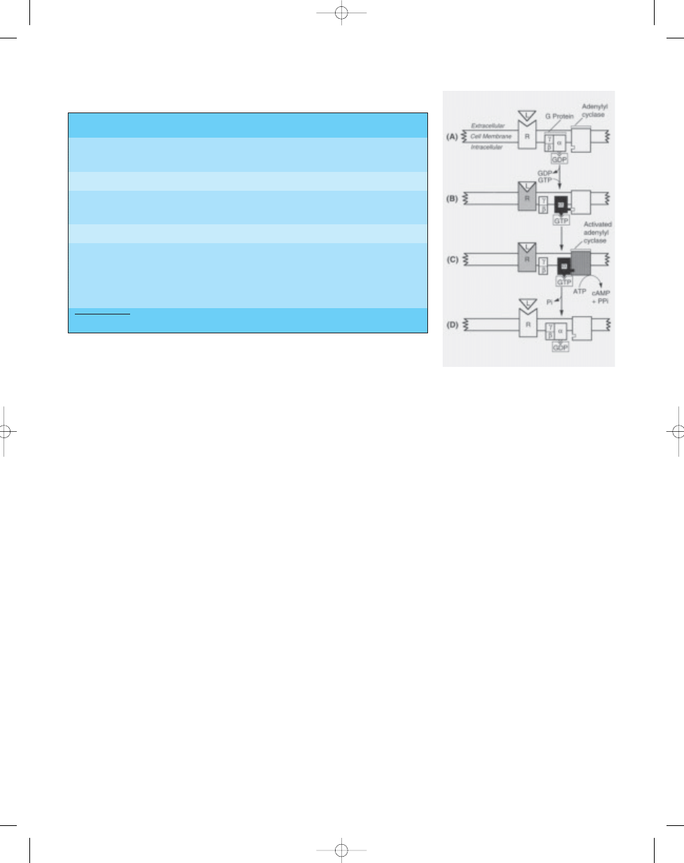
Gene function
G proteins (guanine nucleotide proteins)
are a family of molecules composed of
three subunits designated
α, β, and γ.
The function and specificity of each G
protein is determined by the
α subunit,
which is unique for each type. The
β and
γ subunits tend to be more homoge-
neous. Like all G proteins, the inactive
form of G
s
α contains bound GDP
(guanosine diphosphate). A GPCR (G
protein-coupled receptor) facilitates the
exchange of bound GTP (guanosine
triphosphate) for GDP producing the
active form {382,383}.
Adenylyl cyclase is activated following
ligand-binding to G-protein-coupled
receptor. Ligand-binding (B) produces
a conformational change in the receptor
and GDP is replaced by GTP, which
results in dissociation of the a subunit.
Binding of the active form of the
α sub-
unit to adenylyl cyclase (C) activates
this enzyme, resulting in the formation of
cAMP from ATP. Hydrolysis of GTP to
GDP is catalysed within seconds by the
intrinsic GTPase (guanosine triphos-
phatase) activity of G
s
α which causes
dissociation of the a subunit from
adenylyl cyclase and binding to the
β,
and
γ subunits, resulting in the inactive
form {382,383}.
Mutations
MAS, polyostotic fibrous dysplasia
(PFD), monostotic fibrous dysplasia
(MFD), and solitary pituitary adenoma
(PA) have the same causal genesis – a
ligand-independent, activating
GNAS1
mutation in the a subunit of stimulatory
G protein (G
s
α). Mutations are located
near the site which interacts with the
γ-
Table 21.03
Mutations in the
GNAS1 gene.
Fig. 21.08 (A) G protein composed of
α , β, and γ
subunits. This is the inactive form. B) Ligand (L)
binding produces conformational change in
receptor (R) and guanosine diphosphate (GDP) is
replaced by guanosine triphosphate (GTP), result-
ing in dissociation of the
α subunit. (C) Binding of
α subunit to adenylyl cyclase activates 3',5'-
cyclic adenosine monophosphate (cAMP) from
adenosine triphosphate (ATP). (D) Hydrolysis of
GTP to GDP by GTPase, causing dissociation of
the
α subunit from adenylyl cyclase and binding
to the
β and γ subunits, the inactive form. Ligand
binding causes repetition of the cycle {383}.
358
Congenital and inherited syndromes associated with bone and soft tissue tumours
Disorder
Exon
Nucleotide Change
Amino Acid Substitution
From Cohen and Howell {383}
Polyostotic fibrous dysplasia
8
C --> T
Arg201Cys
Panostotic fibrous dysplasia
8
C --> A
Arg201Ser
McCune-Albright syndrome
8
C --> T
Arg201Cys
8
G --> A
Arg201His
Monostotic fibrous dysplasia
8
C --> T
Arg201Cys
8
G --> A
Arg201His
Solitary pituitary adenoma
8
C --> T
Arg201Cys
8
G --> A
Arg201His
8
C --> A
Arg201Ser
9
A --> G
Gln227Arg
9
G --> T
Gln227His
bb5_29.qxd 13.9.2006 13:59 Page 358
phosphate of GTP, thus interfering with
hydrolysis of GTP to GDP. Because G
s
α
cannot dissociate from adenylyl cyclase
and bind to G
β
γ, adenylyl cyclase
remains active, producing increased
cAMP activity which results in the
pathology of MAS, PFD, MFD, and PA
{382,383,1934,1936,1937,2230}.
GNAS
mutations have also been recorded in
various solitary tumours {382}.
MAS, PFD, MFD, and PA occur sporadi-
cally. Mutations in the
GNAS1 gene
occur postzygotically in a somatic cell.
Clinical manifestations are variable in
distribution and appearance. More gen-
eralized vs. more localized expression
depends on (a) how small or how large
the cell mass is during embryogenesis
when the mutation occurs, and (b)
where in the cell mass the mutation
occurs {382, 383}.
GNAS1 mutations for MAS, PFD, MFD,
and PA are of the gain-of-function type.
It should be carefully noted that
GNAS1
mutations of the loss-of-function type
are found in endocrine disorders char-
acterized by hormone resistance, such
as type 1a pseudohypothyroidism, glu-
cocorticoid deficiency, and nephro-
genic diabetes insipidus {1934}.
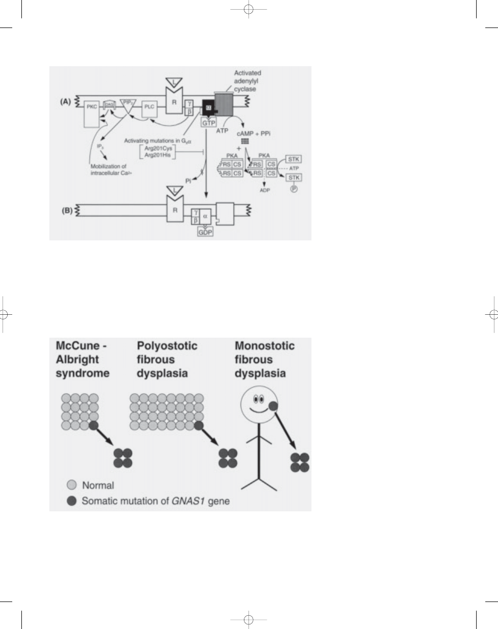
Fig. 21.09 (A) Activating mutations (Arg201Cys or Arg201His) in the gene encoding the
α subunit of stimu-
latory G protein (G
s
α), causing inappropriate stimulation of adenylyl cyclase interfering with hydrolysis of
GTP by GTPase to GDP. The PKA pathway (protein kinase A or cAMP-dependent protein kinase pathway)
is shown on the right. The PKC pathway (protein kinase C pathway) is shown on the left. Because the
α sub-
unit (G
s
α) cannot dissociate from adenylyl cyclase, cAMP is overproduced which, in turn, overactivates the
PKA pathway. PKA is composed of two regulatory subunits (RS) that have binding sites for cAMP, and two
catalytic subunits (CS) that, when dissociated, phosphorylate serine/threonine kinases (STK). The dissoci-
ated
βγ subunit overactivates the PKC pathway. PLC (phospholipase C) cleaves PIP2 (phosphatidylinositol
bisphosphate) into two intracellular messengers: DAG (diacylglycerol) and IP3 (inositol trisphosphate). The
latter triggers the release of sequestered calcium ions (Ca2+) which together with DAG activate PKC {383}.
359
McCune-Albright syndrome
Fig. 21.10 How mutations cause McCune-Albright syndrome, polyostotic fibrous dysplasia, and monostotic
fibrous dysplasia depend on when during embryonic development or during postnatal life the mutation
occurs. Somatic mutation in a small cell mass is likely to result in McCune-Albright syndrome. Mutation in
a larger cell mass may result in polyostotic fibrous dysplasia. A mutation in postnatal life – during infancy,
childhood, or adult life – may result in monostotic fibrous dysplasia {383}.
bb5_29.qxd 13.9.2006 13:59 Page 359
Definition
Multiple osteochondromas (MO) is an
autosomal dominant condition. It is
genetically heterogeneous and is caused
by mutations in one of the
EXT genes.
OMIM numbers
According to the gene involved, the fol-
lowing OMIM numbers have been
assigned:
EXT1
133700
EXT2
133701
EXT3
600209
TRPS2 / Langer Giedion syndrome 150230
Potocki-Shaffer syndrome
601224
Synonyms
EXT, diaphyseal aclasis, (multiple hered-
itary) osteochondromatosis, multiple car-
tilaginous exostoses, hereditary multiple
exostoses.
Incidence
The solitary (sporadic) form of osteo-
chondroma is approximately 6 times
more common than the occurrence within
the context of MO. The incidence of MO
is approximately 1:50,000 persons within
the general population {1887}. Males are
more often affected (male: female ratio
1.5:1) {1236, 2265}, due in part to an
incomplete penetrance in females {1236}.
Approximately 62% of the patients with
multiple osteochondromas have a posi-
tive family history {1236}.
Diagnostic criteria
A diagnosis of multiple exostoses can be
made when radiologically at least two
osteochondromas of the juxta-epiphy-
seal region of long bones are observed
{1236}. MO is diagnosed in case of a
positive family history and/or a proven
germline mutation in one of the
EXT
genes.
Clinical features
Osteochondromas develop and increase
in size in the first decade of life, ceasing
to grow when the growth plates close at
puberty. They are pedunculated or ses-
sile (broad base) and can vary widely in
size. The majority are asymptomatic and
located in bones that develop from carti-
lage, especially the long bones of the
extremities, predominantly around the
knee. The number of osteochondromas
may vary significantly within and between
families. In addition, in the majority of MO
patients bone remodelling defects are
observed resulting in deformities of the
forearm (shortening of the ulna with se-
condary bowing of radius) (39-60%)
{1887, 1929}, inequality in limb length
(10-50%) {1887, 1929}, varus or valgus
angulation of the knee (8-33%) {1887,
1929}, deformity of the ankle (2-54%)
{1887, 1929} and disproportionate short
stature (37-44%) {1236, 2265}. It has long
been thought that these abnormalities
are the result of skeletal dysplasia,
although recent evidence indicates that
osteochondromas are neoplastic (see
chapter 10), and it has been suggested
that the growth retardation in MO may
result from the local effects of enlarging
osteochondromas {1717}. Moreover,
the severity of angular deformity was
found to be correlated with the number of
sessile osteochondromas {309}.
The most important complication of MO
is malignant transformation of an osteo-
chondroma, which is estimated to occur
in 0.5-3% of MO patients {815, 1236,
1695, 1887, 2265}. The suspicion of se-
condary chondrosarcoma is indicated by
growth of the tumour after puberty, the
presence of pain, or a thickness over 1
cm of the cartilaginous cap in adults. The
size of the cartilaginous cap can be well
established with T2-weighted MR imag-
ing. There are no universally accepted
guidelines for surveillance of individuals
with MO so far.
Other complications of the osteochon-
dromas include osseous and cosmetic
deformities, fracture, bursa formation,
arthritis (14%) {2265}) and impingement
on adjacent tendons, nerves (23%)
{2265}, vessels (11%) {2265} or spinal
cord (<1%) {2187, 2265}.
Bone and soft tissue tumours
Hereditary osteochondromas and sec-
ondary peripheral chondrosarcomas
developing within the cartilaginous cap
of hereditary osteochondromas are
histopathologically similar to their spo-
radic counterparts. Morphologically two
types of osteochondroma can be recog-
nized: broad based sessile cases with
irregular cartilaginous linings and those
with a well defined cartilaginous cap.
Both may occur within and outside the
context of MO. Malignant transformation
of osteochondroma leads to a secondary
peripheral chondrosarcoma in 94% of
the cases {2276}. Very rare cases of
other sarcomas developing in osteo-
chondroma have been described, most
often in solitary cases of osteochon-
droma {56, 1214, 1576, 1902, 1968, 2181}
including osteosarcomas, and spindle
cell sarcomas {1214,1356}. These tu-
mours develop in the stalk of the osteo-
chondroma, in contrast to secondary
peripheral chondrosarcomas, which
develop in the cap of the pre-existing
osteochondroma. A few cases of MO
J.V.M.G. Bovée
P.C.W. Hogendoorn
Multiple osteochondromas
Fig. 21.11 Multiple osteochondromas in a patient
with hereditary multiple osteochondromas.
360
Congenital and inherited syndromes associated with bone and soft tissue tumours
bb5_29.qxd 13.9.2006 13:59 Page 360
patients have been reported to develop
other sarcomas as well {239, 2139}.
These osteosarcomas and spindle cell
sarcomas (malignant fibrous histiocy-
tomas and fibrosarcomas) display an
indistinguishable phenotype from their
non osteochondroma-related counter-
parts. Even more rare is the occurrence
of "conventional" dedifferentiated periph-
eral chondrosarcoma, in which case the
osteochondroma gives rise to peripheral
low grade chondrosarcoma that in turn
"dedifferentiates" into a high grade sarco-
ma that may appear as fibrosarcoma,
malignant fibrous histiocytoma or
osteosarcoma {183}. No soft tissue neo-
plasms are described within the context
of MO.
Genetics
MO is a genetically heterogeneous disor-
der for which two genes,
EXT1 and EXT2
located respectively at 8q24 and 11p11-
p12, have been isolated {20, 395, 2031,
2310}. Additional linkage to chromosome
arm 19p has been found, suggesting the
existence of an
EXT3 gene {1229}. Loss
of heterozygosity however is absent at
this locus {236, 924, 1760} and the gene
has never been identified. Three new
genes,
EXTL1, EXTL2 and EXTL3 have
been identified based on their homology
with the
EXT1 and EXT2 genes {2180,
2283,2309}. However, no association
with disease has been documented.
Both
EXT genes are involved in a con-
tiguous gene deletion syndrome.
Patients carrying a deletion of 8q24
demonstrate the Langer-Giedion syn-
drome (LGS or trichorhinophalangeal
syndrome type II (TRPS2; OMIM
150230), which is characterized by cran-
iofacial dysmorphism and mental retar-
dation in addition to multiple osteochon-
dromas {975,1297,1298,1491}. LGS is
due to loss of functional copies both of
the
TRPS1 gene, encoding a zinc-finger
protein {1491}, and the
EXT1 gene at
8q24 {975,1298}. Trichorhinophalangeal
syndrome type I (TRPS1) (OMIM 190350)
is similar to LGS although multiple osteo-
chondromas are absent. Patients carry-
ing a deletion of 11p11.2-p12 demon-
strate Potocki-Shaffer syndrome (proxi-
mal 11p deletion syndrome {2307},
DEFECT11, 11p11.2 contiguous gene
deletion syndrome). These patients
demonstrate enlarged parietal foramina,
multiple osteochondromas, and some-
times craniofacial dysostosis and mental
retardation {134,1721}. The syndrome is
caused by deletion of
EXT2 and proba-
bly of
ALX4; haploinsufficiency of the lat-
ter was shown to potentially cause
enlarged parietal foramina {134, 2303}.
Gene structure
The
EXT1 gene was identified by posi-
tional cloning {20}. The gene is com-
posed of 11 exons, and spans approxi-
mately 350 kb of genomic DNA {1296}.
The cDNA has a coding region of 2238
bp {20}. The promoter sequence is char-
acteristic of a housekeeping gene
{1296}. A mouse-homologue is found on
mouse chromosome 15 with a very high
level of sequence homology {1266,
1279}. Additional homologues have been
identified in Caenorhabditis elegans
{369} and Drosophila melanogaster
{156}.
The
EXT2 gene was also identified by
positional cloning {2031,2310} and con-
tains 16 exons, two of which (1a and 1b)
are alternatively spliced {369}. The gene
spans approximately 108 kb of genomic
DNA {369}. The cDNA consists of
approximately 3 kb, defining a single
open reading frame of 2154 bp. The
mRNA demonstrates alternative splicing
{2031,2310}. A highly significant similari-
ty with the
EXT1 gene product has been
found, especially in the carboxy terminal
region {2031,2310}. Homologues are
found on mouse chromosome 2 {369,
2032} and in Caenorhabditis elegans
{369}.
Gene expression
Both
EXT1 and EXT2 mRNA is ubiqui-
tously expressed {20, 2031, 2310}. A
high level of expression of
Ext1 and Ext2
mRNA has been found in developing
limb buds of mouse embryos {1265,
2032} and expression was demonstrated
to be confined to the proliferating and
prehypertrophic chondrocytes of the
growth plate {2030}. The gene products,
exostosin-1 (
EXT1) and exostosin-2
(
EXT2), are endoplasmic reticulum local-
ized type II transmembrane glycopro-
teins which form a Golgi-localized het-
ero-oligomeric complex that catalyzes
heparan sulphate (HS) polymerisation
361
Multiple osteochondromas
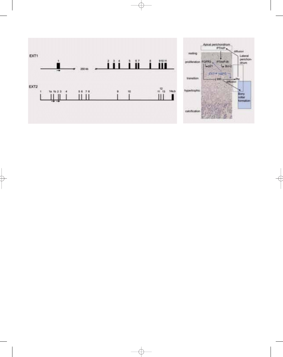
Fig. 21.12 Genomic structure of the EXT1 and EXT2 genes.
Fig. 21.13 706 Hypothesized function of EXT within
the normal early embryonic growth plate.
bb5_29.qxd 13.9.2006 13:59 Page 361

362
Congenital and inherited syndromes associated with bone and soft tissue tumours
{1267,1372,1373,1954}. Heparan sul-
phate proteoglycans (HSPG) are large
macromolecules composed of heparan
sulphate glycosaminoglycan chains
linked to a protein core. Four HSPG fam-
ilies have been identified: syndecan,
glypican, perlecan and isoforms of
CD44. HSPGs are required for high-affin-
ity binding of fibroblast growth factor to
its receptor {1275}. Furthermore, an
EXT1 homologue in Drosophila (tout-
velu, Ttv) has been shown to be required
for diffusion of an important segment
polarity protein called Hedgehog (Hh)
{156, 2107, 2126}, a homologue of mam-
malian Indian Hedgehog (IHh). It is
therefore hypothesized that
EXT muta-
tions affect FGF and IHh signalling within
the normal growth plate.
Mutations
The
EXT1 gene was reported to show
linkage in 44%-66% of the MO families
{1235, 1761}, whereas
EXT2 would be
involved in 27% {1235}. Germline muta-
tions of
EXT1 and EXT2 in MO patients
have been studied extensively in
Caucasian as well as Asian popula-
tions {2306} (For overview see also:
The human gene mutation database
Cardiff www.hgmd.org {1176}). In
EXT1,
mutations are more or less randomly
distributed over the first 6 exons, while
the last 5 exons, containing the con-
served carboxyterminal region, contain
significantly less mutations {2306}.
Similarly, in
EXT2 most mutations are
found in the first exons. No mutational
hotspots are found {2306}. Appro-
ximately 80% of the mutations are
either non-sense, frameshift, or splice-
site mutations leading to premature
termination of EXT proteins {714,1656,
1761,1917,2308,2313}. The majority of
missense mutations also lead to defec-
tive EXT protein function {340}.
Loss of the remaining wildtype allele
has been demonstrated {238}, indica-
ting that the
EXT genes act as tumour
suppressor genes. The limited number
of genotype-fenotype correlational stu-
dies performed so far provide no uniform
data {309,714}. The risk of malignant
transformation would be higher in pa-
tients carrying
EXT1 mutations {714}.
Gene
Chromosomal Associated
Function:
localization
disease
glycosyltransferase activity involved in
heparan sulphate (HS) biosynthesis:
Table 21.04
The
EXT gene family.
EXT2
11p11-p12 {977,961}
MO
HS chain elongation {1267, 1372, 1954}
EXT1
8q24 {907}
MO
HS chain elongation {1267, 1372, 1954}
EXTL1
1p36.1 {971}
Unknown
HS chain elongation {1108}
EXTL2
1p11-p12 {976}
Unknown
HS chain initiation {1127}
EXTL3
8p12-p22 {966}
Unknown
HS chain initiation and elongation {1108}
bb5_29.qxd 13.9.2006 13:59 Page 362
Definition
Retinoblastoma (RB) is a malignant
tumour originating from the embryonic
neural retina. Familial retinoblastoma is
typically bilateral, caused by a germline
mutation in the
RB1 tumour suppressor
gene and is often associated with the
development of second site primary
tumours, including osteosarcoma, fibro-
sarcoma, chondrosarcoma, Ewing sarco-
ma, pinealoblastoma, epithelial tumours,
leukaemia, lymphoma, mela-noma and
brain tumours.
OMIM number
180200 {1376}
Synonym
Retinoblastoma / osteogenic sarcoma
syndrome.
Incidence
Retinoblastoma, the most common
intraocular tumour of children, has a
worldwide incidence between 1/3500
and 1/25000 with no significant differ-
ences between the sexes or races {28,
147,511,1856}.
Diagnostic criteria
Presentation is a white, pink-white, or
yellow-white pupillary reflex termed
"leukocoria'' resulting from replacement
of the vitreous by tumour, or by a large
tumour growing in the macula {718}.
Another common symptom, strabismus
(exotropia or esotropia), can occur alone
or associated with leukocoria. Less
frequent presenting signs include a red,
painful eye with secondary glaucoma,
low-vision orbital cellulitis, unilateral
mydriasis, and heterochromia {2346}.
The tumour can be difficult to differenti-
ate from a variety of simulating lesions
such as persistent hyperplastic primary
vitreous, retrolental fibroplasia, Coats
disease, Toxocara canis infection, retinal
dysplasia, or chronic retinal detach-
ment {582,976}. These can be distin-
guished using CT, MRI, ultrasonography
or fine-needle aspiration biopsy and a
careful history of the family and affected
child {582}.
Clinical features
Retinoblastoma can be unifocal or multi-
focal. In bilateral cases, one eye is usu-
ally in a more advanced stage, while the
contralateral eye has one or more tumour
foci. The average age at diagnosis is 12
months for bilateral and 18 months for
unilateral cases, with 90 percent of the
cases diagnosed before the age of 3 {29,
1157,1860,2123}. Retinoblastoma can
be a part of the 13q-deletion syndrome in
association with moderate growth and
mental retardation, broad prominent
nasal bridge, short nose, ear and dental
abnormalities, and muscular hypotonia
{38, 717}.
Trilateral retinoblastoma describes the
association between bilateral retinoblas-
toma and midline brain tumours, usually
in the pineal region {554}. Pineal tumours
resembling well differentiated retinoblas-
tomas are also called ectopic retinoblas-
toma. CT scanning and MRI have
reduced the misinterpretation of pineal
tumours as intracranial spread of
retinoblastoma {2346}. This is clinically
important since ectopic intracranial
retinoblastoma requires therapy to the
whole neuraxis as well as high-dose
equivalent radiotherapy to the primary
tumour.
Pathology of retinoblastoma
Retinoblastoma occurs as a mass
between the choroid and retina (exo-
phytic) or bulge from the retina toward
the vitreous (endophytic). Most ad-
vanced tumours show both patterns
of growth. The tumour is histologi-
cally characterized by rosettes and
fleurettes, which are believed to rep-
resent maturated or differentiated neo-
plastic cells. Rosettes are spherical
structures (circular in section) of uni-
form cuboidal or short columnar cells
arranged about a small round lumen
(Flexner-Wintersteiner rosette) or with-
out any lumen (Homer-Right rosette).
The latter also appears in other neuro-
ectodermal tumours such as medul-
loblastoma. Fleurettes are arranged
with short, thin stromal axes surroun-
W.K. Cavenee
O. Bögler
T. Hadjistilianou
I.F. Newsham
Retinoblastoma syndrome
Fig. 21.14 Genomic and protein domain organization of the 105kD retinoblastoma protein. Mutational
hotspots for frameshift and nonsense mutations are identified above individual exons. Examples of some of
the known cellular binding proteins and their region of interaction are depicted below the protein domains.
Sites of phosphorylation are also noted.
363
Retinoblastoma syndrome
bb5_29.qxd 13.9.2006 13:59 Page 363
364
Congenital and inherited syndromes associated with bone and soft tissue tumours
ded by differentiated neoplastic cells
with their apical part facing the exter-
num. Tumours can be necrotic, with sur-
viving cells around blood vessels,
creating ``pseudo-rosettes.'' Calcified
foci and debris from nucleic acids can
be found in necrotic areas giving rise
to basophilic vessel walls {29, 1860,
2123}.
Growth patterns and other histological
parameters are not useful for determin-
ing prognosis. The degree of differentia-
tion and number of mitoses show a weak
correlation. Stronger relationships exist
with invasion of the choroids, optic
nerves and sclera. Progressive invasion
of the eye coats, even in the horizontal
plane, is highly informative for determi-
ning prognosis {1157, 2123}.
Bone and soft tissue tumours
Second-site primary malignant tumours
refer to nonmetastatic tumours arising in
"disease-free'' patients treated for initial
disease. Tumours associated with
retinoblastoma include osteosarcoma,
fibrosarcoma, chondrosarcoma, epithe-
lial malignant tumours, Ewing sarcoma,
leukaemia, lymphoma, melanoma, brain
tumours, and pinealoblastoma {12, 502,
550, 1793, 1837}. These second tumours
are classified into five groups: (a)
tumours in the irradiated area, (b)
tumours outside and remote from the
irradiated area, (c) tumours in patients
not receiving radiotherapy, (d) tumours
unable to be determined as primary or
metastases, and (e) tumours in members
of retinoblastoma families who were free
of retinal tumours. Two important obser-
vations have emerged from analysing
these patients: (a) the great majority of
children in whom second neoplasms
develop have or will have bilateral
retinoblastoma, and (b) the incidence of
second neoplasms in this group was
similar whether they received radiation or
not. Osteogenic sarcomas are the most
frequent second site neoplasms in all the
published series {12, 336a, 502, 550,
1720a,1723a,1793,1837}.
Genetics
Retinoblastoma has served as the proto-
typic example of a genetic predisposi-
tion to cancer. It is estimated that 60 per-
cent of cases are nonhereditary and uni-
lateral, 15 percent are hereditary and
unilateral, and 25 percent are hereditary
and bilateral. In the latter two types,
autosomal dominant inheritance with
nearly complete penetrance is observed.
Analysis of such cases by epidemiolo-
gical / cytogenetic {716,941, 1145,2006,
2017, 2044}, molecular genetic {317,
318} and molecular biological {558, 969}
methods suggests as few as two
required stochastic mutational events in
the
RB1 locus for tumour formation. The
first mutation can be inherited through
the germ line or somatically acquired,
whereas the second occurs somatically
in either case.
RB1 locus inactivation is
also found in non-hereditary retinoblas-
toma {552}, osteogenic and other sarco-
mas occurring as second primary
tumours in retinoblastoma patients and
some primary sarcomas in the absence
of retinoblastoma involvement {724,
882}.
Gene structure and expression
The
RB1 locus in chromosome band
13q14.1 {317, 716, 2006, 2044} encom-
passes 200 kb of genomic DNA organ-
ized into 27 exons {228, 744, 1233}. The
105 kD RB1 protein is ubiquitously
expressed in normal human and rodent
tissues, including brain, kidney, ovary,
spleen, liver, placenta, and retina. RB1 is
differentially phosphorylated {1234}, with
the unphosphorylated form predomi-
nantly found in the G1 stage of the cell
cycle, and an initial phosphorylation
occurring at the G1/S boundary {284,
482}. Viral proteins bind the p105RB pro-
tein {481, 564, 2262} using regions nec-
essary for their transforming function.
Over 100 intracellular pRB binding pro-
teins have also been identified including
E2F transcription factors, tumour sup-
pressor BRCA1 and the RB-like proteins
p107 and p130 {1508}. Complexing of
the two latter factors also oscillates in a
cell-cycle-dependent manner linking the
tumour-suppressing function of RB1 with
transcriptional regulation.
Mutations
Mutations that result in loss of RB1 func-
tion have been described for retinoblas-
toma patients and their tumours at the
DNA, RNA, and protein levels. RB1 alter-
ations have also been detected in a vari-
ety of clinically related second-site pri-
mary tumours including osteosarcoma,
as well as other non-secondary tumours
such as breast and small-cell lung car-
cinoma. Detection of
RB1 mutations pro-
vides for accurate prenatal risk assess-
ment {319, 970, 2267, 2318}.
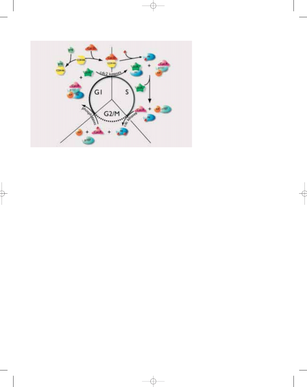
Fig. 21.15 Regulation of the cell cycle through oscillating phosphorylation of the p105 retinoblastoma
protein.
bb5_29.qxd 13.9.2006 13:59 Page 364
Definition
Rothmund-Thomson syndrome (RTS) is
a constellation of various skin abnormal-
ities, skeletal defects, juvenile cataracts,
premature ageing, and a predisposition
to osteosarcoma, skin cancer, and other
tumours. At least a subset of cases are
caused by inherited mutations in the
RECQL4 helicase gene.
OMIM number
268400
Synonym
Poikiloderma congenitale.
Incidence
RTS is a rare, autosomal recessive dis-
order. The exact incidence is unknown,
but more than 250 cases have been
reported in the world literature from a
variety of ethnic backgrounds. A slight
male preponderance (M:F = 2:1) has
been reported {2212}.
Diagnostic criteria
Specific criteria for the diagnosis of RTS
have not been established. The diagno-
sis is based upon clinical findings, the
identification of
RECQL4 in a subset of
cases, and laboratory tests that can
exclude some other, similar disorders.
Clinical features
The cardinal feature of RTS is a sun-sen-
sitive erythematous rash that typically
appears during the first 6 months. It
usually starts in the face and then
spreads to the buttocks and extremities.
With time, the rash enters a chronic
phase resulting in skin atrophy, telang-
iectasias, and marbleized mixed hyper-
and hypopigmentation (poikiloderma)
{1735, 2198, 2199, 2212}. Other fea-
tures associated with RTS include short
stature (~2/3 of the cases), premature
greying and loss of hair (50-65%),
sparse eyebrows/lashes (60-75%), juve-
nile cataracts (7-50%), photosensitivity
(35%), radial ray anomalies (>20%) and
other bony abnormalities, dystrophic
nails and teeth, hypogonadism, and
hypersensitivity to cytotoxic drugs and
radiotherapy {1735, 2198, 2199, 2212}.
RTS does not seem to be associated
with intellectual or immunological
impairment. There are no specific or
consistently identifiable laboratory fea-
tures in RTS. There have been several
reports of acquired, clonal somatic
mosaicism for chromosome abnormali-
ties, especially trisomies, isochromo-
somes, and translocations frequently
involving chromosome 8, often found in
fibroblast cultures {1269}. There is no
evidence of mismatch repair deficiency
in the form of tumour microsatellite
instability, as seen in tumours associat-
ed with the hereditary non-polyposis
colon cancer syndrome, due to
germline mutations in genes of the DNA
mismatch repair complex). Furthermore,
there is no increase in chromosomal sis-
ter-chromatid exchange rates (as seen
in Bloom syndrome), no excess of
bleomycin-induced chromosome break-
age (as seen in ataxia telangiectasia),
and no chromosomal radial formation
with mitomycin-C exposure (as seen in
Fanconi anaemia). Ultraviolet sensitivity
studies have yielded inconsistent
results.
Bone and soft tissue tumours
Osteosarcomas, involving any bone and
especially in non-common sites, have
been reported to occur in up to one third
of the patients, with a median age of
diagnosis at 11.5 years {2212}. Also
cutaneous malignancies, in particular
squamous cell carcinomas, have been
reported to be overrepresented in RTS
{1735, 2212}.
Genetics
At least a subset of the cases of RTS are
caused by mutations in the
RECQL4
(also known as
RECQ4) helicase gene
in chromosome band 8q24.3 {1128}.
Only a small number of patients has as
yet been investigated, with mutations
being detected in approximately 40% of
the cases {749}. The
RECQL4 gene
has a predicted protein product of
1208 amino acids. It is highly ex-
pressed in the thymus and testis with
low levels of intranuclear expression
in multiple other tissues.
RECQL4 mu-
tation analysis is available only in
specialized centres. Mutations have
included frameshift mutations, non-
sense mutations, and deletions in-
cluding part of the consensus heli-
case domain. This gene is homologous
to the genes that cause Bloom syn-
drome and Werner syndrome, which
might explain some of the clinical over-
lap {749}.
N.M. Lindor
Rothmund-Thomson syndrome
365
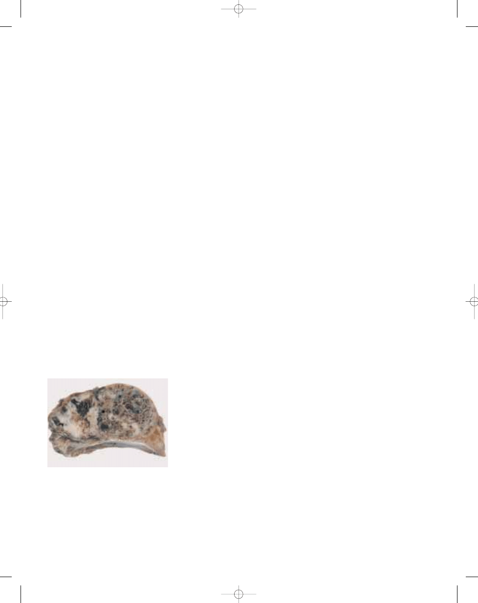
Rothmund-Thomson syndrome
Fig. 21.16 Osteosarcoma of the rib in a patient with
Rothmund-Thomson syndrome.
bb5_29.qxd 13.9.2006 13:59 Page 365
366
Congenital and inherited syndromes associated with bone and soft tissue tumours
Definition
Werner syndrome (WS) is a rare, autoso-
mal recessive genetic instability syn-
drome and is caused by mutations in the
WRN gene. Affected patients develop a
prematurely aged appearance in the
second and third decades of life, and are
at increased risk of developing both neo-
plastic and non-neoplastic diseases.
Tumours include soft tissue sarcomas,
thyroid carcinoma, malignant melanoma,
meningioma, haematological neoplasms,
and osteosarcoma. The most common
causes of death are cancer and athero-
sclerotic cardiovascular disease.
OMIM number
277700
Synonym
Progeria of the adult.
Incidence
WS patients have been identified world-
wide {819}. Estimates of the frequency or
prevalence of WS, obtained by case
counting and from consanguinity data,
range from 1/22,000 to 1/10
6
(reviewed in
{1883}). The frequency of WS in different
countries is strongly influenced by the
presence of founder mutations and the
frequency of consanguinity or inbreed-
ing. The range of frequency estimates
also undoubtedly reflects the variable
and delayed development of the WS clin-
ical phenotype {604, 819}, with conse-
quent underdiagnosis.
Clinical features and diagnostic criteria
The most consistent clinical findings
develop after age 10. These include bi-
lateral cataracts, dermatological patho-
logy resembling scleroderma, short
stature and premature greying and loss
of scalp hair {604,819}. There may be
affected siblings as well as evidence
of parental consanguinity (3rd cousin or
closer). Additional, less consistent find-
ings include diabetes mellitus, hypogo-
nadism, osteoporosis, soft tissue calcifi-
cation, premature atherosclerotic cardio-
vascular disease, high pitched, ‘squeeky’,
or hoarse voice and flat feet.
A definite diagnosis can be established
on clinical grounds when all of the con-
sistent features and at least two addi-
tional findings are present. Additional
diagnostic aids include evidence of ele-
vated 24 hr urinary hyaluronic acid
secretion; loss of WRN protein from
fibroblasts or peripheral blood lympho-
cytes; and mutations in the
WRN gene
on chromosome arm 8p.
A clinical scoring system has been
devised to identify more reliably definite,
probable or possible WS patients.
Additional information on this scoring
system and the clinical diagnosis of
WS can be found on the Internatio-
nal Registry of Werner Syndrome
Web site:
www.pathology.washington.edu/
research/werner/registry/diagnostic.html
R.J. Monnat, Jr.
Werner syndrome
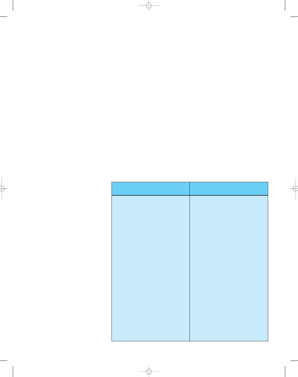
Table 21.05
Histopathological spectrum of neoplasia in Werner syndrome.
A wide spectrum of neoplasms has been identified in Werner syndrome (WS) patients, who are clearly at
elevated risk of developing one or more of the neoplasms listed in the left column (‘frequent’). These neo-
plasms represent 71% of all neoplasms reported in WS patients. WS patients may be at elevated risk of
developing neoplasms listed in the right column, although the number of affected patients is too small in
most cases to firmly establish this suspicion. A total of 257 neoplasms were represented in this analysis
{820, 1494} (Y. Ishikawa, personal communication). The percentage of neoplasms from this analysis in each
column or tumour type is indicated in parentheses.
Soft tissue sarcomas (15.5% of cases)
malignant fibrous histiocytoma
leiomyosarcoma
fibrosarcoma
malignant schwannoma
synovial sarcoma
rhabdomyosarcoma
Thyroid carcinomas (14%)
follicular
papillary
anaplastic
Malignant melanoma (12.6%)
acral lentigenous melanoma
mucosal malignant melanoma
Meningioma (11.1%)
benign
multiple / malignant
Haematological (11.1%)
acute myelogenous
leukaemias (M1-5)
erythroleukaemia (M6)
megakaryocytic leukaemia (M7)
myelofibrosis/myelodysplasia
aplastic anaemia
Osteosarcoma (6.3%)
Non-melanoma skin cancer (5.8%)
Hepatobiliary carcinomas (5.3%)
hepatocellular
cholangiocarcinoma
gallbladder
Genito-urinary (4.8%)
bladder carcinoma
uterine/ovarian carcinoma
renal cell carcinoma
prostate carcinoma
seminoma
Gastro-intestinal carcinoma (4.3%)
gastric
oesophagus
pancreas
colon
Breast carcinoma (3.9%)
Oro-pharyngeal carcinoma (2.4%)
Frequent (71%)
Less common (29%)
bb5_29.qxd 13.9.2006 13:59 Page 366

367
Werner syndrome
Neoplastic disease spectrum
WS patients are at increased risk of
developing both sarcomas and epithe-
lial neoplasms {820, 1494}. The elevat-
ed risk of neoplasia is selective, and
includes the following neoplasms in
order of decreasing frequency: soft tis-
sue sarcomas, thyroid carcinoma,
meningioma, malignant melanoma,
malignant or pre-neoplastic haemato-
logical disease and osteosarcoma.
Many other neoplasms, including com-
mon adult epithelial malignancies, have
been observed in WS patients.
However, it is not clear whether the risk
of developing these neoplasms is ele-
vated above population controls. This
histo-pathological spectrum of neo-
plasms overlaps with, though is distinct
from, that observed in patients with two
other RecQ helicase deficiency syn-
dromes, Bloom syndrome and
Rothmund-Thomson syndrome {1494}.
Several features of neoplasia in WS
patients indicates that this human RecQ
helicase deficiency syndrome is a heri-
table cancer predisposition: patients
develop neoplasms at a comparatively
early age; often have unusual sites of
presentation (e.g., osteosarcoma of the
patella) or less common histopathologic
subtypes (e.g., follicular as opposed to
papillary thyroid carcinoma); and can
have multiple concurrent or sequential
neoplasms, e.g., thyroid carcinoma and
osteosarcoma. Estimates of the
increased risk of neoplasia in WS
patients range from 30-fold elevated
overall lifetime risk across all tumour
types to 1000-fold elevated risk for acral
lentigenous melanoma.
Soft tissue sarcomas that have been
identified in WS patients include malig-
nant fibrous histiocytoma, malignant
peripheral nerve sheath tumour, fibro-
sarcoma, rhabdomyosarcoma, lipo-sar-
coma, and synovial sarcoma. Three his-
tological subtypes of thyroid carcinoma
have been reported in WS patients (fol-
licular, papillary and anaplastic), with a
predominance of the less common fol-
licular variant. There has been no
reported case of medullary thyroid car-
cinoma in a WS patient. The risk of
malignant melanoma is confined almost
exclusively to the relatively rare variants
that arise on the palms and soles (acral
lentigenous melanoma) or in mucosa
of the nasal cavity or esophagus.
Melanoma risk is most clearly elevated
in Japanese WS patients {820}.
The spectrum of haematological dis-
ease in WS includes acute myelogenous
leukaemia (M1-5), erythroleukaemia
(M6) and megakaryocytic leukaemia
(M7); atypical leukemia arising in the
context of myelodysplasia; and the pre-
malignant conditions myelodysplasia,
myelofibrosis, and aplastic anaemia.
The elevated risk of developing marrow-
associated pre-malignant or malignant
disease may be related to the progres-
sive accumulation of genetic damage in
bone marrow cell lineages {1509}.
Genetics
WS is an autosomal recessive disease:
no cases are known to have been
acquired or to have been caused by
other agents. WS constitutes, together
with Bloom syndrome and Rothmund-
Thomson syndrome, a group of inherit-
ed human genetic instability / cancer
predisposition syndromes that result
from loss of function of a human RecQ
helicase protein.
Gene structure and expression
The
WRN gene consists of 35 exons in a
165 kb region of chromosome region
8p11-12 {2331}.
Two stable RNAs are encoded by the
WRN gene, and the shorter, of 5.8 kb, is
ubiquitously expressed at varying levels
in many cell types, tissues and organs
{2331}. The 162 kDa WRN protein is
readily detectable in cell lines and tis-
sue samples from normal individuals
and heterozygous carriers of single
mutant copies of the
WRN gene by
Western blot analysis {1510}. No sys-
tematic study of the level of expression
of WRN protein as a function of cell type
or of development has as yet been pub-
lished. The WRN protein encodes both
DNA helicase and exonuclease activi-
ties {1931}, and is likely to play an
important physiologic role in homolo-
gous recombinational repair in human
somatic cells {1728}.
Mutations
WS is an autosomal recessive disease,
and thus patients have mutations in both
WRN alleles. Virtually all of the WRN
patient mutations thus far identified
truncate the
WRN open reading frame,
lead to protein reduction or loss from
patient cells and thus can be detected
by Western blot analysis {821,1510}.
Further mutation characterization can
be performed by a combination of muta-
tion-specific allele identification and / or
DNA sequencing. Mutation analysis can
be especially helpful in the diagnosis of
WS in young patients, where the diag-
nosis is suspected but the clinical phe-
notype may be incompletely developed.
A HUGO Locus-Specific
WRN Muta-
tional Database summarizes patient
mutation data and mutation designa-
tions, polymorphism data, and re-
lated clinical data and cross-refere-
nces these to the primary literature
( w w w. p a t h o l o g y. w a s h i n g t o n . e d u /
r e s e a r c h / w e r n e r / w s
_
w r n . h t m l )
{1511}. Additional information on
WRN
mutation analysis for the purpose of
confirming a diagnosis of Werner
syndrome can be obtained through
the International Registry of Werner
Syndrome Web site (www.pathology.
washington.edu/research/werner/reg-
istry/diagnostic.html).
bb5_29.qxd 13.9.2006 13:59 Page 367
Wyszukiwarka
Podobne podstrony:
bb5 chap3
bb5 chap8
bb5 chap1
BB5 BOX
bb5 chap16
bb5 chap15
bb5 contents
bb5 chap12
bb5 chap4
bb5 references
bb5 chap6
bb5 chap17
bb5 chap20
bb5 chap5
Lista wszystkich dostępnych polskich Product Code dla telefonów platformy BB5
bb5 source
bb5 chap13
bb5 chap19
więcej podobnych podstron