CHAPTER 19
Myogenic, Lipogenic, Neural,
and Epithelial Tumours
Smooth muscle tumours of bone, usually leiomyosarcoma, are
very rare. A metastasis from a distant site, especially the uterus,
has to be excluded before accepting the diagnosis of primary
leiomyosarcoma of bone.
Lipomas are not uncommon in bone and are incidental findings
on X-rays and frequently involve the calcaneus.
Roentgenograms show a well-circumscribed area of lucency
with a central area of calcification. CT and MRI help to confirm
the fatty nature of the tumour.
Neurilemmomas (schwannomas) occur rarely in bone. Along the
spine, especially in the sacrum, they may involve bone second-
arily. The most common location for an intraosseous neurilem-
moma is the mandible. The histological features are similar to
those of schwannomas elsewhere. Malignant peripheral nerve
sheath tumours (MPNST) do not occur in bone.
Adamantinoma has an epithelial phenotype and almost exclu-
sively involves the tibia. It is a low-grade malignancy with a
favourable clinical course. The roentgenographic, morphologic
and genetic features are often similar to those of osteofibrous
dysplasia.
Metastatic carcinoma is by far the commonest malignancy in the
skeleton, the most frequent primary tumours being carcinomas
of the lung, breast, prostate and kidney. Haematogeneous
metastasis of sarcomas to bone is a rare event.
bb5_27.qxd 13.9.2006 13:53 Page 325
Leiomyoma of bone
E. McCarthy
Definition
A benign spindle cell tumour of bone
with smooth muscle differentiation.
Epidemiology
Leiomyomas of bone are very rare. Most
patients are adults over age 30, although
a child age 3 years has been reported.
Males and females are equally affected
{2166}.
Sites of involvement
The facial bones are most commonly
affected by primary leiomyoma. The
most common site is the mandible. In the
extragnathic skeleton, the tibia is the
most common site {2166}.
Clinical features
Patients present with pain. Radiologi-
cally, lesions are radiolytic, often multi-
locular. A sclerotic rim may be present.
Occasionally there may be cortical
expansion.
Macroscopy
Primary leiomyomas of bone are firm
gray tan tumours. Most lesions are 3 cm
or smaller in maximum dimension.
Histopathology
Histologically, leiomyomas of bone are
identical to leiomyomas in other loca-
tions. Uniform spindle cells are present
in interlacing bundles. Mitotic figures are
extremely rare. The cells are positive with
immunohistochemical stains for smooth
muscle actin and desmin. Occasionally,
thick-walled blood vessels are present in
a pattern identical to angioleiomyoma
{2166}.
Prognostic factors
Local excision results in a complete
cure.
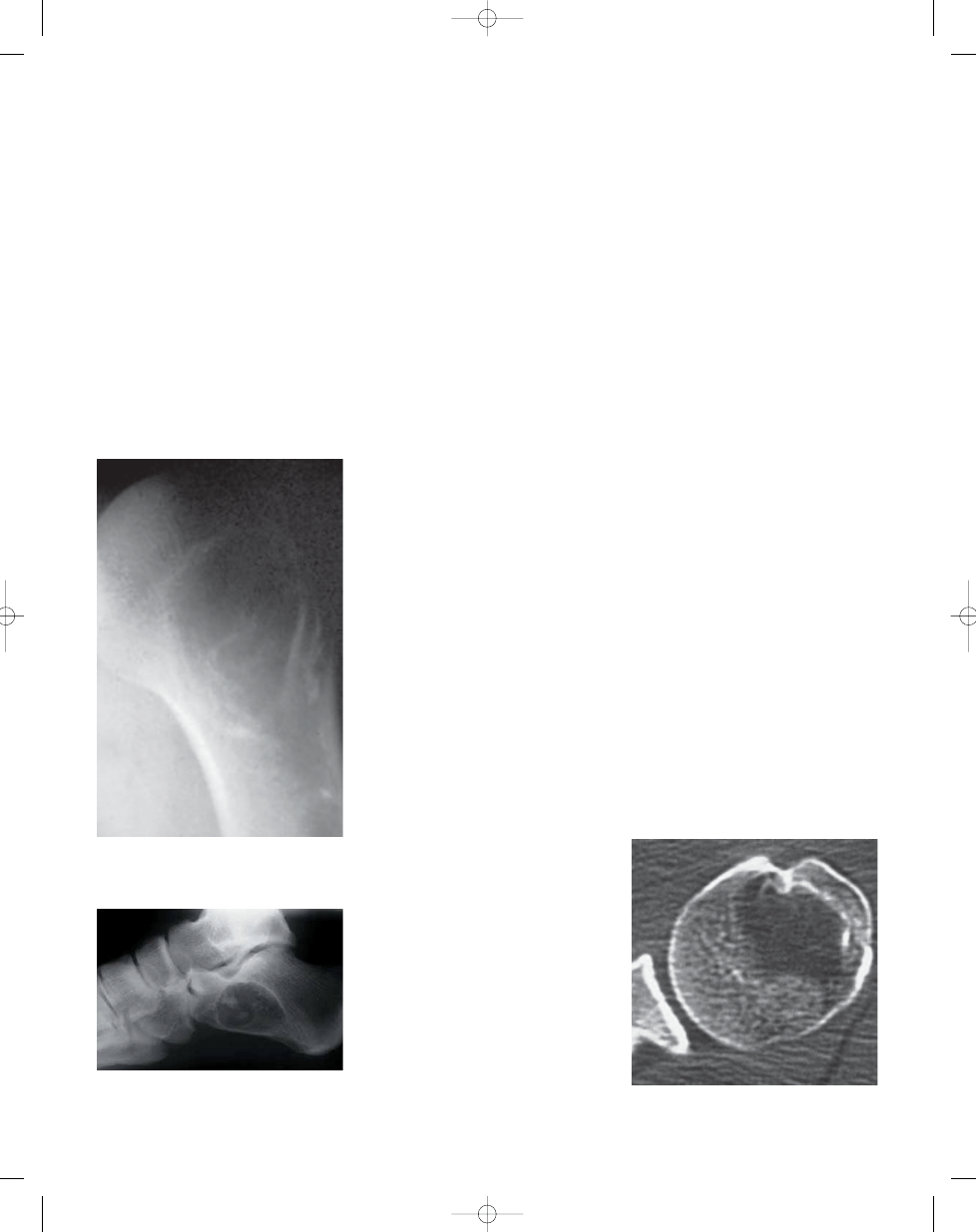
Fig. 19.01
Leiomyoma. CT scan showing a well
defined lytic lesion with a sclerotic rim in the ilium.
326
Smooth muscle tumours
Fig. 19.02 Leiomyoma. A Low power view showing bundles of uniform spindle cells. B Thick walled blood ves-
sels admixed with spindle cells in a pattern of angiomyoleiomyoma.
A
B
bb5_27.qxd 13.9.2006 13:53 Page 326
Definition
A very rare malignant spindle cell sarco-
ma of bone which shows smooth muscle
differentiation with immunohistochemical
or electron microscopic studies.
Epidemiology
Although the reported age range is from
9 to 87 years, the mean age is 44 years
{165,1049,1932}. There is a slight male
predominance.
Sites of involvement
Most lesions occur in the lower extremity
around the knee, either in the distal
femoral metaphysis or proximal tibial
metaphysis. The craniofacial skeleton is
the next most common area to be
involved {68}.
Clinical features
Pain, present from 2 weeks to 1 year prior
to diagnosis, is the most common symp-
tom. Approximately 15% of patients pres-
ent with pathological fracture.
Radiographically, it is an aggressive radi-
olytic lesion, with poorly defined margins,
a permeative growth pattern, and cortical
destruction. MRI shows a hypointense
lesion on T1 and an iso-or hypointense
lesion on T2 weighted studies {2056}.
Macroscopy
Lesions are grey to tan, firm or creamy
masses, often with areas of necrosis or
cystic degeneration. Despite a broad
range in size, lesions average 6 cm in
greatest dimension {68}. Cortical pene-
tration is common.
Histopathology
Histologically, lesions are identical to
leiomyosarcomas in other locations.
Plump and pleomorphic spindle cells are
arranged in bundles or fascicles. Mitotic
figures are common. Often areas of
necrosis are present. Smooth muscle dif-
ferentiation is demonstrable by positive
immunohistochemical staining for
smooth muscle actin and desmin.
Electron microscopic studies demon-
strate fine filamentous actin fibrils in the
cytoplasm.
Prognostic factors
Approximately 50% of patients develop
metastases to the lungs within 5 years
{68}. Ultimately, 50% of patients die from
leiomyosarcoma of bone {1099}.
E. McCarthy
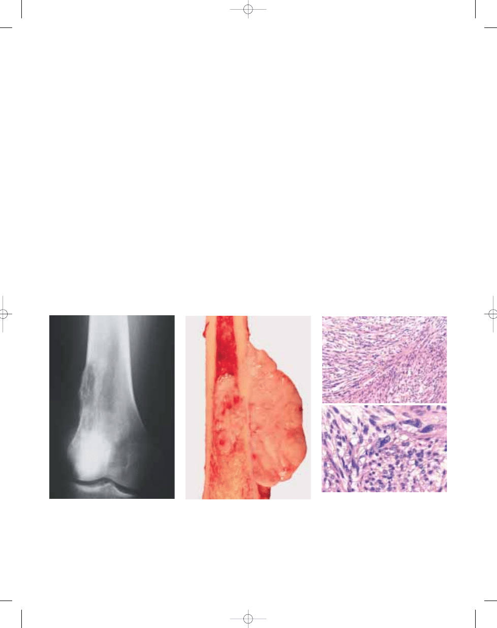
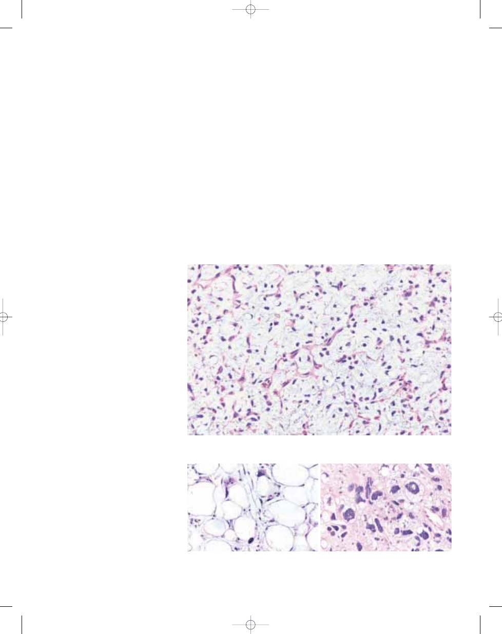
Leiomyosarcoma of bone
Fig. 19.03 Leiomyosarcoma. X-ray of a tumour in
distal femur showing an aggressive, permeative
lytic lesion with cortical destruction.
Fig. 19.04
Leiomyosarcoma. Macroscopy of the
femoral lesion. Note both an intraosseous and an
extraosseous component of the white fleshy tumour.
Fig. 19.05 Leiomyosarcoma. A Low power photomi-
crograph showing bundles of spindle cells.
B On high power magnification, note the cellular
pleomorphism of the tumour cells.
A
B
327
Leiomyosarcoma of bone
bb5_27.qxd 13.9.2006 13:53 Page 327
Lipoma of bone
A.E. Rosenberg
J.A. Bridge
Definition
Lipoma of bone is a benign neoplasm of
adipocytes that arises within the
medullary cavity, cortex, or on the sur-
face of bone.
Synonyms
Intramedullary lipoma, intracortical lipo-
ma, ossifying lipoma, parosteal lipoma.
Epidemiology
Lipoma of bone is rare and accounts for
less than 0.1% of primary bone tumours;
their incidence is not known.
Intramedullary lipoma has a wide age
range (2nd-8th decades) but most
patients are approximately 40 years old
at the time of diagnosis {1458}. Males are
affected more frequently than females at
a ratio of approximately 1.6:1 {1458}.
Parosteal lipoma usually develops dur-
ing adulthood and most patients are in
their 5th-6th decade of life at the time of
diagnosis {1462}. There is a slight male
predominance (9:7) {1462}.
Sites of Involvement
The vast majority of intraosseous lipomas
arise within the medullary cavity and
rarely develop in the cortex {2317}. They
most commonly affect the metaphyseal
regions of the long tubular bones, espe-
cially the femur, tibia and fibula and the
calcaneus. However, they have also
been described in many bones including
the pelvis, vertebrae, sacrum, skull,
mandible, maxilla, and ribs.
Parosteal lipomas generally develop on
the diaphyseal surface of long tubular
bones, especially the femur, humerus,
and tibia {1462}.
Clinical features / Imaging
Intramedullary lipoma may be asympto-
matic or produce achy pain. Rarely, it
presents as a pathological fracture {822,
951, 1458}. Radiographically, intra-
medullary lipoma usually produces a
well defined lytic mass that is surround-
ed by a thin rim of sclerosis. The lesion
may also contain trabeculations or cen-
tral calcifications. Bony expansion may
occur in small caliber bones {822,951,
1458,1732}. CT shows that the fatty com-
ponent has a low attenuation value simi-
lar to that of subcutaneus fat and on MRI
the fat has high signal intensity on both
T1 and T2 weighted images {1732}.
Parosteal lipoma is frequently asympto-
matic and may present as a visible or
palpable mass. Radiographs may reveal
a radiolucent mass adjacent to the corti-
cal surface that may show thickening or
a periosteal reaction. Similar to
intraosseous lipoma, the CT and MRI
findings have the same features as sub-
cutaneous fat except if there is calcifica-
tion, cartilage or ossification within the
lesion {1079,1752}.
Macroscopy
Intramedullary lipoma is usually 3-5 cm
in size, is well defined, soft, and yellow.
The surrounding bone is often sclerotic.
Parosteal lipoma is usually 4-10 cm in
greatest dimension, is well defined, soft
and yellow. Some cases contain gritty
spicules of bone or firm nodules of carti-
lage in the base or scattered throughout
the mass.
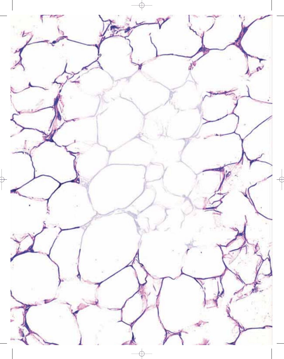
Histopathology
Intramedullary lipoma is well defined and
consists of lobules of mature-appearing
adipocytes that may replace the marrow
and encase preexisting bony trabeculae.
The adipocytes have a single large clear
cytoplasmic vacuole that displaces the
crescent shaped nucleus to the periph-
ery. Some tumours may demonstrate fat
necrosis with foamy macrophages and
fibrosis. In ossifying lipomas delicate tra-
beculae of woven and lamellar bone may
be present throughout the tumour {121,
346}.
Fig. 19.06 Lipoma. Radiograph of intramedullary
lipoma of humerus demonstrating an oval lytic
lesion with a sclerotic rim.
Fig. 19.08 Lipoma. Axial CT showing that the lipoma
has the tissue density of fat.
Fig. 19.07 Lipoma of calcaneous producing a well
defined lytic lesion with central mineralization. Axial
CT confirms the fatty nature of the lesion.
328
Lipogenic tumours
bb5_27.qxd 13.9.2006 13:53 Page 328

329
Lipoma of bone
Parosteal lipoma is also well defined and
consists of lobules of mature appearing
white adipocytes. The adipocytes have a
single large clear cytoplasmic vacuole
that displaces the crescent shaped
nucleus to the periphery. Some cases
may have bone with or without a hyaline
cartilage in the base of the lesion or scat-
tered throughout the mass in small
islands {1462}.
Immunophenotype
The neoplastic fat expresses vimentin
and S100 protein.
Genetics
The translocation t(3;12)(q28;q14) and
its associated fusion transcript
HMGIC/
LPP characteristic of subcutaneous lipo-
ma has been detected in a case of
parosteal lipoma {255,1698}.
Prognostic factors
Lipoma of bone has an excellent progno-
sis and rarely recurs.
B
A
Fig. 19.09 A Well defined intramedullary lipoma composed of sheets of white adipocytes. B Parosteal lipoma composed of lobules of white fat cells.
bb5_27.qxd 13.9.2006 13:53 Page 329
Definition
Liposarcoma of bone is a malignant neo-
plasm whose phenotype recapitulates
fat.
Epidemiology
Liposarcoma of bone is an extraordinari-
ly rare neoplasm. Most cases are
described in the form of single case
reports in older literature and the validity
of the diagnosis in some cases has been
questioned {457}. Liposarcoma of bone
occurs in all age groups although the
majority of patients are adults {15,457,
1090,2121}. Men are affected slightly
more frequently than women.
Sites of involvement
Liposarcoma of bone usually develops in
the long tubular bones especially the
tibia and femur and has been reported to
arise in the diaphysis, metaphysis, and
epiphysis {15,457,1090,2121}.
Clinical features / Imaging
Liposarcoma of bone presents as a
painful mass. Radiographically, the
tumour manifests as a well defined or
poorly defined mass {15,457,1090,
2121}.
Macroscopy
Most liposarcomas are large, lobulated,
soft to firm and are yellow to tan-white in
colour. Myxoid tumours may be glisten-
ing, slimy and mucinous.
Histopathology
The histological variants of liposarcoma
reported in bone include well differentiat-
ed lipoma-like, myxoid and pleomorphic
types {15,457,1090,2121}. Well differenti-
ated lipoma-like liposarcoma consists of
sheets of mature appearing adipocytes
with scattered tumour cells having
enlarged hyperchromatic nuclei. Some
of these atypical cells are lipoblasts and
are distinguished by cytoplasmic vac-
uoles that are round, clear, and scallop
the nucleus. Myxoid liposarcoma con-
sists of mildly atypical stellate and spin-
dle cells enmeshed in a myxoid stroma
that contains a finely arborizing vascular
tree that has a plexiform pattern. Also
scattered throughout the tumour are
lipoblasts. Sheets of large pleomorphic
cells in which the cytoplasm is either
eosinophilic or filled with round clear vac-
uoles characterize pleomorphic liposar-
coma. Mitoses are usually numerous.
Immunophenotype
There are no data regarding the immuno-
phenotype of liposarcoma of bone.
Ultrastructure
The cytoplasm of the neoplastic cells
contains membrane bound lipid droplets
of varying size. Dilated rough endoplas-
mic reticulum and scattered mitochon-
dria are also present {1650}.
Prognostic factors
Prognostic information regarding liposar-
coma of bone is scant. Generally, the
behaviour of the tumour should correlate
with its histological grade.
A.E. Rosenberg
Liposarcoma of bone
330
Lipogenic / neural tumours
Fig. 19.10 Myxoid liposarcoma consisting of scattered spindle and stellate cells and occasional lipoblasts
enmeshed in a frothy myxoid stroma that contains branching small caliber capillaries.
B
A
Fig. 19.11 A Well differentiated liposarcoma, lipoma-like type, containing mature appearing white fat cells
and scattered adipocytes that have enlarged hyperchromatic nuclei. B Sheets of pleomorphic cells includ-
ing lipoblasts characterize pleomorphic liposarcoma.
bb5_27.qxd 13.9.2006 13:53 Page 330
331
Schwannoma
Schwannoma
K.K. Unni
Definition
Schwannoma is a benign neoplasm of
Schwann cell origin arising within bone.
ICD-O code
9560/0
Synonyms
Neurilemmoma, neurinoma.
Epidemiology
Neurogenic tumours of bone are extre-
mely uncommon. Although roentge-
nographic abnormalities may be found
involving the skeleton in patients with
neurofibromatosis, there are no well rec-
ognized examples of neurofibroma in
bone. All benign neurogenic tumours in
the skeleton are Schwannomas. They
compose less than 1% of all benign
tumours in the Mayo Clinic files (unpub-
lished statistics, Unni, K. K.).
Sites of involvement
The mandible and the sacrum are the
most common sites of involvement with
neurilemmoma. In the mandible, the
lesion almost always involves the mental
foramen. When neurilemmoma involves
the spine or the sacrum, it is frequently
difficult to know whether the tumour is
truly of bony origin.
Clinical features / Imaging
Most neurilemmomas are asymptomatic,
incidental findings on roentgenograms.
Occasionally, they produce pain and/or
swelling.
Macroscopy
Schwannomas of bone are extremely
well circumscribed and may show a
fibrous capsule. They are tan to white
and glistening. Foci of yellow discoloura-
tion may be seen.
Histopathology
Schwannoma is composed of spindle
cells with wavy appearing nuclei. The
nuclei frequently are arranged in a pal-
isading fashion. Areas of hypocellularity
may alternate with areas of hypercellular-
ity. Focally, the nuclei may be enlarged
and pleomorphic appearing. Mitotic
activity is rare. Schwannomas are always
diffusely and strongly positive with S100
protein.
Prognostic factors
Schwannomas are benign lesions and
complete, but conservative surgical
removal leads to cure. There are no
examples of malignant transformation of
neurilemmomas in bone.
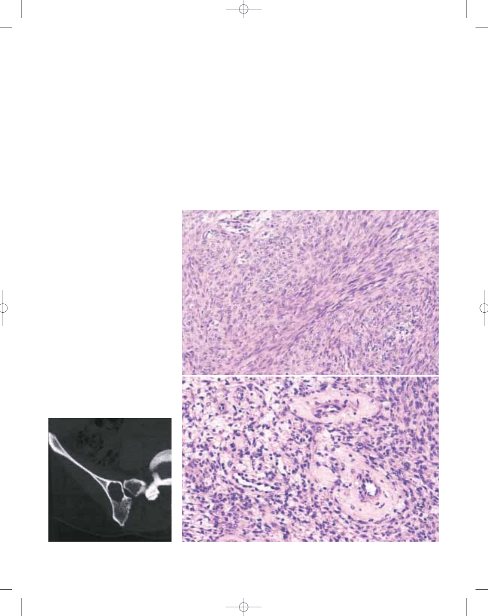
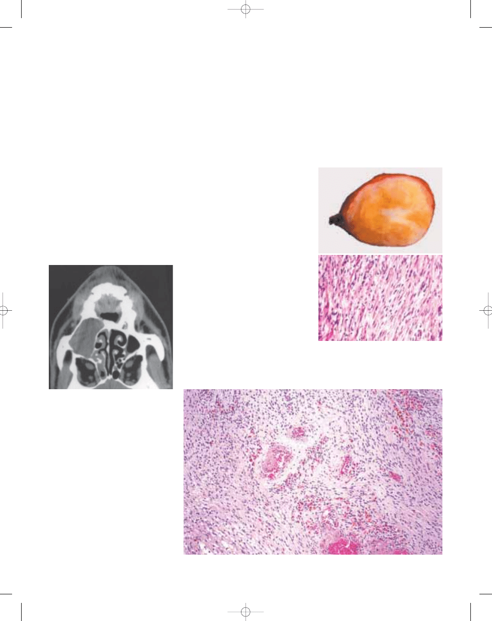
Fig. 19.12 CT of a well-demarcated Schwannoma
of the maxilla.
Fig. 19.13 A Encapsulated mandibular Schwannoma
with tan and white areas. B Note the discrete ten-
dency of spindle cell nuclei to palisade. The nuclei do
not show cytological atypia.
A
B
Fig. 19.14 Schwannoma. Note the hyalinization of vascular walls.
bb5_27.qxd 13.9.2006 13:53 Page 331
Adamantinoma
P.C.W. Hogendoorn
H. Hashimoto
Definition
A low grade, malignant biphasic tumour
characterized by a variety of morpholog-
ical patterns, most commonly epithelial
cells, surrounded by a relatively bland
spindle-cell osteo-fibrous component.
ICD-O code
9261/3
Synonyms
Adamantinoma of long bones, extrag-
nathic adamantinoma, differentiated
adamantinoma, juvenile intracortical
adamantinoma.
Epidemiology
Adamantinoma comprises about 0.4% of
all primary bone tumours {987,1503,
1518}. Patients present with this tumour
from 3 up to 86 years, with a median age
of 25-35 years. The youngest age group
predominantly includes patients with
osteofibrous dysplasia-like adamantino-
ma, but very young patients with classic
adamantinoma (age 3) and older ones
with the osteofibrous dysplasia-like sub-
type (age 38) have been reported {918,
1502,2069}. There is a slight predomi-
nance in males.
Sites of involvement
The tibia, in particular the anterior
(meta-) diaphysis, is involved in 85-90%
of cases. In up to 10% this is combined
with one or more lesions in the ipsilateral
fibula as well. Rare other sites have been
reported, especially the ulna.
Clinical features / Imaging
The main complaint is swelling with or
without pain. Adamantinoma often dis-
plays a protracted clinical behaviour.
Clinical symptoms like swelling or radi-
ographic abnormality may last for more
than 30 years prior to diagnosis, where-
as local recurrences or metastases may
develop years after primary, intralesional
or marginal surgical treatment. On X-ray,
typically a well circumscribed, cortical,
(multi-)lobulated osteolytic lesion with
intralesional opacities, septation and
peripheral sclerosis is seen {217,987}.
Multifocality within the same bone is reg-
ularly observed. The lesion commonly
seems to remain intracortical and
extends longitudinally, but may also
destroy the cortex and invade the
medullary cavity or surrounding perios-
teum and soft tissue. This is usually
accompanied by lamellar or solid
periosteal reaction. Aggressive tumours
occasionally present as single large lytic
lesions. MRI is useful to document multi-
centricity, the extension of the lesion, and
eventual soft tissue involvement.
Macroscopy
Classic adamantinoma usually presents
as a cortical, well-demarcated, yellow-
ish-grey, lobulated tumour of firm to bony
consistency with peripheral sclerosis. It
may be a single lesion, but its multifocal
appearance with apparently normal cor-
tical bone lying in between is occasion-
ally striking. Small lesions remain intra-
cortical, and are usually white and gritty.
Larger tumours show intramedullary
extension and cortical breakthrough with
soft tissue invasion in a minority of cases.
Macroscopically detectable cystic
spaces are common, filled with straw-
coloured or blood-like fluid.
Histopathology
Classic adamantinomas are character-
ized by an epithelial and an osteofibrous
component, that may be intermingled
with each other in various proportions
and differentiation patterns. The four
main differentiation patterns of classic
adamantinoma are basaloid, tubular,
spindle cell, and squamous {2235}. The
first two patterns are encountered most
commonly, but all patterns may be pres-
ent in one lesion. The spindle cell com-
ponent is more often observed in recur-
rences, lining cystic spaces, and in
metastases. The osteofibrous compo-
332
Epithelial tumours
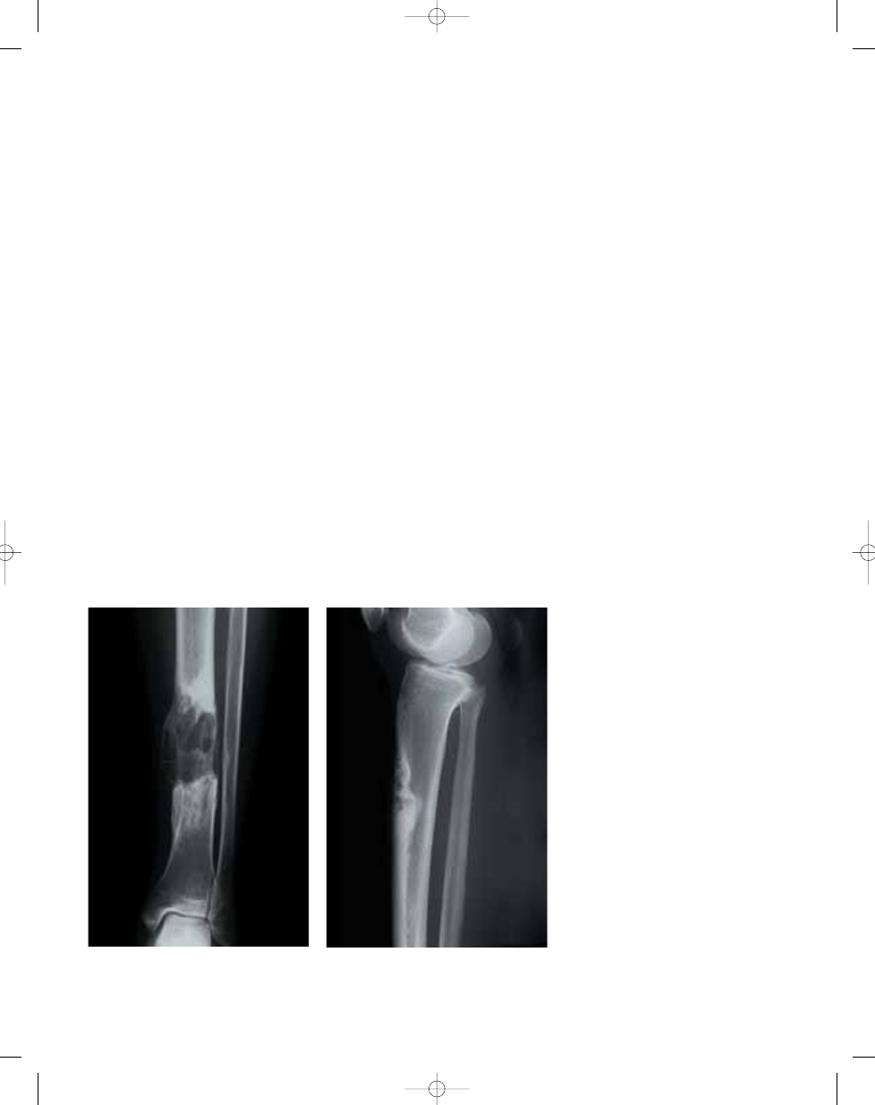
Fig. 19.15 Classic adamantinoma. The radiograph
of the distal tibia shows an expansive, lobulated,
lytic lesion with a defect of the outer surface of the
cortex.
Fig. 19.16 Osteofibrous dysplasia-like adamantino-
ma. The lateral radiograph of the proximal aspect of
the tibia shows a multilocular, lytic lesion with sur-
rounding osteosclerosis of the anterior cortex.
bb5_27.qxd 13.9.2006 13:53 Page 332
333
Adamantinoma
nent is composed of storiform oriented
spindle cells. Woven bone trabeculae
are usually present in or next to the cen-
tre of the lesion, prominently rimmed by
osteoblasts, and with varying amounts of
transformation to lamellar bone at the
periphery of the tumour. Foam cells or
myxoid change may be present, and
mast cells or multinucleated giant cells
are occasionally detected. Mitotic activi-
ty is usually low. A fifth histological pat-
tern, the so-called osteofibrous dyspla-
sia-like variant, is characterized by pre-
dominance of osteofibrous tissue, in
which small groups of epithelial cells are
only encountered by careful search or
immunohistochemistry. The majority of
classic and osteofibrous dysplasia-like
adamantinomas display a "zonal" archi-
tecture. In classic adamantinoma, the
centre is usually dominated by the
epithelial component, and only few, small
immature bone trabeculae are present in
the fibrous tissue. Towards the periphery,
the epithelial islands decrease to incon-
spicuous elements and the osteofibrous
component gradually takes over with
increasing amounts of woven bone tra-
beculae, transforming to lamellar bone.
In osteofibrous dysplasia-like adamantin-
oma, the centre is occupied by fibrous
tissue with scanty and thin immature
woven bone trabeculae with epithelial
elements. Small clusters of epithelial
cells are the only feature which differenti-
ate osteofibrous dysplasia-like adaman-
tinoma from osteofibrous dysplasia.
Immunophenotype
The fibrous tissue is vimentin-positive.
The epithelial cells show co-expression
for keratin, EMA and vimentin. Chain-
specific keratin expression {917,1050}
revealed a predominantly basal epithelial
cell differentiation, regardless of subtype,
with widespread presence of basal
epithelial cell keratins 5, 14, and 19. Also
keratins 1, 13 and 17 are variably pres-
ent. Keratins 8 and 18 are virtually
absent. In classic adamantinomas, the
epithelial component is surrounded by a
continuous basement membrane, where-
as less distinct epithelial islands show
multiple interruptions or no surrounding
basement membrane at all {919}.
EGF/EGFR expression is restricted to the
epithelial component. FGF2/FGFR1 is
present in both components {242}.
Ultrastructure
Electron microscopic studies have con-
firmed the epithelial nature of adamantin-
oma, showing intracytoplasmic hemi-
desmosomes, tonofilaments, and micro-
filaments. Irrespective of histological
subtype, the epithelial cells are bound by
desmosomes and basement membranes
have been found to surround the epithe-
lial nests.
B
A
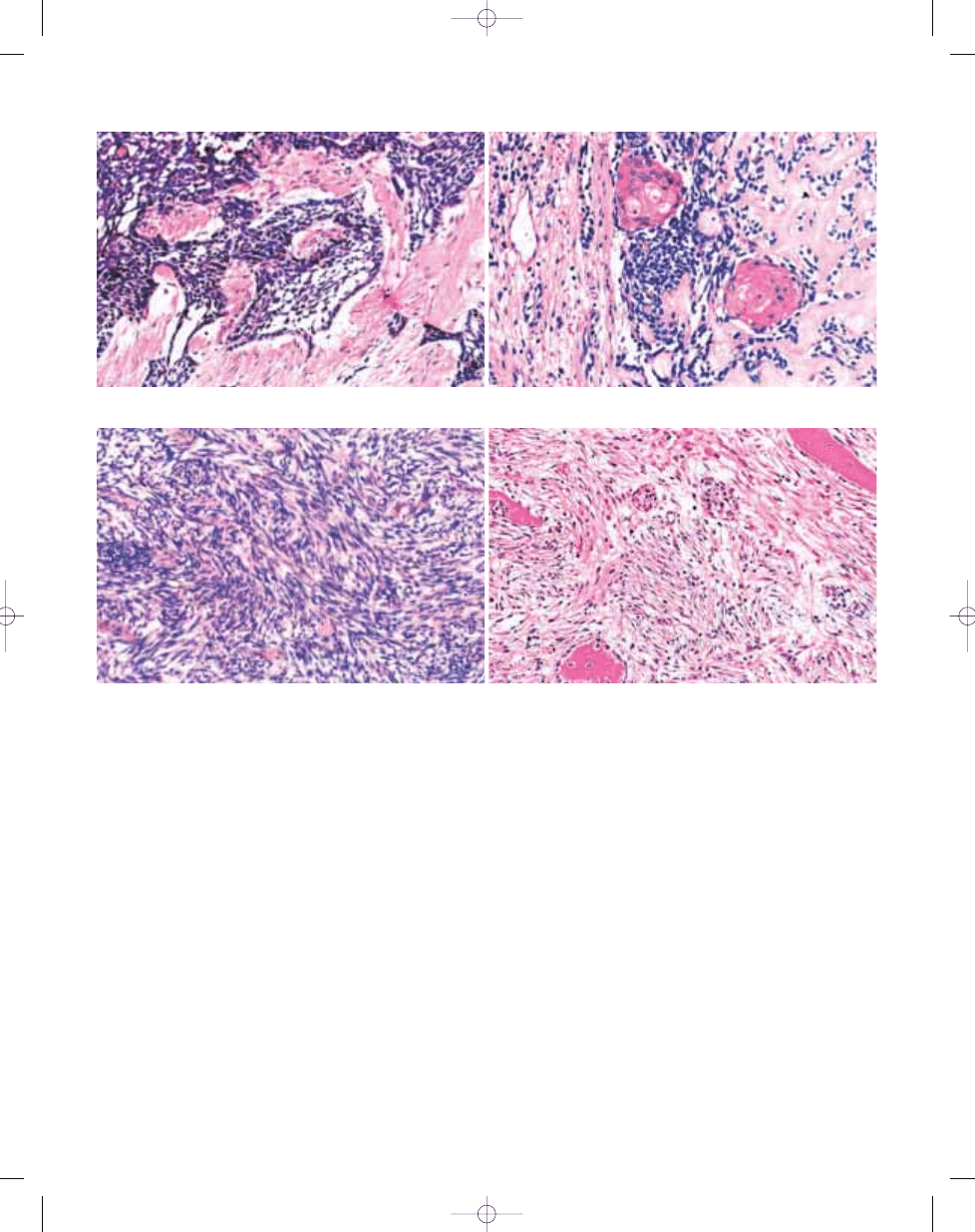
Fig. 19.17 Adamantinoma. A Basaloid pattern. Easily distuingishable epithelial fields without clear pallisading. B Squamoid pattern.
B
A
Fig. 19.18 Adamantinoma. A Spindle cell pattern. B Osteofibrous dysplasia like adamantinoma. Small epithelial clusters in a fibro-osseous stroma.
bb5_27.qxd 13.9.2006 13:53 Page 333
Genetics
Adamantinomas, classic as well as
osteofibrous dysplasia-like, show recur-
rent numerical chromosomal abnormal-
ities, mainly gain of chromosomes 7, 8,
12, and 19 {920,1058,1318,2004}. DNA
flow cytometric and image cytometric
studies showed that in aneuploid
tumours, the aneuploid population was
always restricted to the epithelial com-
ponent {916}.
TP53 gene aberrations –
as detected immunohistochemically or
by loss of heterozygosity analysis - are
restricted to the epithelial component of
adamantinoma. There have been some
cases reported with histological fea-
tures of adamantinoma as well as
Ewing sarcoma, sometimes called
'atypical' or 'Ewing-like' adamantinoma
{741,1013, 1273,1400,1891,2178}.
Cytogenetic ana-lysis combined with
FISH and RT-PCR of two cases formerly
described as atypical or Ewing-like
adamantinoma revealed an (11;22)
translocation, typical for Ewing sarco-
ma {257}. Because of these findings
these tumours were labelled "adaman-
tinoma-like Ewing sarcoma". The
t(11;22) translocation is not present in
adamantinoma {908,1318}.
Prognostic factors
Risk factors for recurrence are intrale-
sional or marginal surgery and extra-
compartmental growth {918,1050,1084,
1739}. Recurrence percentages after
non-radical surgery may rise up to 90%
{918,1050,1084}. Recurrence is associ-
ated with an increase in epithelium-to-
stroma ratio and more aggressive
behaviour {918,1084,1503}. Besides,
male sex {1050,1084}, females at
young age {1503}, pain at presentation
{1084}, short duration of symptoms
{918,1084}, young age (<20 years)
{918}, and lack of squamous differenti-
ation of the tumour {918, 1084} have
been associated with increased rates
of recurrence or metastasis.
Adamantinomas metastasise in 12-29%
of patients with comparable mortality
rates {918,1084,1503,1739}. Metastatic
tumours are all classic adamantinomas,
although rarely osteofibrous dysplasia-
like adamantinomas may metastasise
after recurrence and subsequent pro-
gression to classic adamantinoma
{918}. The tumour spreads to regional
lymph nodes and the lungs, and infre-
quently to skeleton, liver, and brain.
Definition
A tumour (usually malignant) involving
bone, which has originated from another
(distant) site.
Synonyms
Metastatic carcinoma, skeletal deposits,
osseus metastasis, secondaries in bone,
bony implants.
Epidemiology
The skeletal system is the third most
common site to be involved by metastat-
ic tumour after the lungs and liver {174}.
Metastatic carcinomas are the most com-
mon malignant tumour affecting the
skeleton {2154}. Over two-thirds of
patients with bone metastasis are
between 40-60 years of age {504}. Most
metastases originate from common can-
cers namely breast, lung, prostate, kid-
ney and thyroid gland which account for
93% of all deposits {504}. A complete
radiographic and clinical search will
identify the primary site in up to 85% of
cases {1812}.
Although metastases are rare in children,
when they occur, they most often include
neuroblastoma, rhabdomyosarcoma and
clear cell sarcoma of kidney.
Sites of Involvement
Metastatic carcinomas involve bones
with persistent red marrow such as verte-
bra, proximal femur ribs, sternum, pelvis,
skull and shoulder girdle. Out of 114 his-
tologically evaluated lesions 44.3%
involved axial skeleton, 28.8% the
appendicular skeleton and 26.9%
involved multiple bones {504}. The lum-
bar spine {757,1872} and proximal femur
{757} are favoured sites. Bones of the
hands and feet are rarely involved {923,
1252,1433,1507,1925}.
Clinical features
Pain, swelling, fracture and neurological
symptoms (spine) are common {278}.
Skull base metastasis may cause Collet-
Sicard syndrome {1865}; hypercal-
caemia may accompany osteolysis
{1520}.
Plain radiographs reveal lytic, blastic or
N.A. Jambhekar
A. Borges
Metastases involving bone
334
Epithelial tumours
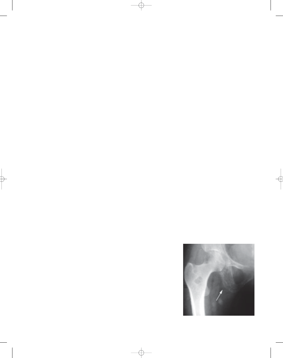
Fig. 19.19
Permeative destruction of bone by a
metastasis (primary tumour unknown).
bb5_27.qxd 13.9.2006 13:53 Page 334
335
Bone metastases
mixed patterns {756}. Lung and breast
deposits cause irregular lytic destruction,
but are occasionally osteoblastic {1460,
1514}. Thyroid and kidney deposits are
purely lytic; prostatic deposits are
osteoblastic. Solitary metastasis {2120},
or an irregular periosteal reaction {1238,
1581} may simulate a primary bone sar-
coma.
Plain radiographs are unreliable to detect
vertebral deposits {707,1872} and
despite gross evidence of spinal
deposits in 36% of 832 autopsied
patients dying of cancer, 26% had nega-
tive plain X-rays {1872}.
Bone scintigraphy is a sensitive method
for the detection of skeletal metastases,
because it covers the whole skeleton,
making it valuable for identifying the
extent of the disease. CT scan is useful
for guiding needle biopsies. MRI has
also been used in some cases to detect
and delineate metastases.
Aetiology
The location of the primary tumour and
the local pattern of blood flow determine
involvement of skeletal sites. The verte-
bral venous plexus (Batson’s plexus) is a
high volume, low pressure, valveless
venous system independent of the pul-
monary, portal and caval systems; it com-
municates directly with veins of the
pelvis, proximal half of lower extremity,
proximal half of upper extremity and head
and neck {140}. Any increase in intrab-
dominal or intrathoracic pressure during
exhalation or straining causes a backflow
into the vertebral plexus bypassing the
heart and lungs. This explains the prefer-
ential involvement of the vertebral and the
proximal appendicular bones, and the
occassional occurrence of extensive
skeletal deposits despite lack of visceral
involvement {1470}.
Macroscopy
The macroscopic appearance of skele-
tal metastasis varies depending upon
the amount of bone produced in
response to the tumour. Thus, osteoblas-
tic metastases from the breast are grey-
ish white firm, whereas renal cell carci-
noma produces soft haemorrhagic
deposits.
Morphology
Metastatic tumours attempt to recapitu-
late the original tumour. Squamous car-
cinomas from most sites look alike, how-
ever, many adenocarcinomas such as
renal cell, prostate and thyroid retain
morphological similarities to the primary
tumour. An accompanying fibroblastic,
vascular, osteoblastic and osteoclastic
response may be present. Sarcomatoid
(spindle cell) carcinomas originating in
the kidney or the lung may simulate a
primary bone sarcoma.
Immunophenotype
Immunohistochemistry is useful when
the diagnosis of metastatic carcinoma is
straightforward but not distinctive
enough to identify the primary site, or,
when the differential is broad and
includes sarcoma, carcinoma and
melanoma {514}.
Prognostic factors
Bone metastasis usually heralds incur-
ability and treatment is palliative. The
outcome depends upon the primary site
and the extent of disease.
B
A
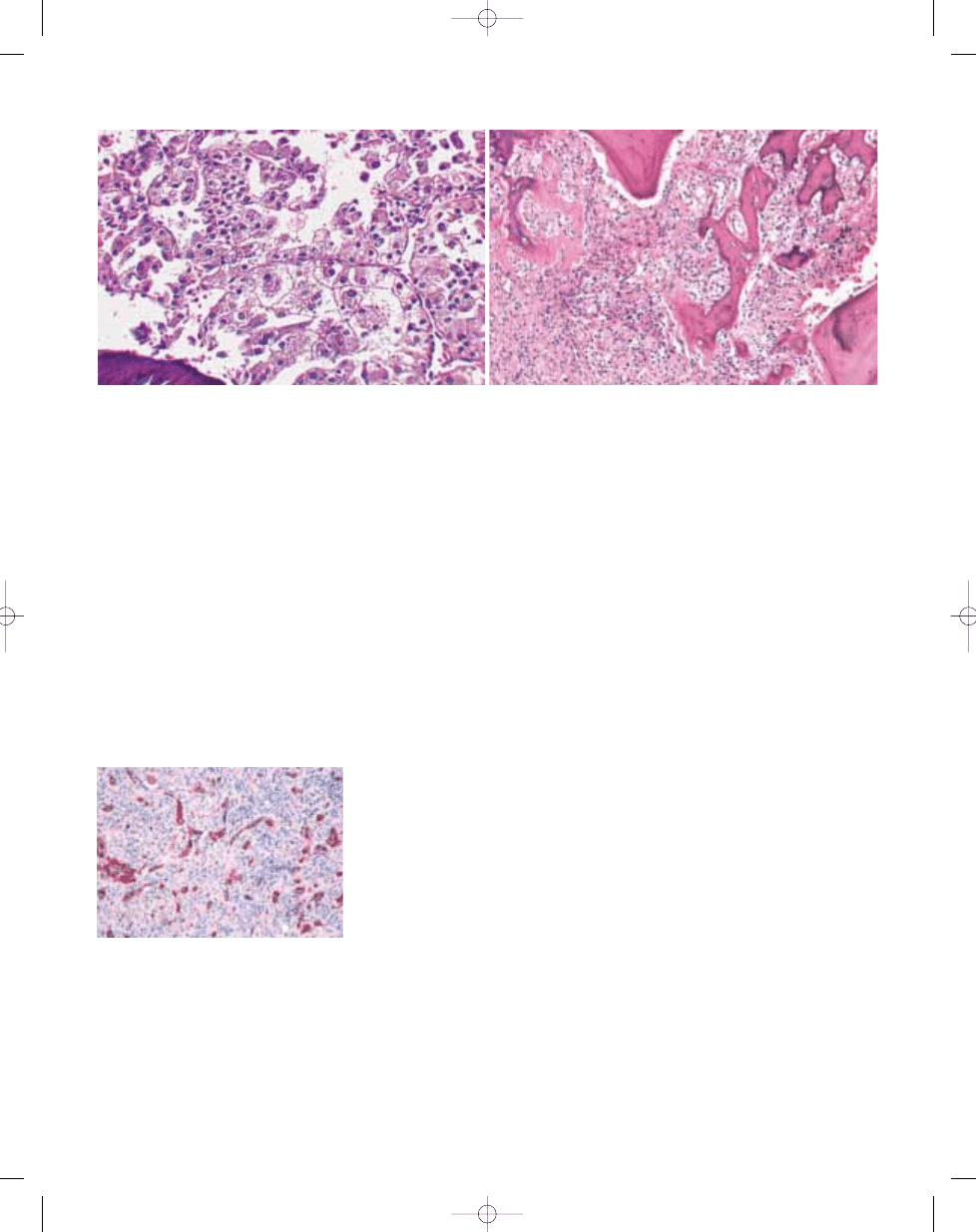
Fig. 19.20 A Metastatic renal cell carcinoma showing an alveolar and nesting pattern. B Metastatic prostate carcinoma; note monotonous small cells and irregular
osteoid deposition.
Fig. 19.21 Metastatic carcinoma. Scattered cytoker-
atin-positive tumour cells confirm the epithelial char-
acter of the lesion.
bb5_27.qxd 13.9.2006 13:53 Page 335
Wyszukiwarka
Podobne podstrony:
bb5 chap3
bb5 chap8
bb5 chap1
BB5 BOX
bb5 chap16
bb5 chap15
bb5 contents
bb5 chap12
bb5 chap4
bb5 references
bb5 chap6
bb5 chap17
bb5 chap20
bb5 chap5
Lista wszystkich dostępnych polskich Product Code dla telefonów platformy BB5
bb5 chap21
bb5 source
bb5 chap13
więcej podobnych podstron