
6
S-Layers
Uwe B. Sleytr, Eva-Maria Egelseer, Dietmar Pum, and Bernhard Schuster
6.1
Overview
The fabrication of supramolecular structures and devices requires molecules that are cap-
able of interlocking in a predictable, well-defined manner. Thus, molecular self- assembly
systems which exploit the molecular-scale manufacturing precision of biological systems
are prime candidates for supramolecular engineering. Although self-assembly of mole-
cules is an ubiquitous strategy of morphogenesis in nature, in molecular nanotechnology
these unique features of molecules are not yet fully exploited for the functionalization of
surfaces and interfaces and for hierarchical self-assembly systems as required for the pro-
duction of biomimetic membranes and encapsulating systems.
Crystalline bacterial cell-surface layers (S-layers) have been optimized during billions of
years of biological evolution as one of the simplest biological membranes [1–3]. S-layers
are composed of a single protein or glycoprotein species endowed with the ability to
assemble into monomolecular arrays on the supporting cell envelope component of pro-
karyotic organisms (bacteria and archaea). The wealth of information accumulated on
the structure, chemistry, assembly, genetics, and function of S-layers has led to a broad
spectrum of applications for life and material sciences [3–7].
Abbreviations
Bet v1
major birch pollen allergen
BLMs
bilayer lipid membranes
cAB
camel antibody sequence recognizing lysozyme
CdS
cadmium sulfide
DNA
deoxyribonucleic acid
H
2
S
hydrogen sulfide
IgE
immunoglobulin E
IL-8
interleukin 8
MFMs
microfiltration membranes
MPL
main phospholipid of Thermoplasma acidophilum
77
Nanobiotechnology. Edited by Christof Niemeyer, Chad Mirkin
Copyright
c 2004 WILEY-VCH Verlag GmbH & Co. K aA, Weinheim
ISBN 3-527-30658-7
G

rSbpA
recombinant S-layer protein of Bacillus sphaericus CCM 2177
SbpA
S-layer protein of Bacillus sphaericus CCM 2177
SbsB
S-layer protein of Geobacillus stearothermophilus PV72/p2
SbsC
S-layer protein of Geobacillus stearothermophilus ATCC 12980
SCWP
secondary cell wall polymer
S-layer
surface layer
SLH
S-layer homology domain
SPR
surface plasmon resonance
SUMs
S-layer ultrafiltration membranes
t-PA
tissue type plasminogen activator
6.1.1
Chemistry and Structure
With few exceptions, S-layers are composed of a single homogeneous protein or glycopro-
tein species with molecular weights ranging from 40 to 200 kDa (Table 6.1). The results of
amino acid analysis of various S-layer proteins and the secondary structure estimated by
protein sequence data and circular dichroism measurements on S-layer proteins are sum-
marized in Table 6.1. Few posttranslational modifications are known to occur in S-layer
proteins, including cleavage of amino- or carboxy-terminal fragments, phosphorylation,
and glycosylation of amino acid residues (Table 6.1) [3, 7]. The latter is a remarkable char-
acteristic of many archaeal and some bacterial S-layer proteins, and in this way the glycan
chains and linkages differ significantly from those of eukaryotes [3, 8–10].
Electron microscopy studies on the mass distribution of the lattices were generally per-
78
6 S-Layers
Table 6.1
Properties of S-layers
x
The relative molecular mass of constituent subunits in the range of 40 kDa to 200 kDa
x
These are weakly acidic proteins (pI
Z4–6), except Methanothermus fervidus (pI = 8.4) and lactobacilli
(pI
i 9.5)
x
Large amounts of glutamic acid, aspartic acid (
Z15 mol. %) and hydrophobic amino acids
(
Z40–60 mol. %), and a high lysine content (Z10 mol. %)
x
Hydrophilic and hydrophobic amino acids do not form extended clusters
x
No or low content of sulfur-containing amino acids
x
In most S-layer proteins,
Z20 % of the amino acids are organized as a-helix, and about 40 % occur as
b
-sheets
x
Aperiodic foldings and b-turn content may vary between 5 and 45 %
x
S-layer lattices can have oblique (p1, p2), square (p4), or hexagonal (p3, p6) symmetry
x
The center-to-center spacing of the morphological unit can range from 3 nm to 35 nm
x
The lattices are generally 5 nm to 20 nm thick (in archaea, up to
Z70 nm)
x
S-layer lattices exhibit pores of identical size and morphology
x
The pore sizes range from approximately 2 nm to 8 nm
x
In many S-layers, two or even more distinct classes of pores are present
x
The pores can occupy 30–70 % of the surface area
x
The outer surface is generally less corrugated than the inner surface
x
Posttranslational modifications of S-proteins include: (i) cleavage of N- or C-terminal fragments;
(ii) glycosylation; and (iii) phosphorylation of amino acid residues.
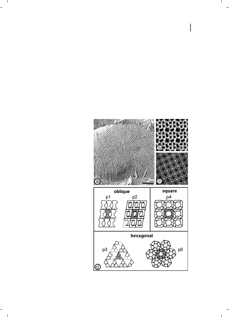
formed on negatively stained preparations or unstained, thin, frozen foils (Figure 6.1a).
Two- and three-dimensional analysis, including computer image enhancement, revealed
structural information down to a range of 0.5–1.5 nm (Figure 6.1b) [11–14]. High-resolu-
tion images of the surface topography of S-layers were also obtained using underwater
atomic force microscopy (Figure 6.1c) [3, 15–17]. A common feature of S-layers is their
smooth outer surface and more corrugated inner surface.
The proteinaceous subunits of S-layers can be aligned in lattices with oblique, square, or
hexagonal symmetry (Figure 6.1d) with center-to-center spacing of the morphological
units of between 3 and 35 nm. Hexagonal lattice symmetry is predominant among
archaea [18, 19]. S-layers are very porous membranes, with pores occupying between 30
and 70 % of their surface area (see Table 6.1). Since S-layers are in most cases assemblies
of identical subunits, they exhibit pores of identical size and morphology. However, in
many protein lattices two or more distinct classes of pores with diameters in the range
of 2 to 8 nm have been identified [19–21].
79
6.1 Overview
Figure 6.1
(a) Freeze-etching prepara-
tion of whole cells of
Thermoanaero-
bacter thermohydrosulfuricus L111-69
revealing a hexagonally ordered array.
Scale bar = 100nm. (b) Three-dimen-
sional model of the S-layer of
Bacillus
stearothermophilus NRS 2004/3a/V2 ex-
hibiting oblique lattice symmetry. The
protein meshwork shows one square-
shaped, two elongated, and four
small pores per morphological unit.
(c) Computer image reconstruction of
scanning force microscopic images of
the topography of the square S-layer
lattice from
Bacillus sphaericus CCM
2177. The images were taken under
water. The surface corrugation corre-
sponding to a gray scale tram black to
white is 1.8 nm. Scale bars in (b) and
(c) = 10 nm. (d) Schematic drawing of
the different S-layer lattice types. The
regular arrays exhibit either oblique (p1,
p2), square (p4), or hexagonal lattice
symmetry (p3, p6). The morphological
units are composed of one, two, three,
four, or six identical subunits. (Repro-
duced from Ref. [3], with permission
from Wiley-VCH.)

In both Gram-positive bacteria and archaea, the lattice assembles on the surface of the
wall matrix (e. g., peptidoglycan or pseudomurein), whereas in Gram-negative bacteria the
S-layer is attached to components of the outer membrane (e. g., lipopolysaccharides). In
most archaea the S-layer represents the exclusive cell-wall component external to the
cytoplasmic membrane.
6.1.2
Genetics and Secondary Cell-Wall Polymers
During the past decade, numerous S-layer genes from organisms of quite different taxo-
nomic affiliations have been cloned and sequenced [1, 7, 22, 23]. Considering the fre-
quently highly competitive situation of closely related organisms in their natural habitats,
it is obvious that the S-layer surface must contribute to diversification rather than to con-
servation. This can be achieved by S-layer variation leading to the expression of different
types of S-layer genes, or to the recombination of partial coding sequences. S-layer varia-
tion was studied in detail for Campylobacter fetus, an important pathogen for humans and
ungulates [24, 25], but was also observed for nonpathogens such as Geobacillus stearother-
mophilus [26–28]. Although it was proposed for several years that sequence identities
among S-layer proteins are extremely rare, or do not even exist, it is now apparent that
high sequence identities are limited to the N-terminal region that is responsible for
anchoring the protein to the cell surface by binding to an accessory secondary cell-wall
polymer (SCWP), and which is covalently linked to the peptidoglycan backbone. In this
context, three repeats of S-layer homology (SLH) motifs, consisting of 50–60 amino
acids each [29], have been identified at the N-terminal part of many S-layer proteins
[22]. If present, SLH motifs are involved in SCWP-mediated anchoring of the S-layer pro-
tein to the peptidoglycan layer [22, 30–37]. During the past few years, a considerable
amount of information on the chemical composition and structure of SCWPs from differ-
ent organisms has been accumulated [8, 30, 33, 38–40], indicating a highly specific lectin-
type recognition mechanism between the S-layer protein and a distinct type of SCWP.
In a recent study, the interaction of the S-layer protein SbsB of G. stearothermophilus
PV72/p2 and the corresponding SCWP was assessed by surface plasmon resonance
(SPR) biosensor technology [41]. By using two truncated forms consisting either of the
three SLH motifs or the residual part of SbsB, the exclusive and complete responsibility
of a functional domain formed by the three SLH motifs of the S-layer protein SbsB for
SCWP recognition was clearly confirmed. The interaction proved to be highly specific
for the carbohydrate component, and strong evidence for glycan pyruvylation was
provided [41]. In contrast to most S-layer proteins of Gram-positive bacteria, those of
G. stearothermophilus wild-type strains [34, 42] and Lactobacillus [31, 43] do not possess
SLH-motifs. Nevertheless, the N-terminal part of G. stearothermophilus wild-type strains
is highly conserved and recognizes a net negatively charged SCWP as the proper bin-
ding site [31, 34]. The production of different truncated forms of the S-layer protein
SbsC of G. stearothermophilus ATCC 12980 confirmed that the N-terminal part is exclu-
sively responsible for cell-wall binding, but this positively charged segment is not involved
in the self-assembly process [35] and seems to fold independently of the remainder of the
protein sequence.
80
6 S-Layers
81
6.1 Overview
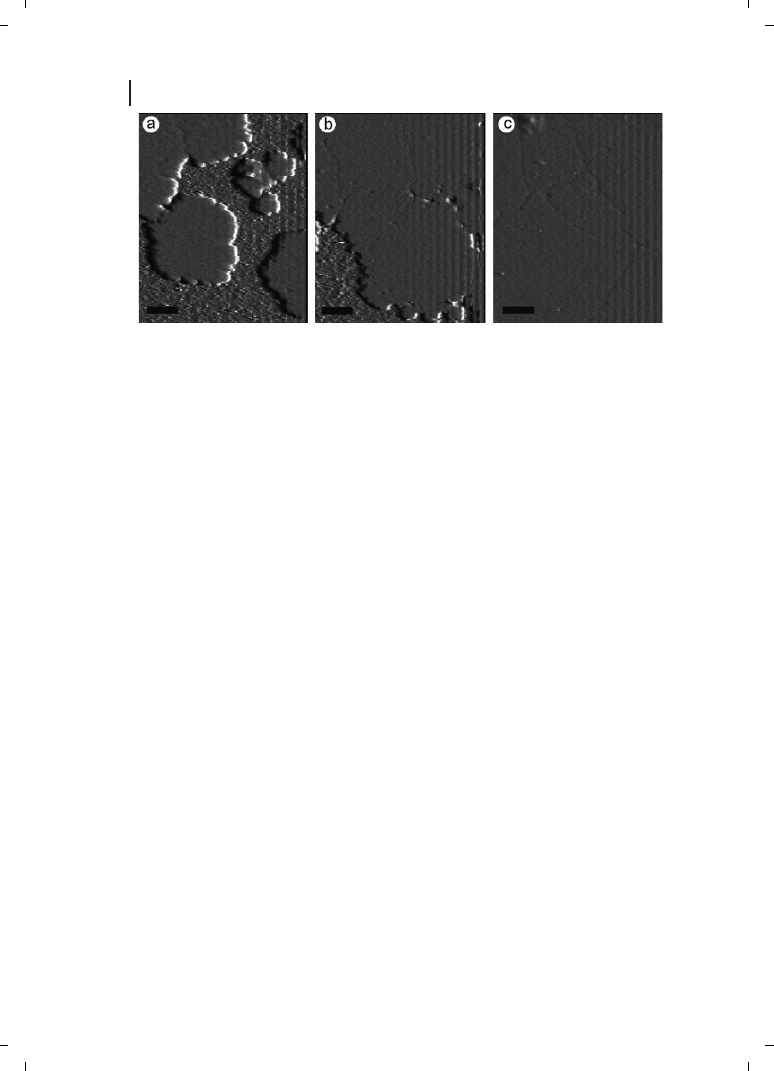
Figure 6.2
Cell wall fragments carrying a
chimeric S-layer formed by the fusion pro-
tein BS1(S1)
3
(a) were capable of binding
biotinylated ferritin (b). At BS1(S1)
3
, one
core streptavidin is fused to the C-termi-
nus of the S-layer protein SbsB of
Geoba-
cillus stearothermophilus PV72/p2. The pro-
teins were refolded to heterotetramers
consisting of one chain of fusion protein
and three chains of streptavidin. (a) Self-
assembly was enabled by the specific in-
teraction between an accessory cell-wall
polymer that is part of the cell wall of
G.
stearothermophilus PV72/p2, and the SLH-
domain of the fusion protein. (b) Bound
biotinylated ferritin reflected the underlying
S-layer lattice. The preparations were ne-
gatively stained with uranyl acetate for
TEM. The arrows indicate the base vectors
of the oblique p1 lattice; scale bars =
100 nm. (c) The cartoon shows the orien-
tation of BS1(S1)
3
after SLH-enabled self-
assembly with the streptavidin carrying
outer face of the S-layer exposed.
(Reproduced from Ref. [45]; copyright
(2002) National Academy of Sciences,
USA.)

In order to determine at which amino acid positions of the S-layer proteins foreign pep-
tide sequences could be fused without interfering with the self-assembly and recrystalliza-
tion properties, the structure–function relationship of distinct segments of different
S-layer proteins had to be elucidated. In the case of the S-layer protein SbpA of Bacillus
sphaericus CCM 2177, it could be demonstrated that the C-terminal end of the full-length
form of recombinant rSbpA (rSbpA
31-1268
) was only available to a limited extent, but was
fully accessible in the C-terminally truncated form rSbpA
31-1068
[37]. Based on these
results, the C-terminally truncated form was exploited as base form for the construction
of further S-layer fusions proteins, incorporating either the major birch pollen allergen
Bet v1 (rSbpA
31-1068
/Bet v1) or a camel antibody sequence recognizing lysozyme as an epi-
tope (rSbpA
31-1068
/cAB) [37, 44]. Owing to the versatile applications of the streptavidin–
biotin interaction as a biomolecular coupling system, minimum-sized core-streptavidin
(118 amino acids) was fused either to N-terminal positions of the S-layer protein SbsB
or attached to the C-terminus of this S-layer protein (Figure 6.2) [45]. The fusion proteins
and core-streptavidin were produced independently in Escherichia coli, isolated and
refolded to heterotetramers consisting of one chain of fusion protein and three chains
of streptavidin. As determined by a fluorescence titration method, the biotin binding
capacity of the heterotetramers was 80 % in comparison to homotetrameric streptavidin,
indicating that at least three of the four core streptavidin residues were accessible
and active. Due to the ability of the heterotetramers to recrystallize in suspension, on
liposomes, and on silicon wafers, this chimeric S-layer can be used as self-assembling
nanopatterned molecular affinity matrix to arrange biotinylated compounds on a surface
(Figure 6.2) [45].
6.1.3
Assembly
A complete solubilization of S-layers composed of native or recombinant proteins into
their constituent subunits can generally be achieved with high concentrations of hydrogen
bond-breaking agents (e. g., guanidine hydrochloride). In summarizing the results from
different disintegration procedures, it was concluded that: (i) in general, bacterial
S-layer proteins are not covalently linked to each other or the supporting cell wall compo-
nent; (ii) different combinations of weak bonds (hydrophobic bonds, ionic bonds, and
hydrogen bonds) are responsible for the structural integrity of S-layers; and (iii) bonds
holding the S-layer subunits together are stronger than those binding the S-layer lattices
to the underlying envelope layer or membrane [5, 6, 46, 47].
6.1.3.1
Self-Assembly in Suspension
S-layers isolated from a broad spectrum of prokaryotic organism have shown the inherent
ability to reassemble into two-dimensional arrays after removal of the disrupting agent
used in the dissolution procedure (Figure 6.3). High-resolution electron microscopical
studies in combination with digital image processing have shown that crystal growth is
initiated simultaneously at many randomly distributed nucleation points and proceeds
in-plane until the crystalline domains meet, thus leading to a closed, coherent mosaic
of individual several micrometer large S-layer domains [48–50]. Most important for ap-
82
6 S-Layers
plied S-layer research, the formation of these self-assembled arrays is only determined by
the amino-acid sequence of the polypeptide chains and, consequently, the tertiary struc-
ture of the S-layer protein species [51, 52]. The self-assembly products may have the
form of flat sheets, open-ended cylinders or closed vesicles [46, 53, 54]. The shape and
size of the self-assembly products depends strongly on the environmental parameters dur-
ing crystallization such as temperature, pH, ion composition, and/or ionic strength.
6.1.3.2
Recrystallization at Solid Supports
Reassembly of isolated S-layer proteins into larger crystalline arrays can be also induced
on solid surfaces. In particular, the recrystallization of S-layer proteins on technologically
relevant substrates such as silicon wafers (Figure 6.4), carbon-, platinum- or gold electro-
des and on synthetic polymers already revealed a broad application potential for the crys-
talline arrays in micro- and nanotechnology [14, 48, 55, 56]. The formation of coherent
crystalline arrays depends strongly on the S-layer protein species, the environmental
conditions of the bulk phase and, in particular, on the surface properties of the sub-
strate.
6.1.3.3
Recrystallization at the Air/Water Interface and on Langmuir Lipid Films
Reassembly of isolated S-layer subunits at the air/water interface and on Langmuir–Blod-
gett lipid films (see below) has proven to be an easy and reproducible way to generate co-
herent S-layer lattices on a large scale. In accordance with S-layers recrystallized on solid
surfaces the orientation of the protein arrays at liquid interfaces is determined by the an-
isotropy in the physico-chemical surface properties of the protein lattice. Electron micro-
scopical examinations revealed that recrystallized S-layers were oriented with their outer
charge neutral, more hydrophobic face against the air/water interface and with their ne-
gatively charged, more hydrophilic inner face against charge neutral, charged or zwitter-
ionic headgroups of phospho- or tetraether lipid films [57]. As with S-layer lattices recrys-
83
6.1 Overview
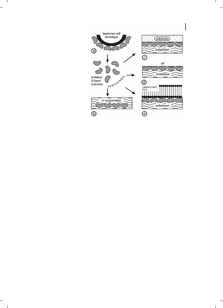
Figure 6.3
(a) Schematic illustration of
the recrystallization of isolated S-layer
subunits into crystalline arrays. The self
assembly process can occur in sus-
pension (b), on solid supports (c),
at the air/water interface (d), and on
Langmuir lipid films (e). (Reproduced
from Ref. [3], with permission from
Wiley-VCH.)
tallized on solid surfaces, S-layer protein monolayers consist of a closed mosaic of indivi-
dual monocrystalline domains.
6.2
Methods
6.2.1
Diagnostics
Studies on the structure, morphogenesis, genetics, and function of S-layers revealed that
these isoporous monomolecular arrays have a considerable application potential in bio-
technology, molecular nanotechnology, and biomimetics. The repetitive features of
S-layers have led to their applications in the production of S-layer ultrafiltration mem-
branes (SUMs), as supports for a defined covalent attachment of functional molecules
(e. g., enzymes, antibodies, antigens, protein A, biotin, and avidin) as required for affinity
and enzyme membranes, in the development of solid-phase immunoassays, or in biosen-
sors [3, 7, 22, 58, 59].
In dipstick-style solid-phase immunoassays, the respective monoclonal antibody was
covalently bound to the carbodiimide-activated carboxylic acid groups of the S-layer lattice
[60]. Proof of principle was demonstrated for different types of SUM-based dipsticks. For
example, for the diagnosis of type I allergies (determination of IgE in whole blood or
serum against the major birch pollen allergen Bet v1), for quantification of tissue type
plasminogen activator (t-PA) in patients’ whole blood or plasma for monitoring t-PA levels
during the course of thrombolytic therapies after myocardial infarction, or for determina-
tion of interleukin 8 (IL-8) in the supernatants of human umbilical vein endothelial cells
induced with lipopolysaccharides [7, 61, 62].
84
6 S-Layers
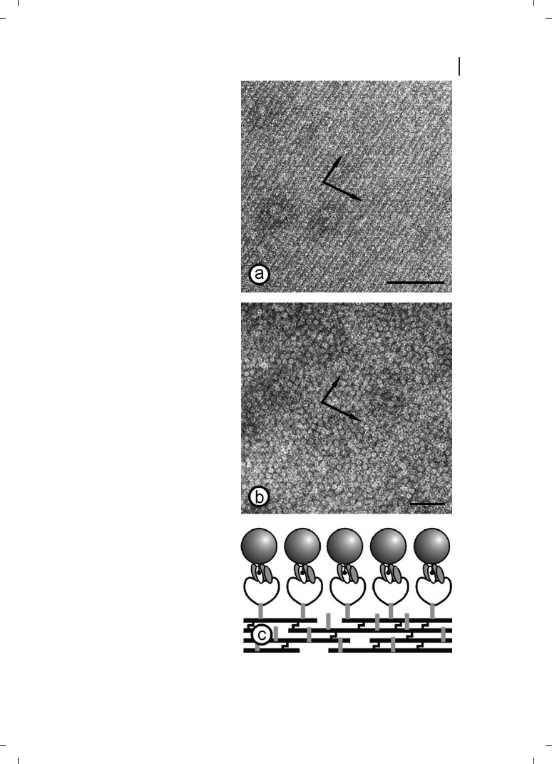
Figure 6.4
Recrystallization of the S-layer protein
SbpA of
Bacillus sphaericus CCM 2177 on a hydro-
philic silicon wafer. The atomic force microscopical
images show that crystal growth is initiated simul-
taneously at many randomly distributed nucleation
points (a) and proceeds in-plane until the crystalline
domains meet (b), thus leading to a closed, co-
herent mosaic of individual several micrometer
large S-layer domains (c). Scale bars = 0.5 mm;
Z-range = 12 nm. (Figure courtesy of E. Györvary
and O. Stein.)

Alternative or complementary to existing S-layer technologies, genetic approaches are
currently used for the construction of chimeric S-layer fusion proteins incorporating bio-
logically active sequences without hindering the self-assembly of S-layer subunits into reg-
ular arrays on surfaces and in suspension. In the chimeric S-layer proteins rSbsC
31-920
/Bet
v1 and rSbpA
31-1068
/Bet v1 carrying the major birch pollen allergen Bet v1 at the C-term-
inal end, the surface location and functionality of the fused allergen was demonstrated by
binding Bet v1-specific IgE [37, 63]. These fusion proteins can be used for building up
arrays for diagnostic test systems to determine the concentration of Bet v1-specific IgE
in patients’ whole blood, plasma, or serum samples [62]. In order to build up functional
monomolecular S-layer protein lattices on solid supports (e. g., gold, silicon, or glass), the
surface must be functionalized with covalently attached chemically modified SCWP, to
which the S-layer fusion proteins bind with their N-terminal part, leaving the C-terminal
part with the fused functional sequence exposed to the ambient environment. Owing to
the versatile applications of the streptavidin–biotin interaction as a biomolecular coupling
system, S-layer-streptavidin fusion proteins were constructed [45]. The two-dimensional
protein lattices displayed streptavidin in defined repetitive spacing, and proved to be cap-
able of binding biotin and also biotinylated functional molecules (see Figure 6.2). Thus,
the chimeric S-layer can be seen as a feasible tool to arrange different biotinylated targets
(e. g., proteins, allergens, antibodies, or oligonucleotides) on a surface which will find ap-
plication in protein, allergy, or DNA-chip technology. Furthermore, chimeric S-layers re-
crystallized on solid supports with a defined orientation are also expected to be a key ele-
ment in the rational design of highly integrated diagnostic devices (Lab-on-Chip). Another
application potential can be seen in the development of label-free detection systems [44].
In the SPR or surface acoustic wave technique, specific binding of functional molecules
(e. g., proteins or antibodies) to the sensor chip functionalized with an oriented chimeric
S-layer can be visualized directly by a mass increase on the chip without the need for any
labeled compound.
To conclude, such supramolecular biomimetic structures consisting of a functional
S-layer fusion protein recrystallized in defined orientation on SCWP-coated solid sup-
ports allow the development of new label-free detection systems as required for biochip
technology.
6.2.2
Lipid Chips
Since it became evident that typically free-standing bilayer lipid membranes (BLMs) sur-
vive for only minutes to hours and are very sensitive toward vibration and mechanical
shocks [64–66], stabilization of BLMs is imperatively necessary to utilize the function
of cell membrane components for practical applications (e. g., as lipid chips). S-layer pro-
teins can be exploited as supporting structures for BLMs (Figure 6.5) since they stabilize
the lipid film and largely retain their physical features (e. g., thickness, fluidity) [57].
In the following section the most promising methods to attach lipid membranes on
porous or solid supports in order to generate attractive lipid chips and membrane protein-
based devices are described. In general, lipid membranes attached to a porous support
combine the advantage of possessing an essentially unlimited ionic reservoir on each
85
6.2 Methods
side of the lipid membrane and of easy manual handling. A new strategy is the applica-
tion of an SUM with the S-layer as stabilizing and biochemical layer between the BLM and
the porous support. SUMs are isoporous structures with very sharp molecular exclusion
limits and were manufactured by depositing S-layer-carrying cell wall fragments under
high pressure on commercial microfiltration membranes (MFMs) with an average pore
size of approximately 0.4 mm [67, 68]. After deposition, the S-layer lattices are chemically
crosslinked to form a coherent smooth surface ideally suited for depositing lipid mem-
branes.
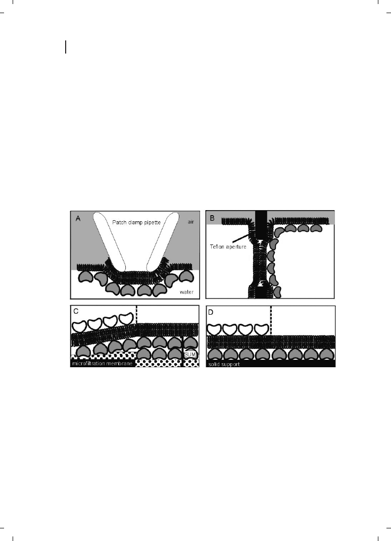
Composite SUM-supported bilayers (Figure 6.5C) are tight structures with breakdown
voltages well above 500 mV during their whole life-time of
Z8 hours [69]. For a compar-
ison, lipid membranes on a plain nylon MFM revealed a life-time of about 3 hours, and
ruptured at breakdown voltages of
Z210 mV. Specific capacitance measurements and
reconstitution experiments revealed functional lipid membranes on the SUM as the
pore-forming protein a-hemolysin could be reconstituted to form lytic channels. For the
first time, the opening and closing behavior of even single a-hemolysin pores (see also
86
6 S-Layers
Figure 6.5
Schematic illustrations of various S-
layer-supported lipid membranes. (A) Bilayer lipid
membranes (BLMs) have been generated across an
aperture of a patch–clamp pipette using the Tip-Dip
method, and a closed S-layer has been recrystallized
from the aqueous subphase. (B) A folded mem-
brane has been generated to span a Teflon aperture
using the method of Montal and Mueller [71].
Subsequently, S-layer protein can be injected into
one or both compartments (not shown), whereby
the protein self-assembles to form closely attached
S-layer lattices on the BLMs. (C) On an S-layer
ultrafiltration membrane (SUM) a BLM can be
generated by a modified Langmuir–Blodgett (LB)
technique. As a further option, a closed S-layer lat-
tice can be attached on the external side of the
SUM-supported BLM (left part). (D) Solid supports
can be covered by a closed S-layer lattice, and
subsequently BLMs can be generated using com-
binations of the LB and Langmuir–Schaefer tech-
niques, and vesicle fusion. As shown in (C), a
closed S-layer lattice can be recrystallized on the
external side of the solid supported BLM (left part).

Chapter 7) could be measured with membranes generated on a porous support [69]. The
main phospholipid of Thermoplasma acidophilum (MPL), a membrane-spanning tetraether
lipid, has also been transferred on an SUM using a modified Langmuir–Blodgett tech-
nique [70, 71]. Again, SUM-supported MPL-membranes allowed reconstitution of
functional molecules, as proven by measurements on single gramicidin pores. Recry-
stallization of an additional monomolecular S-layer protein lattice on the lipid-faced
side of SUM-supported MPL membranes increased the lifetime significantly to
21.2
e 3.1 hours [70].
Solid-supported membranes (Figure 6.5D) were developed in order to overcome the fra-
gility of free-standing BLMs, and also to enable biofunctionalization of inorganic solids
(e. g., semiconductors, gold-covered surfaces) for the use in sensing devices such as
lipid chips [72, 73]. Various types of solid-supported lipid membranes often show consid-
erable drawbacks as there is a limited ionic reservoir at the side facing the solid support,
the membranes often appear to be leaky (noninsulating), and large domains, protruding
from the membrane, may become denatured by the inorganic support [57, 74–78]. Again,
S-layer proteins have been studied to elucidate their potential as stabilizing and separating
ultrathin layer, which maintains also the structural and dynamic properties of the lipid
membranes. Silicon substrates have been covered by a closed S-layer lattice and bilayers
were deposited by the Langmuir–Blodgett technique [79–81]. Lateral diffusion of fluores-
cently labeled lipid molecules in both layers have been investigated by fluorescence recov-
ery after photobleaching studies [82]. In comparison with hybrid lipid bilayers (lipid
monolayer on alkylsilanes) and lipid bilayers on dextran, the mobility of lipids was highest
in S-layer-supported bilayers. Most importantly, the S-layer cover could prevent the forma-
tion of cracks and other inhomogenities in the bilayer [82]. These results have demon-
strated that the biomimetic approach of copying the supramolecular architecture of ar-
chaeal cell envelopes opens new possibilities for exploiting functional lipid membranes
at meso- and macroscopic scale. Moreover, this technology has the potential to initiate
a broad spectrum of lipid chips applicable for sensor technology, diagnostics, electronic
or optical devices, and high-throughput screening for drug discovery.
6.2.3
S-Layers as Templates for the Formation of Regularly Arranged Nanoparticles
The reproducible formation of nanoparticle arrays in large scale with predefined lattice
spacing and symmetries remains a challenge in the development of future generations
of molecular electronic devices (see also Chapter 19). This is particularly true for the rea-
lization of self-assembly and bottom-up approaches, as these strategies acquire the highest
efficiency in a fabrication process. Biomolecular templating has proven to be very attrac-
tive, as the self-assembly of molecules into monomolecular arrays is an intrinsic property
of many biological molecules and has already grown into a scientific and engineering dis-
cipline crossing the boundaries of several established fields (see also Chapters 16 and 17).
The first approach in using S-layers as templates in the generation of perfectly ordered
nanoparticle arrays was developed by Douglas and coworkers [55]. S-layer fragments of
Sulfolobus acidocaldarius were deposited on a smooth carbon surface and metal coated
by evaporation of a
Z1 nm-thick tantalum/tungsten film. Subsequently, this protein–
87
6.2 Methods

metal heterostructure was ion milled, leading to 15 nm-sized holes hexagonally arranged
according to the center-to-center spacing of the S-layer of 22 nm. Later on, this approach
was further optimized using fragments of the same S-layer species on a smooth graphite
surface and titanium oxide for the metal coating [56]. After oxidation in air and fast-atom
beam milling at normal incidence, a thin (
Z3.5 nm) metallic nanoporous mask with
pores in the 10 nm range was obtained. The same group used low-energy electron-en-
hanced etching to pattern the surface properties of a silicon substrate through the regu-
larly arranged pores of the S-layer [83]. After etching and removal of the S-layer, the pat-
terned surface was oxidized in an oxygen plasma, leading to a nanometric array of etched
holes (18 nm diameter) which served as nucleation sites in the formation of an ordered
array of nanometric titanium metal clusters. In a similar approach using argon ion etch-
ing in the final step, the S-layer of Deinococcus radiodurans was used as a nanometric tem-
plate for patterning ferromagnetic films [84]. Uniform hexagonal patterns of 10 nm-wide
dots and lattice spacing of 18 nm were fabricated from 2.5 nm-thick sputter-coated Co,
FeCo, Fe, FeNi, and NiFe films.
More recently, a synthesis pathway for the fabrication of nanoparticles by wet chemical
processes and S-layers as nanometric templates was developed [85–88]. In this approach,
self-assembled S-layer structures were exposed to a metal–salt solution (e. g., [AuCl
4
]
–
,
[PtCl
4
]
2–
), followed by slow reaction with a reducing agent such as hydrogen sulfide
(H
2
S). Nanoparticle superlattices were formed according to the lattice spacing and sym-
metry of the underlying S-layer. Furthermore, since the precipitation of the metals was
confined to the pores of the S-layer, the nanoparticles also resembled the morphology
of the pores. The first example exploiting this technique was the precipitation of cadmium
sulfide (CdS ) on S-layer lattices composed of SbsB and SbpA [85]. After incubation of the
S-layer self-assembly products with a CdCl
2
solution for several hours, the hydrated sam-
ples were exposed towards H
2
S for at least one or two days. The generated CdS nanopar-
ticles were 4–5 nm in size, and their superlattice resembled the oblique lattice symmetry
of SbsB (a = 9.4 nm, b = 7.4 nm, g = 80
h), or the square lattice symmetry of SbpA (a = b =
13.1 nm, g = 90
h), respectively. In a similar approach, a superlattice of 4–5 nm-sized gold
particles was formed by using SbpA (with previously induced thiol groups) as a template
for the precipitation of a tetrachloroauric (III) acid solution [86] (Figure 6.6a). Gold nano-
particles were formed either by reduction of the metal salt with H
2
S or under the electron
beam in a transmission electron microscope. The latter approach is technologically impor-
tant as it allows those areas where nanoparticles are formed to be defined. As determined
by electron diffraction, the gold nanoparticles were crystalline but their ensemble was not
crystallographically aligned. The wet chemical approach was used in the formation of Pd-
(salt: PdCl
2
), Ni- (NiSO
4
), Pt- (KPtCl
6
), Pb- (Pb(NO
3
)
2
) and Fe- (KFe(CN)
6
) nanoparticle
arrays (unpublished results), and for producing platinum nanoparticles on the S-layer
of Sporosarcina ureae [87, 88].
Unfortunately, wet chemical methods do not allow varying size or composition of nano-
particles in the fabrication process. Thus, the binding of preformed nanoparticles into reg-
ular arrays on S-layers has significant advantages in the development of nanoscale electro-
nic devices. Based on the studies of binding biomolecules (e. g. enzymes or antibodies)
onto S-layers, it has already been demonstrated that gold or CdSe nanoparticles can be
electrostatically bound in regular arrangements on S-layers [89–91] (Figure 6.6b). The
88
6 S-Layers
nanoparticles were either negatively charged due to surface citrate ions or positively
charged due to surface coating with poly-l-lysine.
In summary, these experiments have clearly shown that S-layers are perfectly suited to
control the formation of nanoparticle arrays, either by direct precipitation from the vapor
or liquid phase, or by binding preformed nanoparticles. The S-layer approach provides for
the first time a biologically based fabrication technology for the self-assembly of molecular
electronic or optic devices.
6.3
Outlook
At present, most applications developed for using S-layers depend on the in vitro self-
assembly capabilities of native S-layer proteins in suspension, on the surface of solids (e. g.,
silicon wafers, metals, polymers), Langmuir-lipid films, and liposomes. Once the regular
arrays have been formed, a broad spectrum of very precise chemical modifications can be
applied for tailoring the physico-chemical properties of S-layers and for a defined binding
of differently sized functional molecules. In particular, the possibility of immobilizing or
growing other materials (e. g., silicon oxide, metals) on top of recrystallized S-layer lattices
with most accurate spatial controlled architecture opens up many new possibilities in
nanofabrication and supramolecular engineering [6, 7].
An important line of development for the specific tuning of structural and functional
features concerns the genetic manipulation of S-layer proteins. Recent studies have clearly
demonstrated that truncated S-layer proteins incorporating specific functional domains of
other proteins maintain the self-assembly capability into regular arrays [5, 35]. This ap-
89
6.3 Outlook
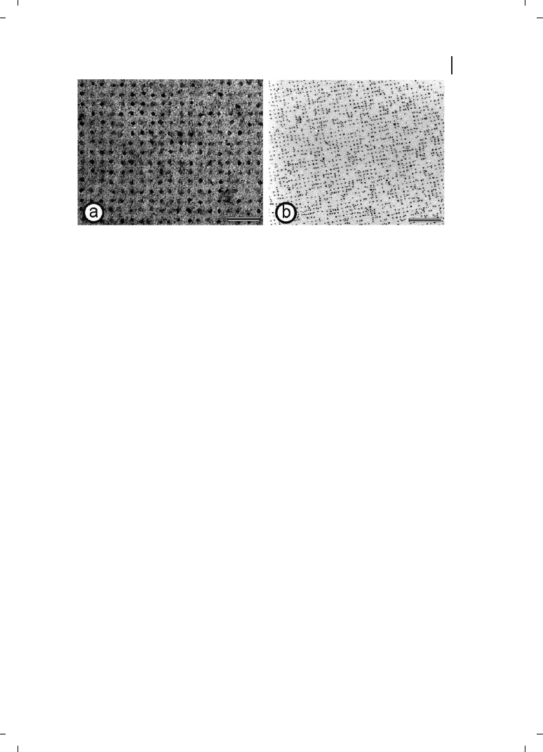
Figure 6.6
(a) Electron microscopical image of
gold nanoparticles (mean diameter 4.5 nm) ob-
tained using wet chemistry. An S-layer with square
lattice symmetry served as template in the precipi-
tation of the metal salt. The gold nanoparticles were
formed in the pore region of the protein meshwork
under the electron beam. Scale bar = 50 nm.
(b) Electron microscopical image of preformed gold
nanoparticles (mean diameter 4 nm) regularly
bound on the surface of an S-layer with square lat-
tice symmetry. Electrostatic interactions between
the surface of the nanoparticles and functional do-
mains on the S-layer are responsible for the binding.
Scale bar = 100 nm. (Reproduced from Ref. [91],
with permission from Elsevier.)

proach can lead to new isoporous ultrafiltration membranes, affinity structures, enzyme
membranes, ion- and metal particle-selective binding matrices, microcarriers, biosensors,
diagnostics, biocompatible surfaces, and vaccines [37, 44, 45, 63, 92].
Moreover, biomimetic approaches copying the supramolecular principle of virus envel-
opes such as S-layer-coated liposomes will provide new strategies for drug targeting and
drug delivery. Preliminary studies have also provided strong evidence that S-layers have
a great potential as patterning elements for non-life science applications (e. g., nonlinear
optics and molecular electronics) [90].
References
90
6 S-Layers
[1]
U. B. Sleytr, T. J. Beveridge, Trends Micro-
biol. 1999, 7, 253–260.
[2]
U. B. Sleytr, P. Messner, D. Pum, M. Sára,
Mol. Microbiol. 1993, 10, 911–916.
[3]
U. B. Sleytr, P. Messner, D. Pum, M. Sára,
Angew. Chem. Int. Ed. 1999, 38, 1034–1054.
[4]
U. B. Sleytr, M. Sára, Trends Biotechnol.
1997, 15, 20–26.
[5]
U. B. Sleytr, M. Sára, D. Pum, B. Schuster,
Prog. Surf. Sci. 2001, 68, 231–278.
[6]
U. B. Sleytr, M. Sára, D. Pum, B. Schuster,
in: M. Rosoff (ed.), Nano-Surface Chemistry,
Marcel Dekker, New York , Basel, 2001,
pp. 333–389.
[7]
U. B. Sleytr, M. Sára, D. Pum, B. Schuster,
P. Messner, C. Schäffer, in: A. Steinbüchel,
S. Fahnestock (eds), Biopolymers, Vol. 7,
Wiley-VCH, Weinheim, Germany, 2003,
pp. 285–338.
[8]
C. Schäffer, P. Messner, in: W. Herz, H.
Falk, G. W. Kirby (eds), Prokaryotic Glyco-
proteins, Springer, Wien, New York, 2003,
pp. 51–124.
[9]
P. Messner, G. Allmaier, C. Schäffer,
T. Wugeditsch, S. Lortal, H. König, R.
Niemetz, M. Dorner, FEMS Microbiol. Rev.
1997, 20, 25–46.
[10]
M. Sumper, F. T. Wieland, in: J. Montreuil,
J. F. G. Vliegenthart, H. Schachter (eds),
Glycoproteins, Elsevier, Amsterdam, 1995,
pp. 455–473.
[11]
W. Baumeister, G. Lembcke, R. Dürr,
B. Phipps, in: J. R. Fryer, D. L. Dorset (eds),
Electron Crystallography of Organic Mole-
cules, Kluwer, Dordrecht, 1990, pp. 283–
296.
[12]
W. Baumeister, G. Lembcke, J. Bioenerg.
Biomembr. 1992, 24, 567–575.
[13]
T. J. Beveridge, Curr. Opin. Struct. Biol.
1994, 4, 204–212.
[14]
D. Pum, U. B. Sleytr, Trends Biotechnol.
1999, 17, 8–12.
[15]
F. Ohnesorge, W. M. Heckl, W. Häberle,
D. Pum, M. Sára, H. Schindler, K. Schil-
cher, A. Kiener, D. P. E. Smith, U. B. Sleytr,
G. Binnig, Ultramicroscopy 1992, 42,
1236–1242.
[16]
S. Karrasch, R. Hegerl, J. Hoh, W. Bau-
meister, A. Engel, Proc. Natl. Acad. Sci.
USA 1994, 91, 836–838.
[17]
D. J. Müller, W. Baumeister, A. Engel,
J. Bacteriol. 1996, 178, 3025–3030.
[18]
H. König, Can. J. Microbiol. 1988, 34,
395–406.
[19]
U. B. Sleytr, P. Messner, D. Pum, M. Sára,
Appendix, in: U. B. Sleytr, P. Messner,
D. Pum, M. Sára (eds), Crystalline Bacterial
Cell Surface Proteins. Landes/Academic
Press, Austin, TX, 1996, pp. 211–225.
[20]
W. Baumeister, I. Wildhaber, B. M. Phipps,
Can. J. Microbiol. 1989, 35, 215–227.
[21]
S. Hovmöller, A. Sjögren, D. N. Wang,
Prog. Biophys. Mol. Biol. 1988, 51, 131–163.
[22]
M. Sára, U. B. Sleytr, J. Bacteriol. 2000, 182,
859–868.
[23]
E. Akca, H. Claus, N. Schultz, G. Karbach,
B. Schlott, T. Debaerdemaeker, J. P.
Declercq, H. König, Extremophiles 2002,
6, 351–358.
[24]
J. Dworkin, M. J. Blaser, Mol. Microbiol.
1997, 26, 433–440.
[25]
S. A. Thompson, M. J. Blaser, in:
I. Nachamkin, M. J. Blaser (eds), Campylo-
bacter, ASM Press, Washington, DC, 2000,
pp. 321–347.

91
References
[26]
M. Sára, B. Kuen, H. F. Mayer, F. Mandl,
K. C. Schuster, U. B. Sleytr, J. Bacteriol.
1996, 178, 2108–2117.
[27]
E. M. Egelseer, T. Danhorn, M. Pleschber-
ger, C. Hotzy, U. B. Sleytr, M. Sára, Arch.
Microbiol. 2001, 177, 70–80.
[28]
H. C. Scholz, E. Riedmann, A. Witte,
W. Lubitz, B. Kuen, J. Bacteriol. 2001,
183, 1672–1679.
[29]
A. Lupas, H. Engelhardt, J. Peters,
U. Santarius, S. Volker, W. Baumeister,
J. Bacteriol. 1994, 176, 1224–1233.
[30]
W. Ries, C. Hotzy, I. Schocher, U. B. Sleytr,
M. Sára, J. Bacteriol. 1997, 179, 3892–3898.
[31]
E. M. Egelseer, K. Leitner, M. Jarosch,
C. Hotzy, S. Zayni, U. B. Sleytr, M. Sára,
J. Bacteriol. 1998, 180, 1488–1495.
[32]
M. Sára, C. Dekitsch, H. F. Mayer, E. M.
Egelseer, U. B. Sleytr, J. Bacteriol. 1998,
180, 4146–4153.
[33]
N. Ilk, P. Kosma, M. Puchberger, E. M.
Egelseer, H. F. Mayer, U. B. Sleytr, M. Sára,
J. Bacteriol. 1999, 181, 7643–7646.
[34]
M. Jarosch, E. M. Egelseer, D. Mattanovich,
U. B. Sleytr, M. Sára, Microbiology. 2000,
146, 273–281.
[35]
M. Jarosch, E. M. Egelseer, C. Huber,
D. Moll, D. Mattanovich, U. B. Sleytr, M.
Sára, Microbiology. 2001, 147, 1353–1363.
[36]
M. Sára, Trends Microbiol. 2001, 9, 47–49.
[37]
N. Ilk, C. Völlenkle, E. M. Egelseer,
A. Breitwieser, U. B. Sleytr, M. Sára, Appl.
Environ. Microbiol. 2002, 68, 3251–3260.
[38]
C. Schäffer, H. Kählig, R. Christian,
G. Schulz, S. Zayni, P. Messner, Micro-
biology. 1999, 145, 1575–1583.
[39]
C. Schäffer, N. Müller, P. K. Mandal, R.
Christian, S. Zayni, P. Messner,
Glycoconjug. J. 2000, 17, 681–690.
[40]
C. Steindl, C. Schäffer, T. Wugeditsch,
M. Graninger, I. Matecko, N. Müller, P.
Messner, Biochem. J. 2002, 368, 483–494.
[41]
C. Mader, D. Moll, C. Hotzy, C. Huber,
U. B. Sleytr, M. Sára, Biochemistry sub-
mitted.
[42]
B. Kuen, U. B. Sleytr, W. Lubitz, Gene.
1994, 145, 115–120.
[43]
H. J. Boot, C. P. Kolen, P. H. Pouwels, Mol.
Microbiol. 1996, 21, 799–809.
[44]
M. Pleschberger, A. Neubauer, E. M. Egel-
seer, S. Weigert, B. Lindner, U. B. Sleytr,
S. Muyldermans, M. Sára, Bioconjug.
Chem. 2003, 14, 440–448.
[45]
D. Moll, C. Huber, B. Schlegel, D. Pum,
U. B. Sleytr, M. Sára, Proc. Natl. Acad. Sci.
USA 2002, 99, 14646–14651.
[46]
U. B. Sleytr, P. Messner, in: H. Plattner
(ed.), Electron Microscopy of Subcellular
Dynamics, CRC Press, Boca Raton, Florida,
1989, pp. 13–31.
[47]
U. B. Sleytr, P. Messner, Annu. Rev. Micro-
biol.1983, 37, 311–339.
[48]
D. Pum, U. B. Sleytr, in: Sleytr, U. B., P.
Messner, D. Pum, M. Sára (eds), Crystalline
Bacterial Cell Surface Layer Proteins
(S-Layers), Academic Press, R. G. Landes
Company, Austin, USA, 1996, pp.175–209.
[49]
D. Pum, U. B. Sleytr, Supramol. Science
1995, 2, 193–197.
[50]
D. Pum, M. Weinhandl, C. Hödl, U. B.
Sleytr, J. Bacteriol. 1993, 175, 2762–2766.
[51]
U. B. Sleytr, Int. Rev. Cytol. 1978, 53, 1–64.
[52]
U. B. Sleytr, Nature 1975, 257, 400–402.
[53]
P. Messner, D. Pum, U. B. Sleytr, J. Ultra-
struct. Mol. Struct. Res. 1986, 97, 73–88.
[54]
U. B. Sleytr, R. Plohberger, in: W. Bau-
meister, W. Vogell (eds), Electron Microscopy
at Molecular Dimensions, Springer-Verlag,
Berlin, Heidelberg, New York, 1980,
pp. 36–47.
[55]
K. Douglas, N. A. Clark, Appl. Phys. Lett.
1986, 48, 676–678.
[56]
K. Douglas, G. Devaud, N. A. Clark, Science
1992, 257, 642–644.
[57]
B. Schuster, U. B. Sleytr, Rev. Mol. Biotech-
nol. 2000, 74, 233–254.
[58]
M. Sára, U. B. Sleytr, Appl. Microbiol.
Biotechnol. 1989, 30, 184–189.
[59]
U. B. Sleytr, M. Sára, D. Pum, in: A. Ciferri
(ed.), Supramolecular Polymers, Marcel
Dekker, Inc., New York, Basel, 2000,
pp. 177–213.
[60]
A. Breitwieser, S. Küpcü, S. Howorka,
S. Weigert, C. Langer, K. Hoffmann-Som-
mergruber, O. Scheiner, U. B. Sleytr,
M. Sára, Biotechniques 1996, 21, 918–925.
[61]
U. B. Sleytr, H. Bayley, M. Sára, A. Breit-
wieser, S. Küpcü, C. Mader, S. Weigert,
F. M. Unger, P. Messner, B. Jahn-Schmid,
B. Schuster, D. Pum, K. Douglas, N. A.
Clark, J. T. Moore, T. A. Winningham,
S. Levy, I. Frithsen, J. Pankovc, P. Beale,
H. P. Gillis, D. A. Choutov, K. P. Martin,
FEMS Microbiol. Rev. 1997, 20, 151–175.
[62]
A. Breitwieser, C. Mader, I. Schocher,
K. Hoffmann-Sommergruber, W. Aberer,

92
6 S-Layers
O. Scheiner, U. B. Sleytr, M. Sára, Allergy
1998, 53, 786–793.
[63]
A. Breitwieser, E. M. Egelseer, D. Moll,
N. Ilk, C. Hotzy, B. Bohle, C. Ebner, U. B.
Sleytr, M. Sára, Protein Eng. 2002, 15,
243–249.
[64]
T. H. Tien, A. L. Ottova, J. Membr. Sci. 2001,
189, 83–117.
[65]
B. Raguse, V. Braach-Maksvytis, B. A. Cor-
nell, L. G. King, P. D. J. Osman, R. J. Pace,
L. Wieczorek, Langmuir 1998, 14, 648–659.
[66]
M. Zviman, H. T. Tien, Biosens. Bioelectron.
1991, 6, 37–42.
[67]
M. Sára, U. B. Sleytr, J. Bacteriol. 1987, 169,
2804–2809.
[68]
S. Weigert, M. Sára, J. Membrane Sci. 1995,
106, 147–159.
[69]
B. Schuster, D. Pum, M. Sára, O. Braha,
H. Bayley, U. B. Sleytr, Langmuir 2001, 17,
499–503.
[70]
B. Schuster, S. Weigert, D. Pum, M. Sára,
U. B. Sleytr, Langmuir 2003, 19, in press.
[71]
M. Montal, P. Mueller, Proc. Natl. Acad.
Sci. USA 1972, 69, 3561–3566.
[72]
E. Sackmann, M. Tanaka, Trends Biotechnol.
2000, 18, 58–64.
[73]
B. A. Cornell, V. L. Braach-Maksvytis, L. G.
King, P. D. Osman, B. Raguse, L. Wieczo-
rek, R. J. Pace, Nature 1997, 387,
580–583.
[74]
W. Knoll, C. W. Frank, C. Heibel, R. Nau-
mann, A. Offenhäusser, J. Rühe, E. K.
Schmidt, W. W. Shen, A. Sinner, Rev. Mol.
Biotechnol. 2000, 74, 137–158.
[75]
D. P. Nikolelis, T. Hianik, U. J. Krull,
Electroanalysis 1999, 11, 7–15.
[76]
S. Heyse, T. Stora, E. Schmid, J. H. Lakely,
H. Vogel, Biochim. Biophys. Acta 1998,
1376, 319–338.
[77]
A. L. Plant, Langmuir 1993, 9, 2764–2767.
[78]
E. Kalb, S. Frey, L. K. Tamm, Biochim.
Biophys. Acta 1992, 1103, 307–316.
[79]
A. Zasadzinski, R. Viswanathan, L. Mad-
son, J. Garnaes, K. D. Schwartz, Science
1994, 263, 1726–1733.
[80]
I. Langmuir, V. J. Schaefer, J. Am. Chem.
Soc. 1937, 59, 1406–1417.
[81]
K. J. Blodgett, J. Am. Chem. Soc. 1935, 57,
1007–1022.
[82]
E. Györvary, B. Wetzer, U. B. Sleytr,
A. Sinner, A. Offenhäusser, W. Knoll,
Langmuir 1999, 15, 1337–1347.
[83]
T. A. Winningham, H. P. Gillis, D. A.
Choutov, K. P. Martin, J. T. Moore,
K. Douglas, Surf. Sci. 1998, 406, 221–228.
[84]
M. Panhorst, H. Brückl, B. Kiefer, G. Reiss,
U. Santarius, R., Guckenberger, J. Vac. Sci.
Technol. B 2001, 19, 722–724.
[85]
W. Shenton, D. Pum, U. B. Sleytr, S. Mann,
Nature 1997, 389, 585–587.
[86]
S. Dieluweit, D. Pum, U. B. Sleytr,
Supramol. Science 1998, 5, 15–19.
[87]
M. Mertig, R. Kirsch, W. Pompe, H.
Engelhardt, Eur. Phys. J. 1999, 9, 45–48.
[88]
W. Pompe, M. Mertig, R. Kirsch, R. Wahl,
L. C. Ciachi, J. Richter, R. Seidel, H. Vin-
zelberg, Z. Metallkd. 1999, 90, 1085–1091.
[89]
S. R. Hall, W. Shenton, H. Engelhardt,
S. Mann, Chem. Phys. Chem. 2001, 3,
184–186.
[90]
E. Györvary, A. Schroedter, D. V. Talapin,
H. Weller, D. Pum, U. B. Sleytr, J. Nanosci.
Nanotechnol. submitted.
[91]
U. B. Sleytr, E. Györvary, D. Pum, Prog.
Organic Coatings 2003, in press.
[92]
V. Weber, S. Weigert, M. Sára, U. B. Sleytr,
D. Falkenhagen, Therapeutic Apheresis 2001,
5, 433–438.
Wyszukiwarka
Podobne podstrony:
MT st w 06
Kosci, kregoslup 28[1][1][1] 10 06 dla studentow
06 Kwestia potencjalności Aid 6191 ppt
06 Podstawy syntezy polimerówid 6357 ppt
06
06 Psych zaburz z somatoformiczne i dysocjacyjne
GbpUsd analysis for July 06 Part 1
Probl inter i kard 06'03
06 K6Z4
06 pamięć proceduralna schematy, skrypty, ramyid 6150 ppt
Sys Inf 03 Manning w 06
Ustawa z dnia 25 06 1999 r o świadcz pien z ubezp społ w razie choroby i macierz
06 ZPIU org prod
więcej podobnych podstron