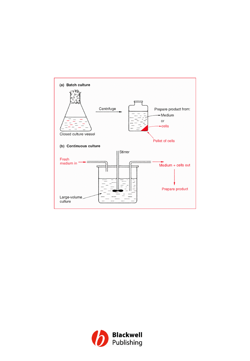
Figure 13.1 Two different systems for the
growth of microorganisms: (a) batch culture;
(b) continuous culture.
Gene Cloning and DNA Analysis by T.A. Brown. © 2006 T.A.
Brown.
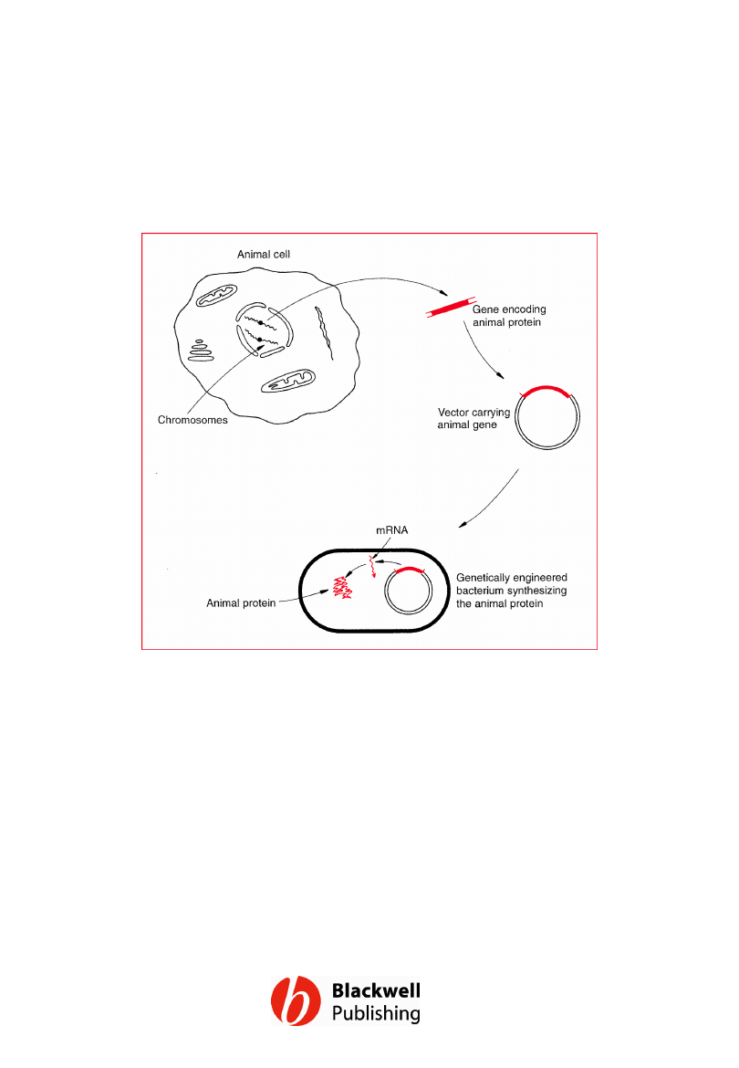
Figure 13.2 A possible scheme for the
production of an animal protein by a
bacterium. mRNA = messenger RNA.
Gene Cloning and DNA Analysis by T.A. Brown. © 2006 T.A.
Brown.
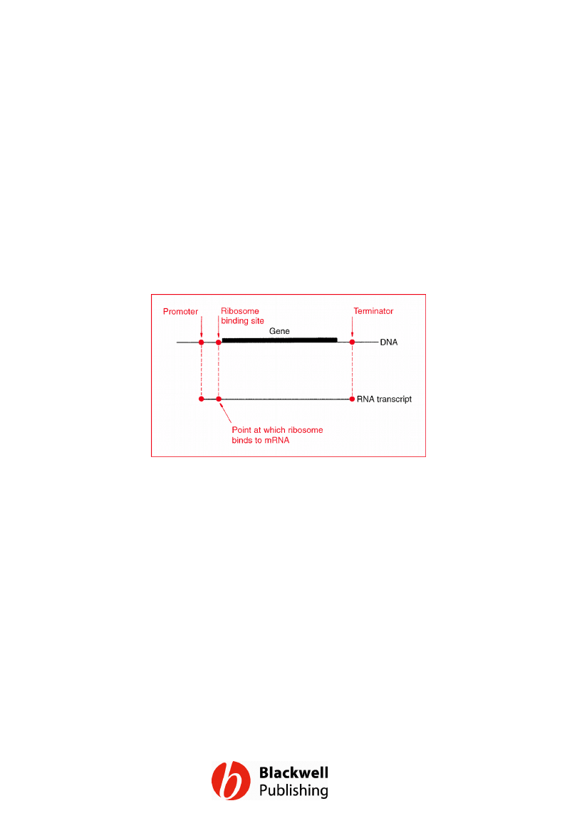
Figure 13.3 The three most important
signals for gene expression in E. coli.
Gene Cloning and DNA Analysis by T.A. Brown. © 2006 T.A.
Brown.
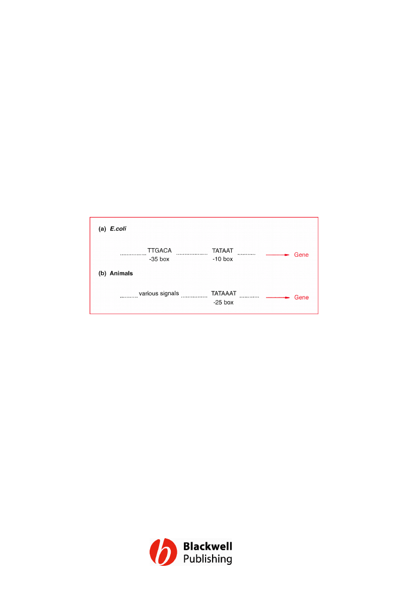
Figure 13.4 Typical promoter sequences for
E. coli and animal genes.
Gene Cloning and DNA Analysis by T.A. Brown. © 2006 T.A.
Brown.
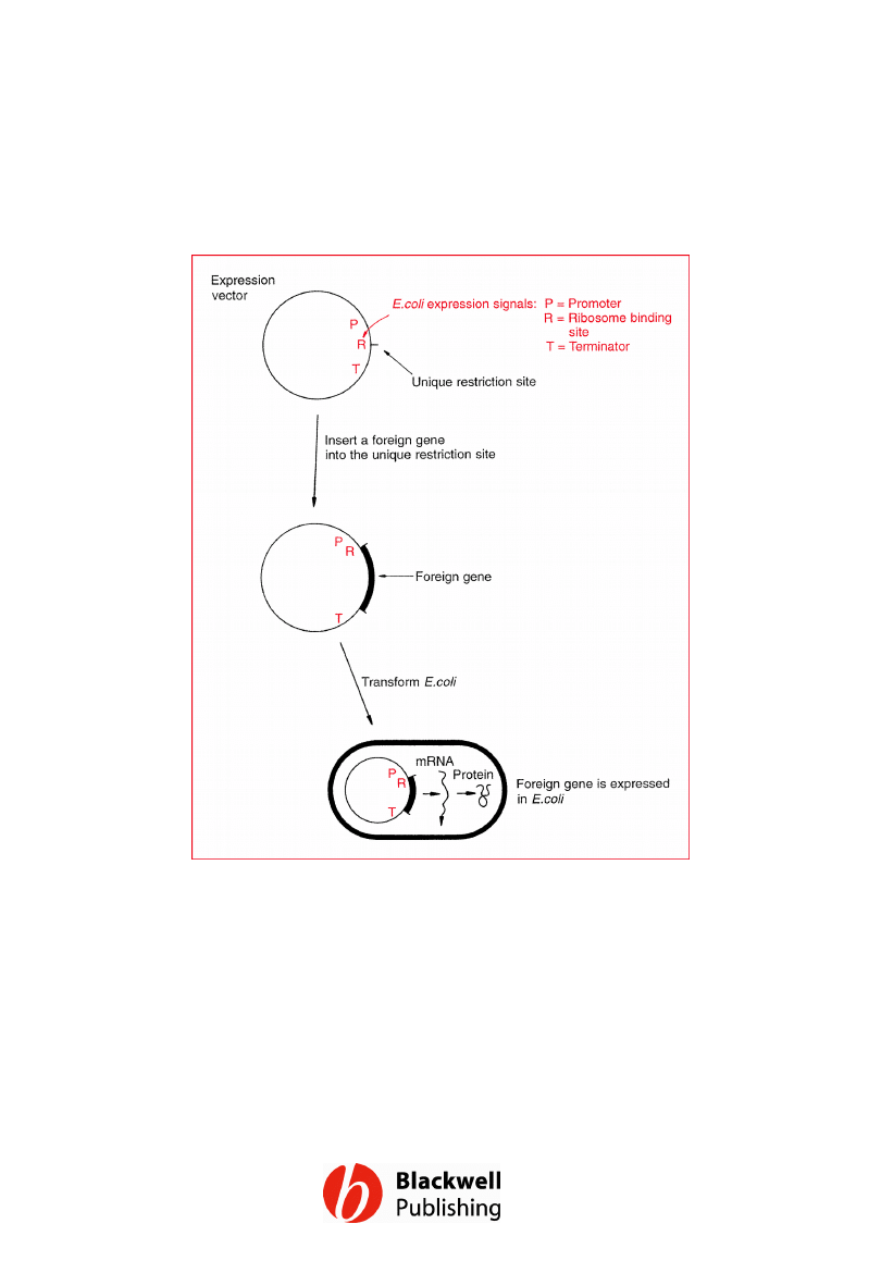
Figure 13.5 The use of an expression vector
to achieve expression of a foreign gene in E.
coli.
Gene Cloning and DNA Analysis by T.A. Brown. © 2006 T.A.
Brown.
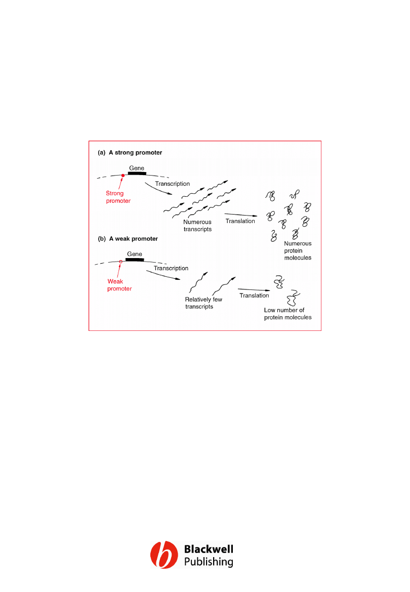
Figure 13.6 Strong and weak promoters.
Gene Cloning and DNA Analysis by T.A. Brown. © 2006 T.A.
Brown.
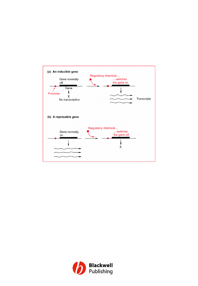
Figure 13.7 Examples of the two major
types of gene regulation that occur in
bacteria: (a) an inducible gene; (b) a
repressible gene.
Gene Cloning and DNA Analysis by T.A. Brown. © 2006 T.A.
Brown.
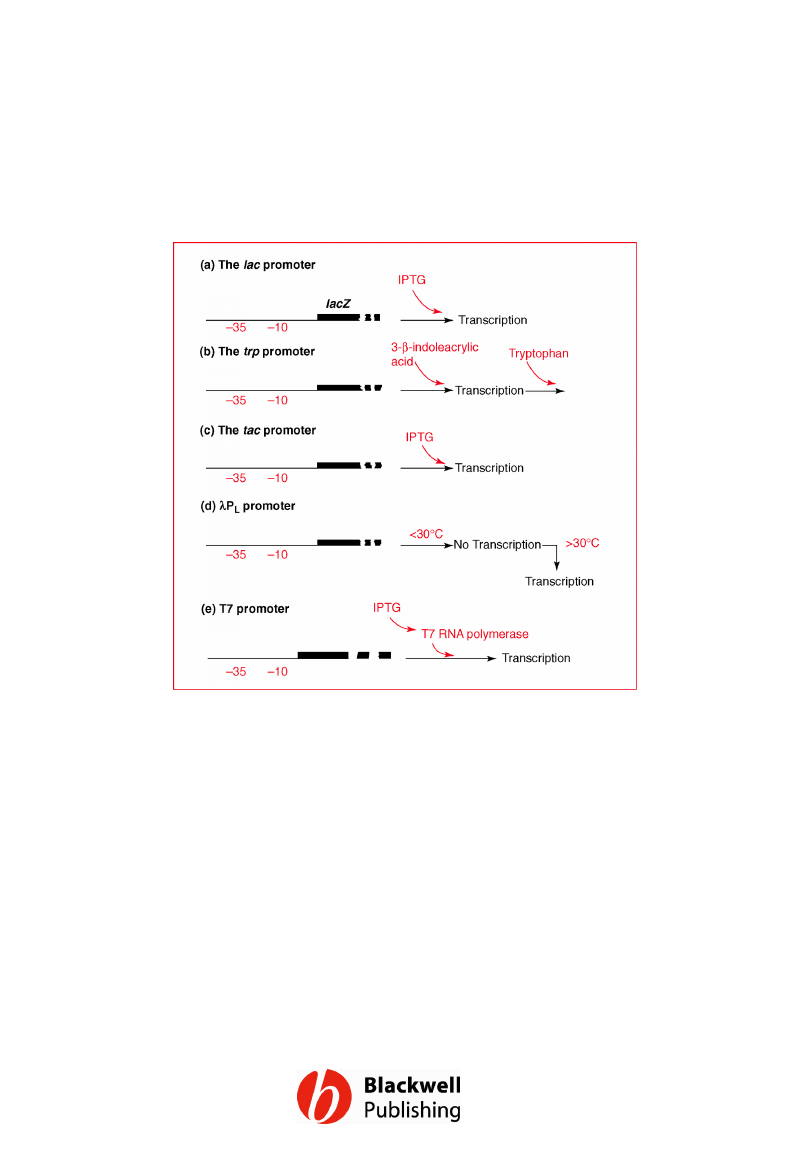
Figure 13.8 Five promoters frequently used
in expression vectors. The lac and trp
promoters are shown upstream of the genes
that they normally control in E. coli.
Gene Cloning and DNA Analysis by T.A. Brown. © 2006 T.A.
Brown.
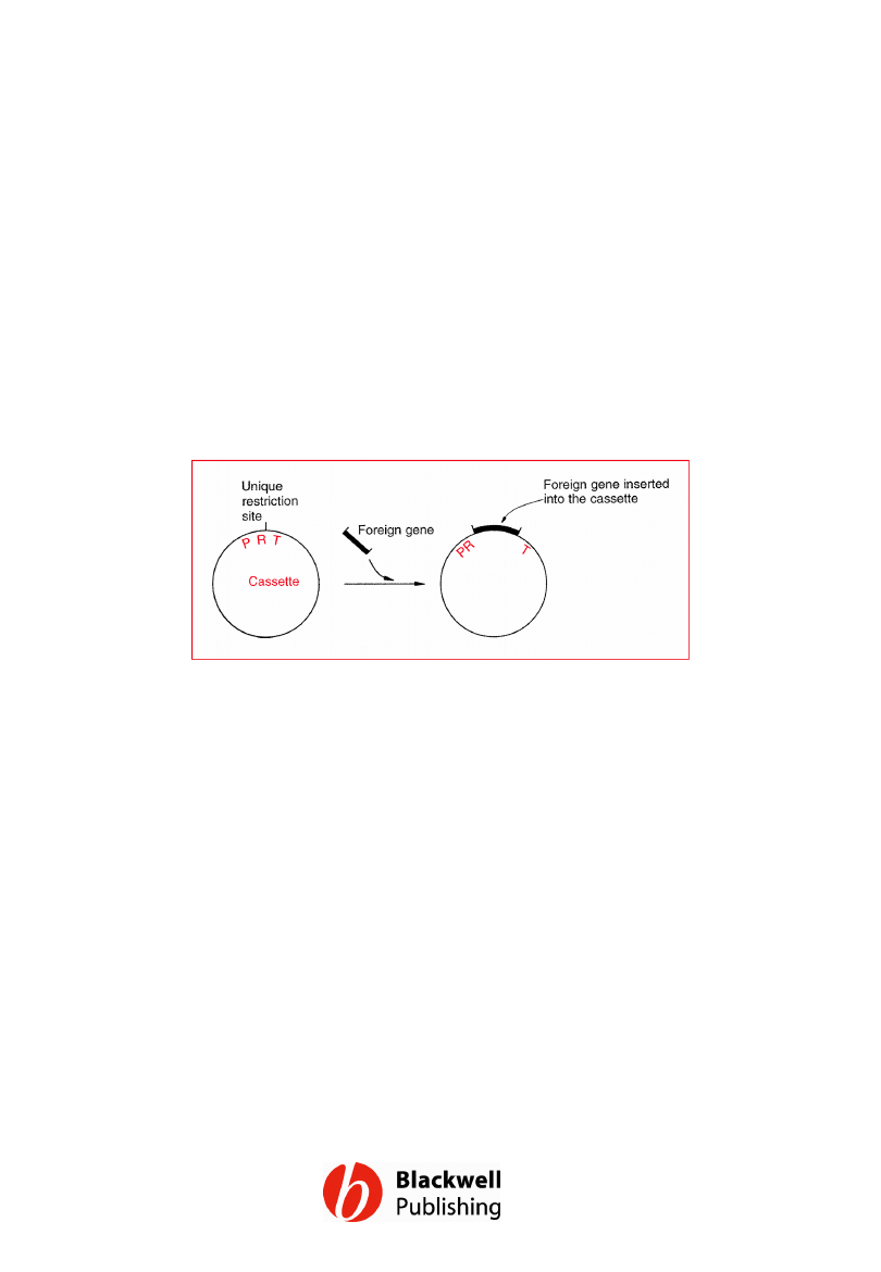
Figure 13.9 A typical cassette vector and
the way it is used. P = promoter, R =
ribosome binding site, T = terminator.
Gene Cloning and DNA Analysis by T.A. Brown. © 2006 T.A.
Brown.
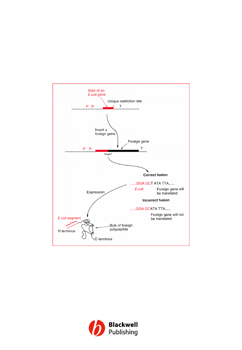
Figure 13.10 The construction of a hybrid
gene and the synthesis of a fusion protein.
Gene Cloning and DNA Analysis by T.A. Brown. © 2006 T.A.
Brown.
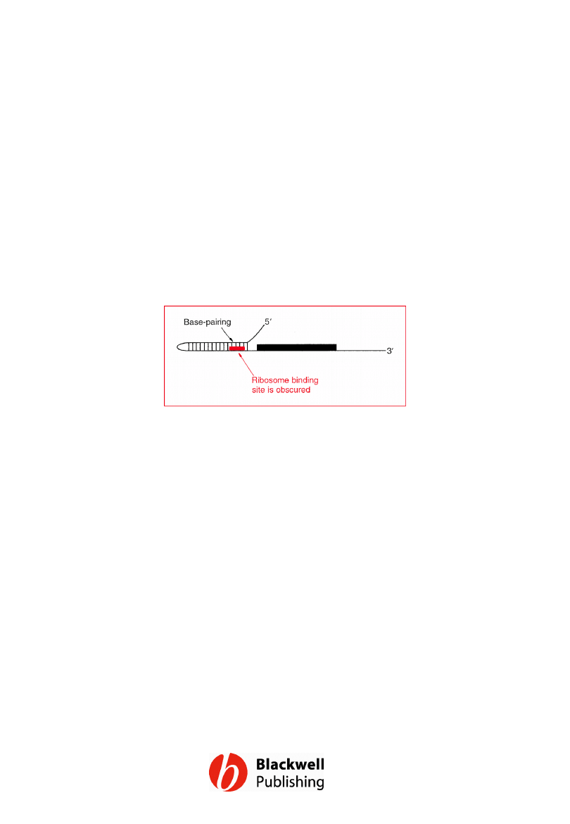
Figure 13.11 A problem caused by
secondary structure at the start of an mRNA.
Gene Cloning and DNA Analysis by T.A. Brown. © 2006 T.A.
Brown.
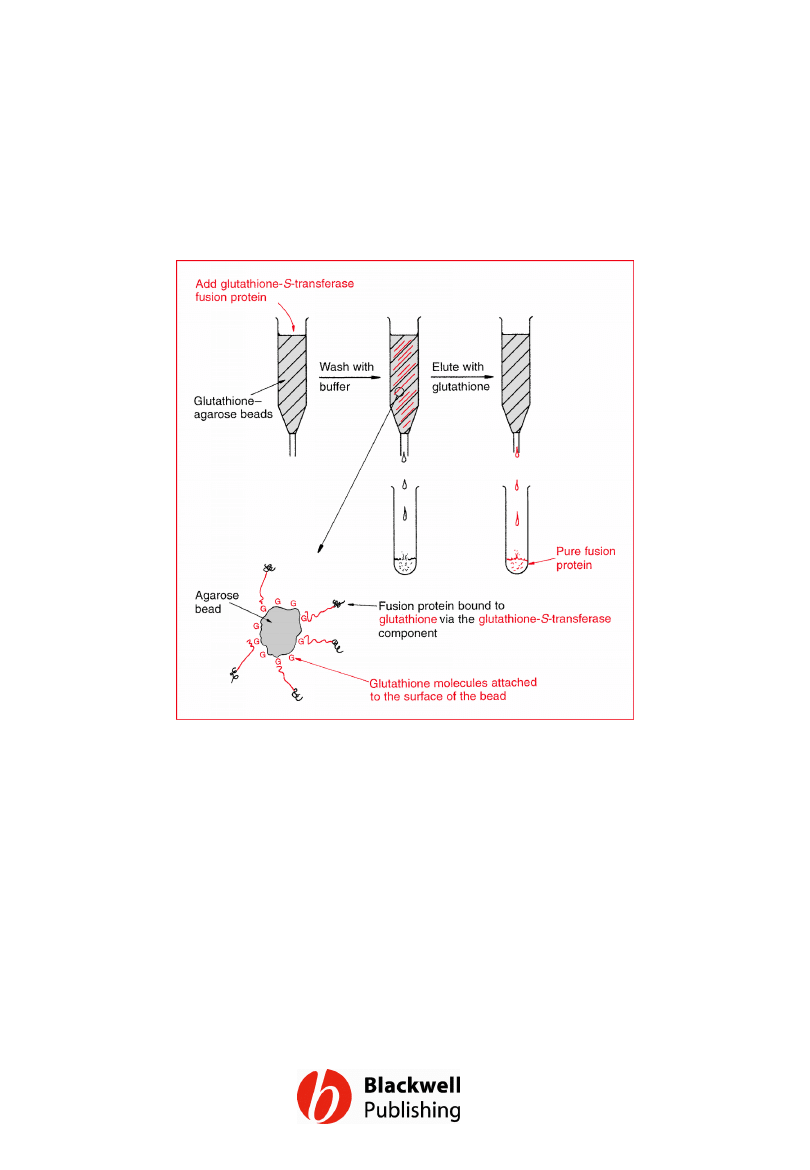
Figure 13.12 The use of affinity
chromatography to purify a glutathione-S-
transferase fusion protein.
Gene Cloning and DNA Analysis by T.A. Brown. © 2006 T.A.
Brown.
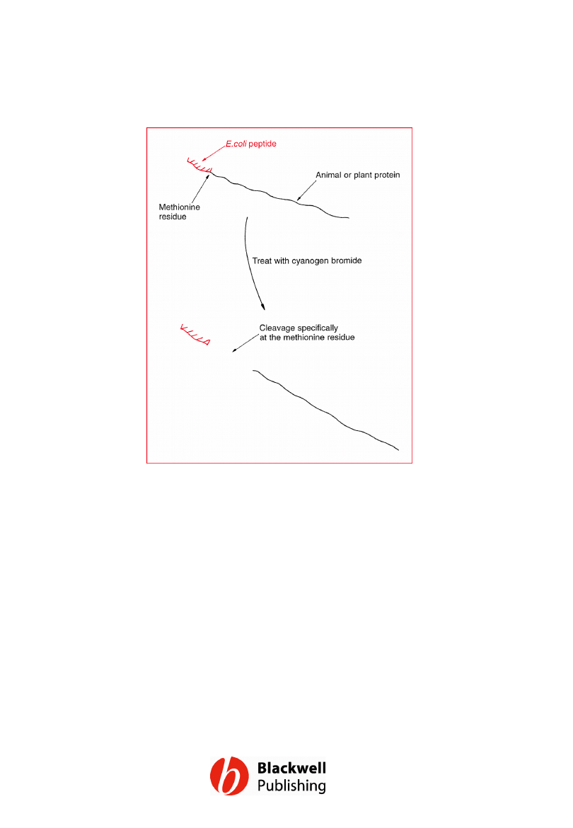
Figure 13.13 One method for the recovery
of the foreign polypeptide from a fusion
protein. The methionine residue at the fusion
junction must be the only one present in the
entire polypeptide: if others are present
cyanogen bromide will cleave the fusion
protein into more than two fragments.
Gene Cloning and DNA Analysis by T.A. Brown. © 2006 T.A.
Brown.
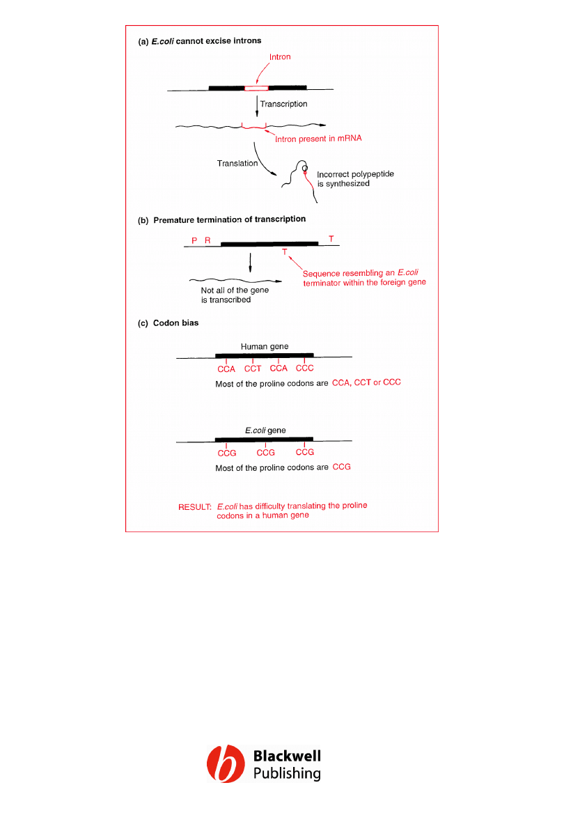
Figure 13.14 Three of the problems that
could be encountered when foreign genes are
expressed in E. coli: (a) introns are not
removed in E. coli; (b) premature termination
of transcription; (c) a problem with codon
bias.
Gene Cloning and DNA Analysis by T.A. Brown. © 2006 T.A.
Brown.
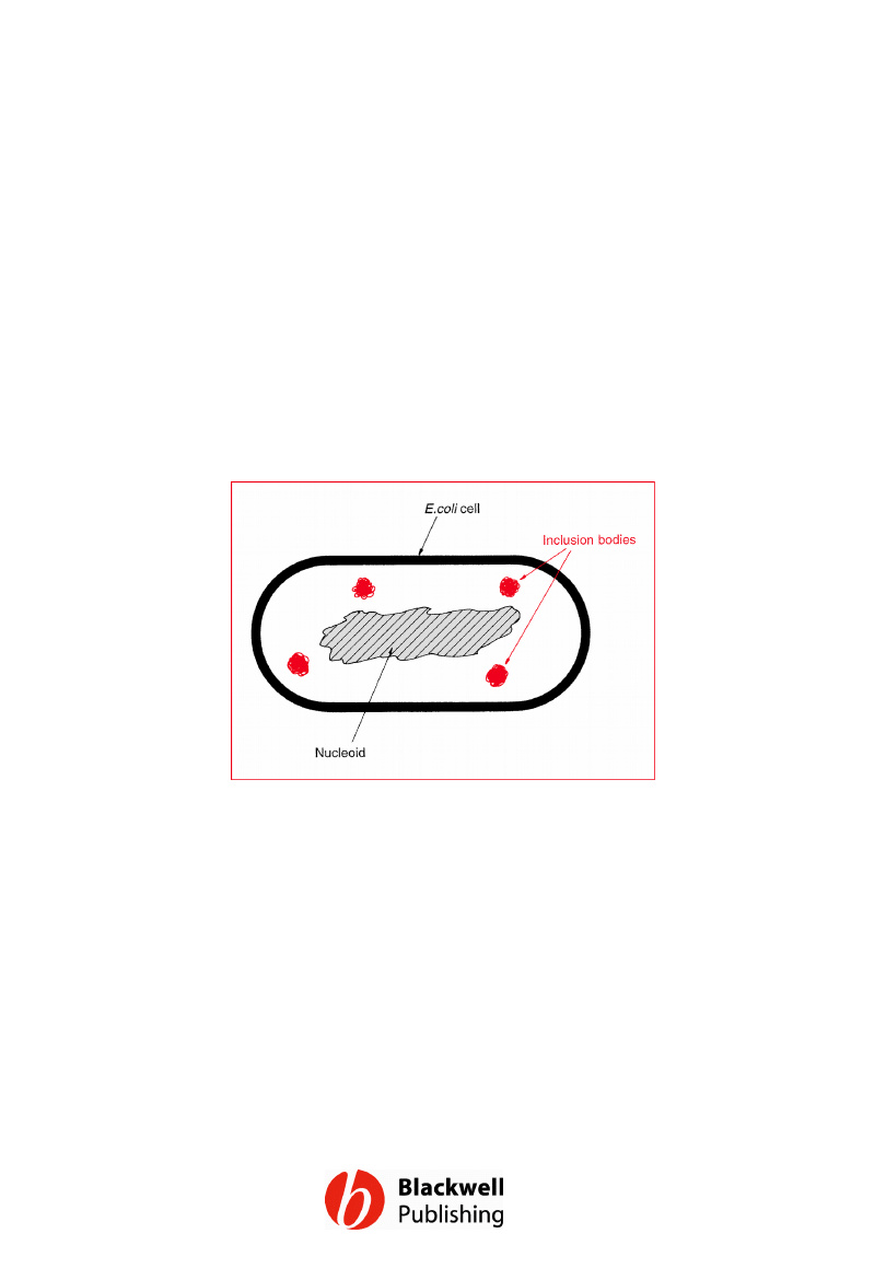
Figure 13.15 Inclusion bodies.
Gene Cloning and DNA Analysis by T.A. Brown. © 2006 T.A.
Brown.
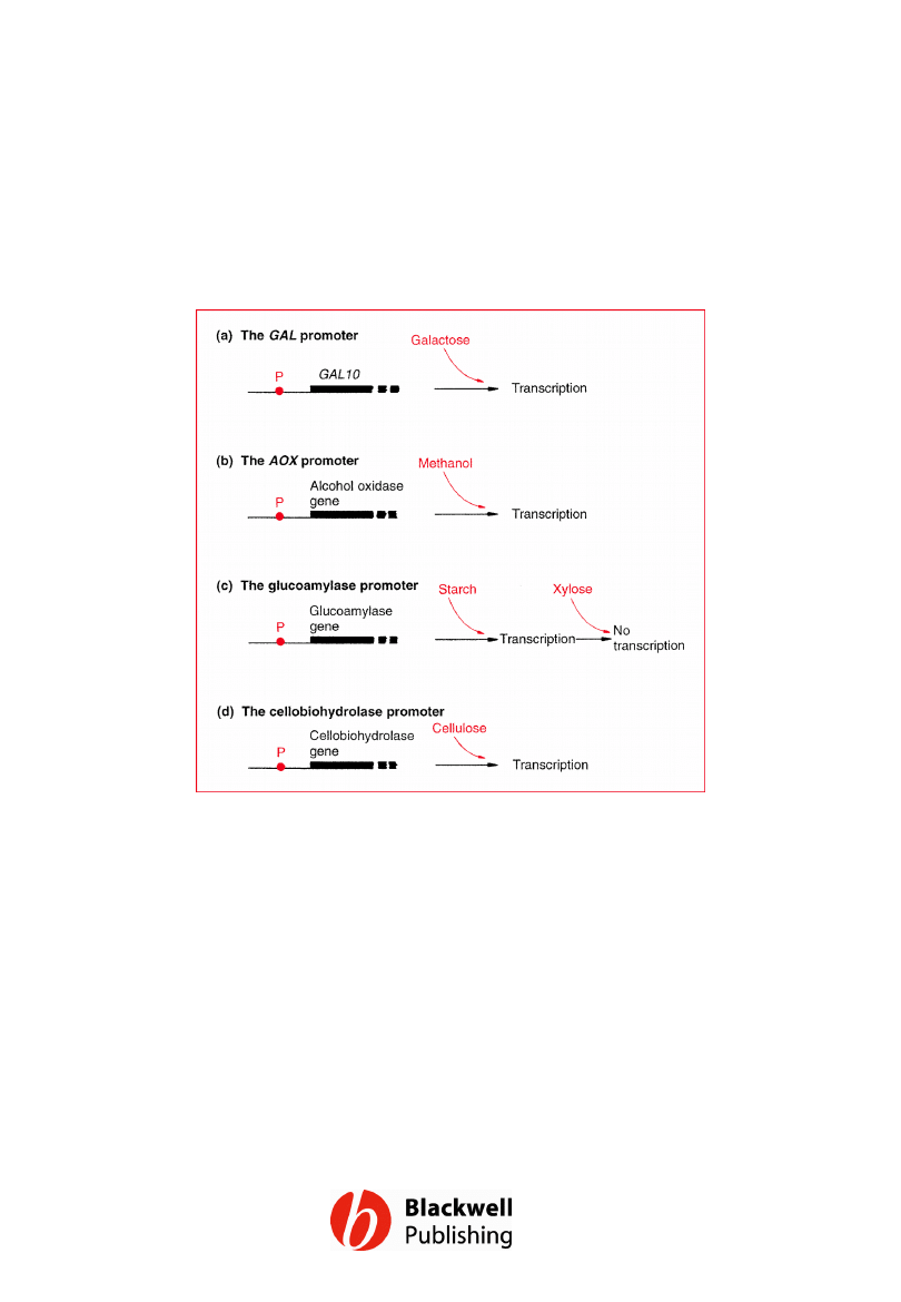
Figure 13.16 Four promoters frequently
used in expression vectors for microbial
eukaryotes. P = promoter.
Gene Cloning and DNA Analysis by T.A. Brown. © 2006 T.A.
Brown.
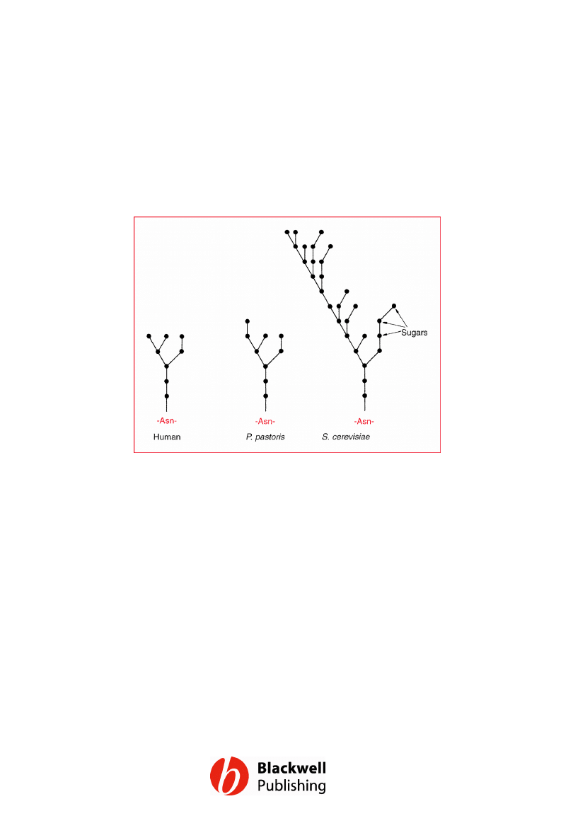
Figure 13.17 Comparison between a typical
glycosylation structure found on an animal
protein and the structures synthesized by P.
pastoris and S. cerevisiae.
Gene Cloning and DNA Analysis by T.A. Brown. © 2006 T.A.
Brown.
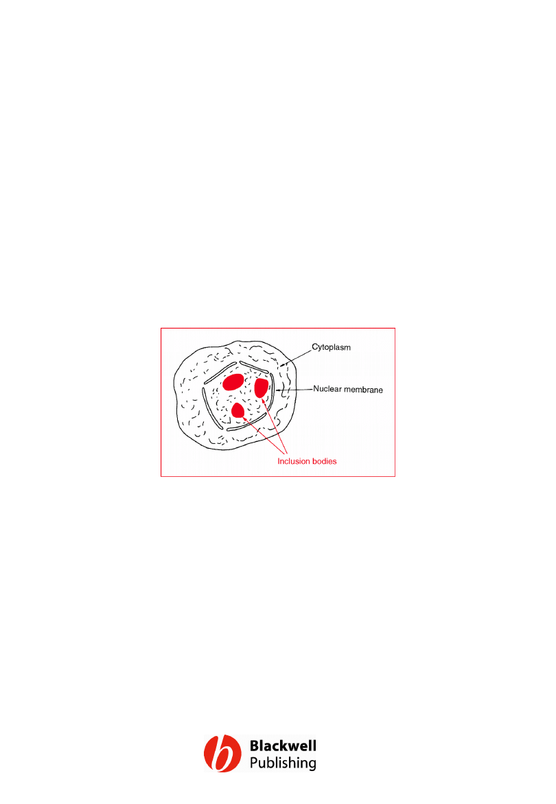
Figure 13.18 Crystalline inclusion bodies in
the nuclei of insect cells infected with a
baculovirus.
Gene Cloning and DNA Analysis by T.A. Brown. © 2006 T.A.
Brown.
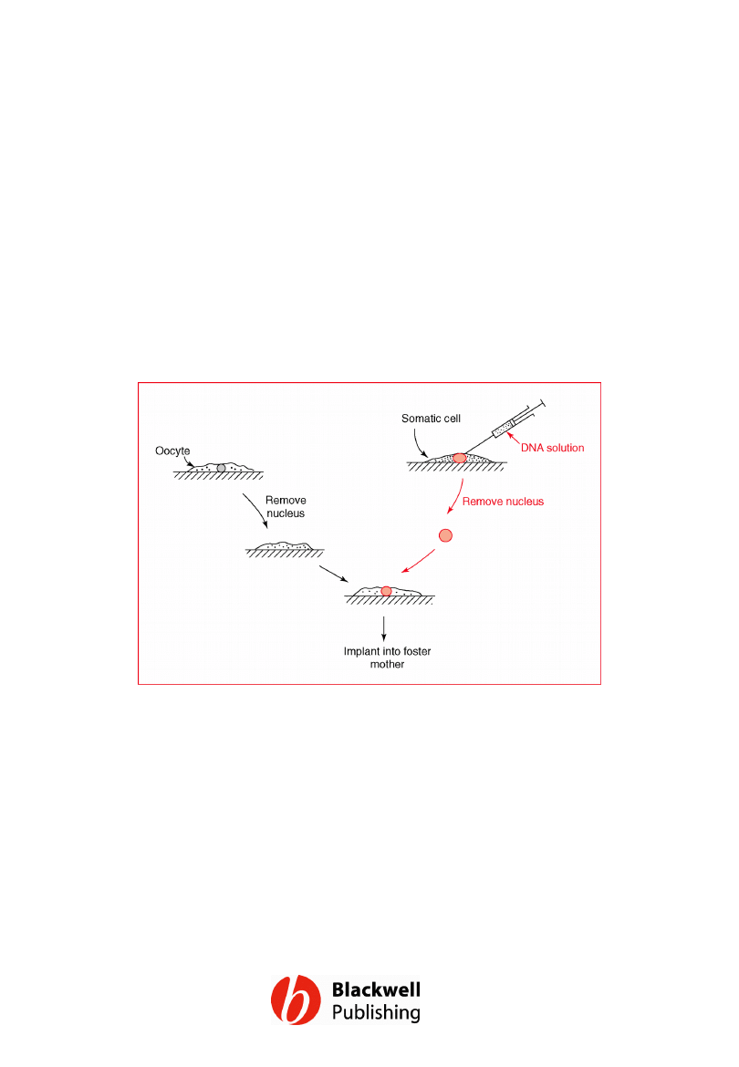
Figure 13.19 Transfer of the nucleus from a
transgenic somatic cell to an oocyte.
Gene Cloning and DNA Analysis by T.A. Brown. © 2006 T.A.
Brown.
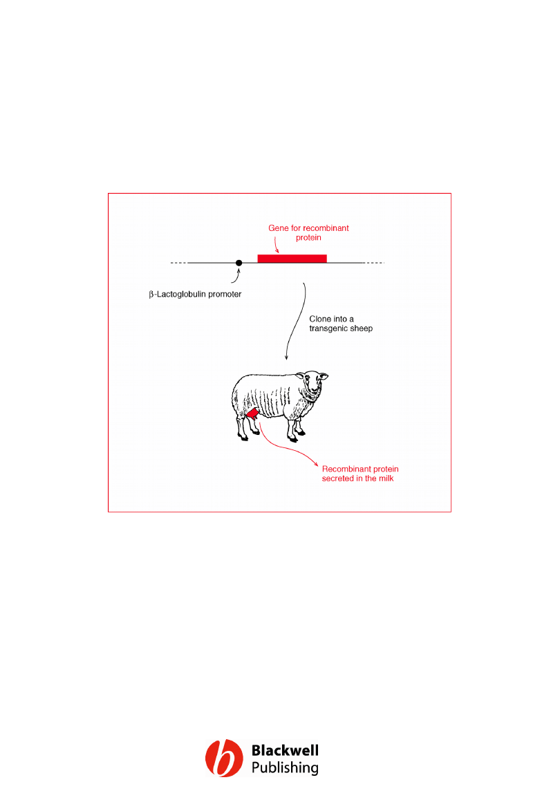
Figure 13.20 Recombinant protein
production in the milk of a transgenic sheep.
Gene Cloning and DNA Analysis by T.A. Brown. © 2006 T.A.
Brown.
Document Outline
- Figure 13.1 Two different systems for the growth of microorganisms: (a) batch culture; (b) continuous culture.
- Figure 13.2 A possible scheme for the production of an animal protein by a bacterium. mRNA = messenger RNA.
- Figure 13.3 The three most important signals for gene expression in E. coli.
- Figure 13.4 Typical promoter sequences for E. coli and animal genes.
- Figure 13.5 The use of an expression vector to achieve expression of a foreign gene in E. coli.
- Figure 13.6 Strong and weak promoters.
- Figure 13.7 Examples of the two major types of gene regulation that occur in bacteria: (a) an inducible gene; (b) a repressible gene.
- Figure 13.8 Five promoters frequently used in expression vectors. The lac and trp promoters are shown upstream of the genes that they normally control in E. coli.
- Figure 13.9 A typical cassette vector and the way it is used. P = promoter, R = ribosome binding site, T = terminator.
- Figure 13.10 The construction of a hybrid gene and the synthesis of a fusion protein.
- Figure 13.11 A problem caused by secondary structure at the start of an mRNA.
- Figure 13.12 The use of affinity chromatography to purify a glutathione-S-transferase fusion protein.
- Figure 13.13 One method for the recovery of the foreign polypeptide from a fusion protein. The methionine residue at the fusion junction must be the only one present in the entire polypeptide: if others are present cyanogen bromide will cleave the fusion protein into more than two fragments.
- Figure 13.14 Three of the problems that could be encountered when foreign genes are expressed in E. coli: (a) introns are not removed in E. coli; (b) premature termination of transcription; (c) a problem with codon bias.
- Figure 13.15 Inclusion bodies.
- Figure 13.16 Four promoters frequently used in expression vectors for microbial eukaryotes. P = promoter.
- Figure 13.17 Comparison between a typical glycosylation structure found on an animal protein and the structures synthesized by P. pastoris and S. cerevisiae.
- Figure 13.18 Crystalline inclusion bodies in the nuclei of insect cells infected with a baculovirus.
- Figure 13.19 Transfer of the nucleus from a transgenic somatic cell to an oocyte.
- Figure 13.20 Recombinant protein production in the milk of a transgenic sheep.
Wyszukiwarka
Podobne podstrony:
Figures for chapter 5
Figures for chapter 12
Figures for chapter 6
Figures for chapter 14
Figures for chapter 10
Figures for chapter 11
Figures for chapter 8
Figures for chapter 9
Figures for chapter 2
Figures for chapter 16
Figures for chapter 3
Figures for chapter 7
Figures for chapter 15
Figures for chapter 1
Figures for chapter 5
Figures for chapter 12
Figures for chapter 6
Figures for chapter 14
Figures for chapter 10
więcej podobnych podstron