NeuroMolecular Medicine
Copyright © 2007 Humana Press Inc.
All rights of any nature whatsoever reserved.
ISSN1535-1084/07/09:83–100/$30.00
(Online) 1559-1174
doi: 10.1385/NMM:9:1:83
NeuroMolecular Medicine
83
Volume 9, 2007
O
RIGINAL
A
RTICLE
Aluminum Adjuvant Linked to Gulf War Illness
Induces Motor Neuron Death in Mice
Michael S. Petrik,*
,1,2
Margaret C. Wong,
1,2
Rena C. Tabata,
1,3
Robert F. Garry,
4
and Christopher A. Shaw
1,5,6
1
Department of Ophthalmology;
2
Program in Neuroscience;
3
Program in Experimental Medicine,
University of British Columbia, Vancouver, British Columbia, Canada;
4
Department of Microbiology and Immunology, Louisiana State University Health Sciences Center,
Tulane University Health Sciences Center, New Orleans, LA;
5
Departments of Physiology;
and
6
Experimental Medicine, University of British Columbia, Vancouver, British Columbia, Canada
Received March 9, 2006; Revised May 3, 2006; Accepted May 9, 2006
Abstract
Gulf War illness (GWI) affects a significant percentage of veterans of the 1991 conflict, but its
origin remains unknown. Associated with some cases of GWI are increased incidences of amyo-
trophic lateral sclerosis and other neurological disorders. Whereas many environmental factors
have been linked to GWI, the role of the anthrax vaccine has come under increasing scrutiny. Among
the vaccine’s potentially toxic components are the adjuvants aluminum hydroxide and squalene.
To examine whether these compounds might contribute to neuronal deficits associated with GWI,
an animal model for examining the potential neurological impact of aluminum hydroxide, squa-
lene, or aluminum hydroxide combined with squalene was developed. Young, male colony CD-1
mice were injected with the adjuvants at doses equivalent to those given to US military service
personnel. All mice were subjected to a battery of motor and cognitive-behavioral tests over a
6-mo period postinjections. Following sacrifice, central nervous system tissues were examined
using immunohistochemistry for evidence of inflammation and cell death. Behavioral testing
showed motor deficits in the aluminum treatment group that expressed as a progressive decrease
in strength measured by the wire-mesh hang test (final deficit at 24 wk; about 50%). Significant
cognitive deficits in water-maze learning were observed in the combined aluminum and squalene
group (4.3 errors per trial) compared with the controls (0.2 errors per trial) after 20 wk. Apoptotic
neurons were identified in aluminum-injected animals that showed significantly increased acti-
vated caspase-3 labeling in lumbar spinal cord (255%) and primary motor cortex (192%) compared
with the controls. Aluminum-treated groups also showed significant motor neuron loss (35%) and
increased numbers of astrocytes (350%) in the lumbar spinal cord. The findings suggest a possible
role for the aluminum adjuvant in some neurological features associated with GWI and possibly
an additional role for the combination of adjuvants.
doi: 10.1385/NMM:9:1:83
*Author to whom all correspondence and reprint requests should be addressed. E-mail: mspetrik@interchange.ubc.ca
M_15 Petrik 9/22/06 10:06 PM Page 83

84
Petrik et al.
NeuroMolecular Medicine
Volume 9, 2007
Introduction
Gulf War illness (GWI), popularly termed “Gulf
War syndrome,” is a spectrum of disorders among
veterans of the Persian Gulf War (1990–1991) char-
acterized by a group of variable and nonspecific
symptoms such as fatigue, muscle and joint pains,
emotional disorders, posttraumatic stress reactions,
headaches, and memory loss (Haley et al., 1997;
Fukuda et al., 1998). Previous studies conducted
on Gulf War veterans by the US Department of
Defense (DOD), the US Department of Veteran
Affairs, and the UK Gulf War Research Illness
Unit have established a strong link between Gulf
War-era service and the occurrence of GWI (Hom
et al., 1997; Unwin et al., 1999; Kang et al., 2002;
Wolfe et al., 2002; Dyer, 2004).
Recent studies have also established a correla-
tion between Gulf War service and a neurological
cluster of amyotrophic lateral sclerosis (ALS)–Gulf
War illness (ALS–GWI; Charatan, 2002; Horner
et al., 2003; Weisskopf et al., 2005). GWI can be par-
tially described as a neurological illness that might
carry an ALS component because of the overlap-
ping symptomatology seen in ALS–GWI and clas-
sical ALS. According to a nationwide study by the
Department of Veteran Affairs, deployed veterans
of the Persian Gulf War are twice more likely to
develop ALS than nondeployed veterans and the
civilian population (Samson, 2002). Overall, GWI,
however, does not appear to distinguish between
troops who were deployed to the Gulf against those
who were not (Steele, 2000). The most unique fea-
ture of this new ALS cluster is that the victims are
younger than typical ALS patients (Haley, 2003).
The only other known ALS cluster involves various
geographical loci in the western Pacific expressing
as a spectrum of neurological disorders termed
ALS–parkinsonism dementia complex (Kurland,
1988; Murakami, 1999). ALS–parkinsonism demen-
tia complex has been linked to environmental fac-
tors (Shaw and Wilson, 2003).
Both ALS clusters offer the possibility to identify
causal environmental and/or genetic factors
involved in sporadic ALS. Regarding ALS–GWI and
GWI in general, epidemiological studies have sug-
gested several potential environmental factors such
as exposure to depleted uranium (Fulco et al., 2000;
Shawky, 2002), nerve gas (Sartin, 2000; Kalra et al.,
2002), organophosphates (Abou-Donia et al., 1996;
Kurt, 1998), vaccines (Hotopf et al., 2000), heavy metals
(Ferguson and Cassaday, 2001–2002), and bacterial
infections (Taylor et al., 1997; Nicolson et al., 2002).
In recent years, increased scrutiny has focused
on vaccines, in particular the anthrax vaccine
absorbed (AVA; Nass, 1999), largely owing to the
observation that nondeployed but vaccinated US
troops have developed GWI symptoms identical to
those who were deployed (Steele, 2000). Soldiers
from the United Kingdom who also received AVA
showed increased psychological distress and
chronic fatigue compared with control cohorts
(Unwin et al., 1999). In contrast, Hunter et al. (2004)
released a study that examined health effects of
Canadian soldiers postanthrax vaccination but
found no apparent link to the AVA vaccine and its
adverse health effects. Notably, however, the study
only monitored health outcomes for a maximum of
8-mo postvaccination; typically, patients with GWI
did not express symptoms until years after the war.
French soldiers participating in the war did not
receive the AVA vaccine but did show some GWI-
related disorders (respiratory, neurocognitive, psy-
chological, and musculoskeletal), but no ALS
symptoms were reported (Salamon et al., 2006).
The anthrax vaccine, in common with many other
vaccines in wide usage, contains one chemical of
particular interest from a neurological perspective:
aluminum hydroxide. A second chemical, the lipid
polymer squalene (a precursor to cholesterol), has been
found in some lots of AVA (Plaisier, 2000); however,
manufacturers of the AVAvaccine along with the DOD
and other government agencies, deny that squalene
was ever part of the formulation of AVA during the
period in question. Antibodies to squalene have been
demonstrated in many personnel expressing GWI (Asa
et al., 2000). The origin of presumed squalene acting
to trigger antibody formation remains uncertain.
Aluminum in various forms is the most common
and currently licensed adjuvant and is generally
regarded by industry and regulatory agencies as safe.
Previous studies have found no adverse or long-
term health effects (Baylor et al., 2002; Kanra et al.,
2003; Jefferson et al., 2004) and the Food and Drug
Index Entries:
Adjuvant; ALS; aluminum hydroxide; anthrax; Gulf War illness; neurotoxicity;
squalene; vaccine.
M_15 Petrik 9/22/06 10:06 PM Page 84

Aluminum Adjuvant Linked to GWI Induces Motor Neuron Death in Mice
85
NeuroMolecular Medicine
Volume 9, 2007
Administration agency has continued its long-stand-
ing approval. However, aluminum in general has
been shown to be neurotoxic under some conditions
(Crapper et al., 1973; Kawahara et al., 2001) and adju-
vants in particular have previously been implicated
in neurological disease (Garruto et al., 1989; Wagner-
Recio et al., 1991; Bilkei-Gorzo, 1993). Squalene has
been intensively investigated as a potential adjuvant
with some reports failing to find any significant health
outcomes (Benisek et al., 2004; Suli et al., 2004; Gabutti
et al., 2005). The potential toxicity of squalene is con-
troversial; however, some reports have demonstrated
both neuropathology (Gajkowska et al., 1999) and
inflammatory responses (Carlson et al., 2000) in
animal tests, albeit at very high concentrations.
Median lethal dose
50
values (for subcutaneous injec-
tion) for either aluminum hydroxide or squalene have
not been published to date to the best of our knowl-
edge (J.T. Baker Material Safety Data Sheets).
The AVAvaccine has been criticized on both safety
and efficacy grounds (Nass, 2002; Schumm et al.,
2002a; Nass et al., 2005) and concerns have been
raised that the Institute of Medicine ignored evi-
dence from studies that implicate vaccine involve-
ment in the epidemiology of GWI (Schumm et al.,
2002b), and a recent publication has raised addi-
tional concerns about the long-term safety of the
anthrax vaccine (Schumm et al., 2005).
Given the controversies surrounding AVAand its
known and suspected vaccine adjuvants, the exper-
iments described in this article were designed in
order to provide an accurate multilevel analysis of
the potential impact of aluminum hydroxide and
squalene on the nervous system over extended time
periods in an outbred strain of young male mice.
The conditions chosen in the model system were
intended to mimic the administration of AVA to
young, predominantly male, US and other coalition
military service personnel.
Methods
Experimental Animals, Diet,
and Tissue Collection
Young adult CD-1 male mice were used in the study
(3 mo old and weight approx 35 g at experiment onset).
Younger animals were deliberately chosen to mimic
the age of service during the Gulf War (Haley, 2003).
Four treatment groups were used; control (n
= 10)
injected with saline/phosphate-buffered saline
(PBS), aluminum hydroxide (n
= 11), squalene (n =
10), and aluminum hydroxide
+ squalene (n = 10).
All animals were housed solitarily at the Jack Bell
Research Center animal care facility in Vancouver,
BC, Canada. An ambient temperature of 22°C and a
12/12 h light cycle were maintained throughout the
experiment. All mice were fed Purina
mouse chow
ad libitum. Mice were subjected at regular intervals
to specific behavioral tests, including wire-mesh
hang (twice a week), open field (once a week), and
water maze (once a week) over a period of 6-mo
postinjection. The order in which the animals were
tested was randomized for each trial. Mice were sac-
rificed with an overdose of halothane and perfused
with 4% paraformadehyde. Central nervous system
(CNS) tissues were collected for histological exam-
ination. Fixed brains and spinal cords from all mice
were transferred to a 30% sucrose/phosphate-
buffered saline (PBS) solution for overnight
incubation and then frozen and stored at –80°C until
sectioning. The CNS sections were cryoprotected in
30% ethylene glycol with 20% glycerol-dibasic and
monobasic sodium phosphate solution and kept
frozen at –20°C until use. All brain tissue blocks were
mounted in Tissue-Tek optimum cutting tempera-
ture (O.C.T) compound (Sakura, Zoeterwoude,
Netherlands), and then sectioned by cryostat into
30-
µm coronal slices. Spinal cords were sectioned at
25
µm in the transverse plane.
Adjuvants
Alhydrogel
, an aluminum hydroxide (Al[OH]
3
)
gel suspension, was used as a source of aluminum
hydroxide. Alhydrogel is manufactured by Super-
fos Biosector a/s (Denmark). MPL
+ TDM + CWS
(Monophosphoryl Lipid A, synthetic Trehalose
Dicorynomycolate, and cell wall skeleton of
Mycobacteria), is a commercial squalene (C
30
H
50
)-
containing adjuvant was manufactured by Corixa
Corporation (Seattle, WA). Both adjuvants were sup-
plied by Sigma, Canada.
Aluminum
To calculate the approximate human dosages of
aluminum hydroxide and squalene for the experi-
ments the following information was used. The
AVA vaccine for human use is made by Bioport
Corporation, Lansing, MI. According to product
data sheets from the Michigan Biologic Products
M_15 Petrik 9/22/06 10:06 PM Page 85

86
Petrik et al.
NeuroMolecular Medicine
Volume 9, 2007
Institute (MBPI, Lansing, MI; Bioport’s predeces-
sor) a single dose of AVA vaccine contains 2.4 mg
of aluminum hydroxide (equivalent to 0.83 mg of
aluminum). Based on an average human body
weight of 70–80 kg, the amount per kilogram body
weight is approx 30–34
µg/kg. Soldiers or civilians
receiving the vaccine would have received between
30 and 34
µg/kg (one injection) up to 120–136 µg/kg
if four injections were received.
Squalene
As noted earlier, both Bioport Corporation
(Lansing, MI) and the MBPI deny the addition of
squalene in AVA formulation. Therefore, MF59 was
calculated based on current vaccines in use outside
the United States that employs a squalene-contain-
ing adjuvant oil emulsion. This adjuvant in experi-
mental influenza vaccines (Chiron Corporation
Emeryville, CA) uses a concentration of 5% squalene.
Based on the total volume of the MF59 injection (0.5
mL), this would be equivalent to 0.025 mL of squa-
lene. Again, based on an average 70–80 kg human,
the amount per injection would be approx 0.31–0.35
µg/kg for one injection, as much as 1.24–1.40 µg/kg
for a full series of four injections. The adjuvant injec-
tions in the mice were calibrated based on average
animal weight for 3-mo-old male CD-1 mice (approx
35 g). Performing two injections as an average (range
1–4) based on US DOD usage during the Gulf War
in 1991 was chosen. Based on the human values cited
earlier, mice receiving aluminum hydroxide
received two doses of 50
µg/kg (suspension) in a
total volume of 200-
µL sterile PBS (0.9%). The mice
in this experiment would, therefore, have received
100
µg/kg against a probable 68 µg/kg in humans.
Mice receiving squalene got the equivalent dose of
2% squalene suspension (MPL
+ TDM + CWS) in
PBS for a total of 0.24–0.28
µg/kg over two injec-
tions compared with the likely human dose of
0.62–0.71
µg/kg at 5% squalene over two injections.
Mice in the aluminum hydroxide
+ squalene group
had both adjuvants administered in the same PBS
volume. Controls were injected with 200-
µL PBS.
Immunization
The injection site for human administration is
typically subcutaneous over the deltoid muscle. For
injections in mice, a subcutaneous injection into the
loose skin behind the neck (the “scruff”) was used
for ease of injection and to minimize discomfort.
Animals received two injections (2 wk apart) of alu-
minum hydroxide, squalene, aluminum hydroxide +
squalene, or PBS. This immunization protocol
mimicked the anthrax vaccine dose schedule set by
the Anthrax Vaccine Immunization Program except
for the route of administration.
Behavioral Tests
In all behavioral tests and histological assays, the
experimenters were blind to the identity of treat-
ment groups of the animals or samples.
Wire-Mesh Hang
A wire-mesh hang test was used three times a
week to test for muscular strength and endurance
(Crawley, 2000). The wire-mesh hang consisted of
a 6-in. wire mesh that was suspended 40-cm in front
of a padded surface. Mice were placed onto the wire
grid and inverted for a maximum period of 60 s.
Latency to fall was measured and recorded.
Open Field
An open-field test was used to evaluate anxiety
(DeFries et al., 1974). The open-field arena consisted
of a brightly lit open-field pool, 1.3 m in diameter,
30-cm high containing mouse bedding approx
5-cm thick. An overhead video camera was used to
record mouse locomotion. The number of squares
crossed in a measured area (outside, inside, and
center perimeters) over a 5-min period was counted.
Anxiety, or fear-related behavior, is seen when the
mouse remains in the corners or near the edges of
the arena (thigmotaxis) rather than moving into the
center of the arena (Crawley et al., 1997). Testing
was conducted once a week for the duration of the
experiment.
Water Maze
The water maze was used to evaluate spatial and
reference memory, both forms of long-term memory
(Morris, 1984). The water-maze setup included a pool,
1.3 m in diameter (Everts and Koolhaas, 1999), five
radial arms 30-cm high, and a rescue platform 5 mm
above the water level. The mice were trained for 4 d,
at three trialsper day before the injection regime. Mice
were placed into the pool at the same start location
M_15 Petrik 9/22/06 10:06 PM Page 86

Aluminum Adjuvant Linked to GWI Induces Motor Neuron Death in Mice
87
NeuroMolecular Medicine
Volume 9, 2007
for each trial and were allowed to explore the pool
for a maximum of 60 s, after which they were guided
to the platform using a ruler. At 90 s, the handler
placed mice on the platform if they still had not
reached it on their own. Training was terminated
when the mice consistently found the platform within
25 s on four consecutive trials. Testing was conducted
once a week for the duration of the experiment.
During testing, an error was scored if the mouse fully
entered an incorrect arm of the maze.
Immunohistochemistry
Neuronal Nuclei and Activated Caspase-3
Labeling
Mouse neuronal nuclei (NeuN) antibody (Chemi-
con International, Temecula, CA, 1:300) a DNA-bind-
ing and neuron-specific nuclear protein was used to
identify neurons (Mullen et al., 1992; Wolf et al., 1996).
Mounted sections were rinsed in 10% Tris-ethylene
diamine tetraacetic acid (EDTA) buffer and micro-
waved for 10 min. After heating, sections were allowed
to cool for 20 min and were then incubated in work-
ing solution of mouse on mouse (MOM
) immuno-
globulin (Ig) blocking reagent (MOM kit, Vector
Laboratories) for 1 h. Sections were immersed in MOM
diluent solution for 5 min and incubated in primary
NeuN antibody for 30 min at room temperature. Sec-
tions were then incubated in MOM Biotinylated Anti-
mouse immunoglobulin (Ig)G reagent for 10 min and
incubated with fluorescein-avidin DCS for 5 min,
then blocked with 10% NGS for 1 h. Sections were
incubated with rabbit-antiactivated caspase-3 anti-
body (Promega; Madison, WI, 1:250) for overnight
and AlexaFluor 546
for 30 min at room tempera-
ture (Molecular Probes; Eugene, OR, 1:500) to detect
cells undergoing apoptosis (Duan et al., 2003). Sec-
tions were mounted with fluorescent DAPI (4’,6
diamidino-2-phenylindole, Vector Laboratories). A
serial approach was used for double-fluorescence
labeling because of having the use of Vector MOM
kit for NeuN. All steps were performed at room tem-
perature unless specified otherwise.
Choline Acetyltransferase Labeling
Choline acetyltransferase (ChAT) antibody
(AB144P, Chemicon International; Temecula, CA,
1:100) was used to identify cholinergic neurons in
the brain and spinal cord. It is used as a specific
marker for spinal motor neurons (Wetts and Vaughn,
1996; Maatkamp et al., 2004). Fluorescent immuno-
labeling was performed on mounted sections pre-
treated with 0.5% Triton X-100 in buffer (PBST) twice
for 15 min. Sections were then blocked in 5% normal
goat serum (NGS) with 5% bovine serum albumin
(BSA) for 3 h, then incubated in goat anti-ChAT IgG
antibody (in PBS with 5% NGS
+ 1% BSA, 1:100)
overnight at 4°C. The sections were incubated
for 2 h each in rabbit antigoat IgG antibody
(DuoLuX
, Elite ABC Kit, Vector Laboratories;
1:200) at room temperature and mounted with flu-
orescent DAPI.
Glial Fibrillary Acidic Protein Labeling
Glial fibrillary acidic protein (GFAP) is a member
of the class III intermediate filament protein family
and stains reactive rodent and normal human brain
astrocytes as well as those induced by a variety of
CNS injuries (Lee et al., 1984; Tohyama et al., 1991).
Antiglial fibrillary acidic protein rat monoclonal
antibody (345860, Calbiochem, San Diego, CA,
1:100) was used to identify astrocytes in lumbar seg-
ment of animal spinal cord. Fluorescent immuno-
labeling was performed on slide-mounted sections
and pretreated in PBST twice for 5 min. Sections were
then blocked in 10% NGS
+ 1% BSA in PBST for 2 h,
then incubated with primary antibody rat anti-
GFAP (in PBST with 1% NGS
+ 1% BSA) at 10 µg/mL
(1:100) in a humidified chamber at room tempera-
ture (23°C) overnight. Sections were then incubated
for 1 h in anti-rat fluorescein isothiocyanate anti-
body (1:200 dilutions in PBS, Serotec Laboratories,
Raleigh, NC) incubate for at room temperature and
mounted with fluorescent DAPI.
Microscopy
Brain and spinal cord sections processed with
fluorescent materials were viewed with a Zeiss
Axiovert (Carl Zeiss Canada Ltd., Toronto, ON)
microscope zoom at
×40 and ×100 (under oil) mag-
nification. DAPI (blue fluorescence) was viewed
with a 359/461 nm absorption/emission filter. Alexa
Fluor 546
(red), and rabbit IgG DuoLuX (red) were
viewed with 556,557/572,573 nm filter; fluorescein
isothiocyanate antibody was viewed with a
490,494/520,525 nm filter. Images were captured
using AxioVision 4.3 software.
M_15 Petrik 9/22/06 10:06 PM Page 87

88
Petrik et al.
NeuroMolecular Medicine
Volume 9, 2007
Histological Measurements
Neuronal Nuclei and Active Caspase-3
Multiple brain (n
= 3) and lumbar spinal cord
(n
= 8) sections from each mouse were examined.
Five mice from each treatment group were used
for assays of both lumbar spinal cord and brain.
Fluorescent intensity levels of NeuN and activated
caspase-3 were used to identify specific antibody
labeling. Stained sections included tissue from
lumbar spinal cord, primary motor cortex, the red
nucleus, substantia nigra, and the dentate gyrus
of the hippocampus. Regions of interest (ROI) were
defined using landmarks from mouse brain and
spinal cord stereotaxic atlases (Sidman et al., 1971;
Paxinos and Franklin, 2001). All sections were
counted in an unbiased manner. Cell counts
included the total number of cells labeled with
either NeuN, activated caspase-3, or both (double
labeling) counted under a
×40 objective lens.
Choline Acetyltransferase
Lumbar spinal cord sections (n
= 8) from each
mouse were captured and ROIs defined using the
methods described earlier. Eight mice from each
treatment group were used for the assay of lumbar
spinal cord. Ventral root motor neurons were
counted under a
×40 objective lens. All motor neu-
rons in the field of view were counted.
Glial Fibrillary Acidic Protein
Lumbar spinal cord sections (n
= 8) from each
mouse were captured and ROIs defined as men-
tioned earlier. Eight mice from each treatment group
were used for the assay of lumbar spinal cord.
Counts were conducted under a
×40 objective lens,
including all astrocytic cells in the field of view.
Squalene Antibody Assay
Serum was collected from animals through tail
bleed and sent to Tulane University Health Sciences
Center for Analysis. Squalene was diluted 10–10
4
-
fold in distilled water, applied to nitrocellulose
membranes using a cotton-tipped applicator, and
allowed to air-dry. The nitrocellulose membranes were
then cut into 4-mm-wide strips, placed in 20-well
trays, and rinsed in wash buffer (tris-buffered saline
containing 0.3% polyoxyethylene sorbitan mono-
laurate and 0.005% thimerosal, pH 7.4). The strips
were incubated in 2-mL blocking buffer (tris-
buffered saline containing 5% powdered instant
milk, 4% goat serum, and 0.008% thimerosal, pH
7.4) for 45 min before the addition of 5
µL of mouse
serum samples (1:100–400 dilution) followed by a
further 90 min incubation. All incubations and
washes were carried out at room temperature on a
rocking platform. The blocking buffer was then
removed and the strips were washed with washing
buffer (three times for 5 min each). After the strips
were washed, 2 mL of blocking buffer containing
biotin conjugated to goat antimouse IgG (Sigma,
St Louis, Mo), diluted 1:1000, was added. After
60 min incubation, the strips were again washed as
above, and 2 mL of blocking buffer containing
avidin-conjugated horseradish peroxidase (Jackson
Immuno Research, West Grove, PA), diluted 1:500,
was added. Following another 60 min incubation,
the strips were washed and 2-mL buffered saline con-
taining 30% methanol and the substrate 0.6 mg/mL
4-chloro-1-napthol, 0.03% hydrogen peroxide (pH 7.4)
was added. The reaction was allowed to proceed for
15 min and was stopped by rinsing the strips in
distilled water. The strips were allowed to air-dry,
then qualitatively scored on a scale of 0–4 (see Asa
et al., 2002).
Statistics
Values for each mouse on the individual tasks
and in the cell counts were used to calculate mean
± S.E.M. for each group and condition. Behavioral
scores and cell counts were normalized to the mean
value of controls. The means were compared using
one-way ANOVA(Statistica, Statsoft Inc., Tulsa, OK;
GraphPad Prism, San Diego, CA).
Results
Behavioral Effects
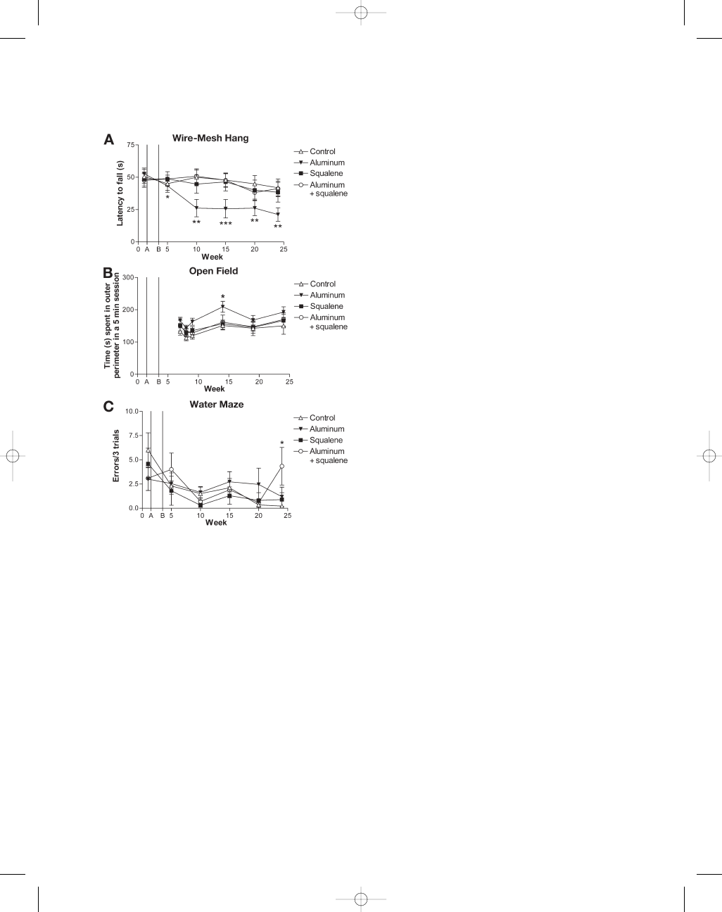
The greatest overall effects were seen in mice
injected with aluminum hydroxide.
These mice showed a progressive and significant
decrease in muscular strength and endurance (50%
at time of sacrifice) compared with the controls
(100% for all data; Fig. 1A). Squalene-injected mice
M_15 Petrik 9/22/06 10:06 PM Page 88
Aluminum Adjuvant Linked to GWI Induces Motor Neuron Death in Mice
89
NeuroMolecular Medicine
Volume 9, 2007
showed a minor decrease in muscular strength that
did not achieve significance. The aluminum hydrox-
ide and squalene (combined) group did not show
any statistically significant differences in muscle
strength and endurance.
Aluminum-injected mice showed a significant
increase in anxiety levels at week 14 (138%) as mea-
sured by the longer time spent in the outer perime-
ter during the open-field tests (Fig. 1B). After 14 wk,
the aluminum group continued to show increased
levels of anxiety compared with the controls but
these values did not reach statistical significance
(p
= 0.018 at week 24). The squalene group also
showed a small increase in anxiety after week 20
but these results did not achieve statistical signif-
icance. There was no difference in anxiety levels
between the combined group and controls.
Assessment of cognitive performance on the water
maze showed that mice injected with aluminum
hydroxide (1.2 errors) or squalene (0.9 errors) showed
an increase in the number of errors after week 20, but
these differences did not reach statistical significance.
Mice injected with both adjuvants had significant
late stage, long-term memory deficits with an increase
in the number of errors after week 20 (4.3 errors) com-
pared with the controls (0.2 errors; Fig. 1C).
CNS Pathology
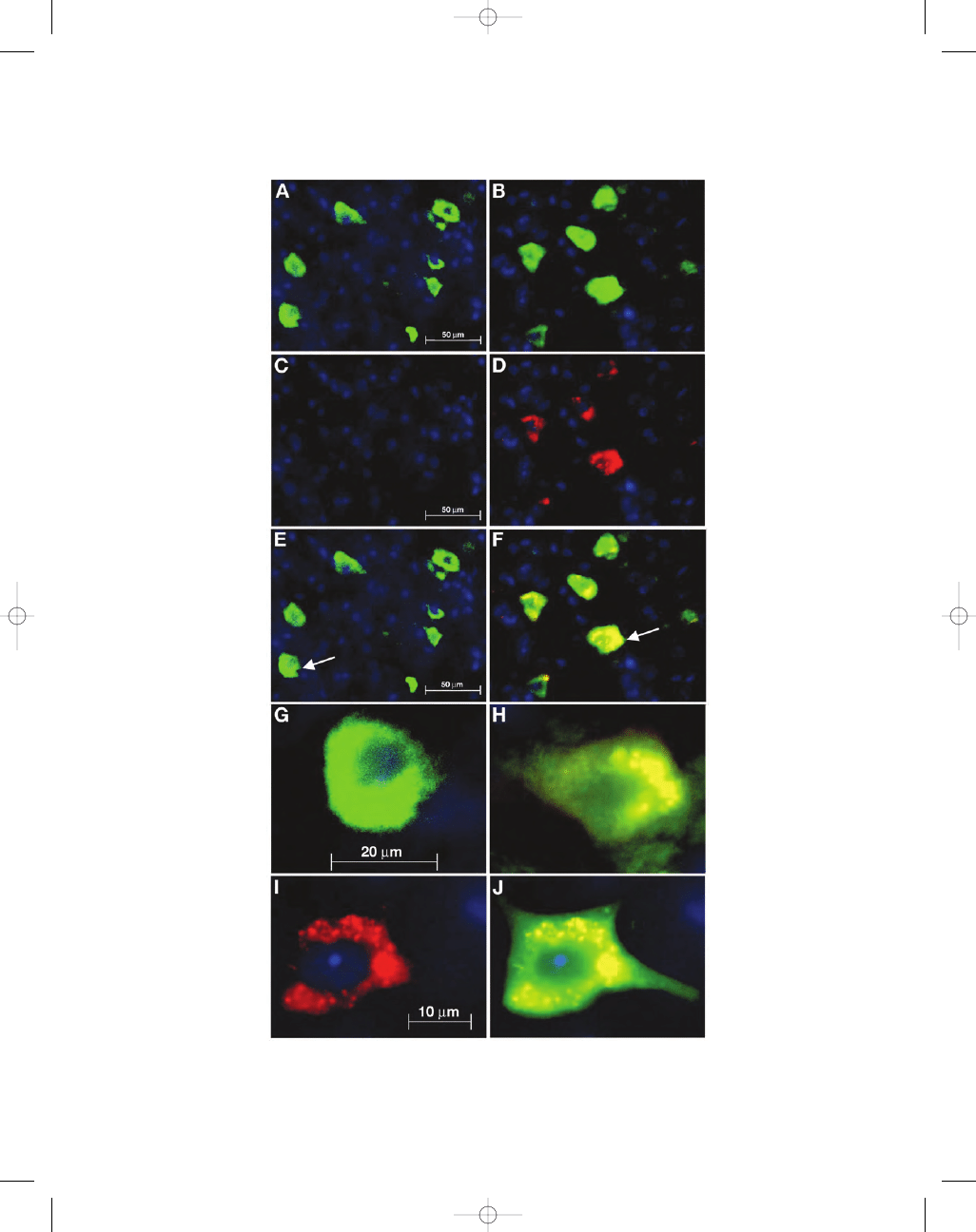
Mice injected with PBS showed little or no acti-
vated caspase-3 labeling in ventral lumbar spinal
cord (Figs. 2C,E,G and 3A). In contrast, mice injected
with aluminum hydroxide showed a significant
255% increase in activated caspase-3 labeling alone
and a significant 233% increase in double labeling
with NeuN (Figs. 2D,F,H–J and 3A). Activated cas-
pase-3 was also increased in the squalene group as
well as the combined aluminum and squalene
group, but quantified cell counts did not reach sta-
tistical significance.
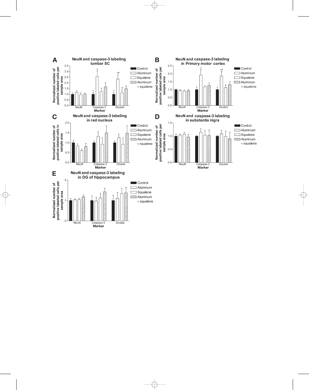
In addition to the spinal cord, other brain struc-
tures involved in motor function were also examined.
NeuN and activated caspase-3 immunohistology
was performed on the primary motor cortex, the red
nucleus, substantia nigra, and hippocampus because
these areas are affected in the human motor diseases
such as ALS and Parkinson’s disease (Sasaki et al.,
1992; Eisen and Weber, 2001; Tsuchiya et al., 2002).
Quantitative analysis of NeuN labeling showed
comparable numbers of labeled neurons in all
Fig. 1. Motor and cognitive effects of known and pre-
sumed AVA adjuvants. (A) Wire-mesh hang test. Mice
injected with aluminum hydroxide showed a significant
decrease in muscular strength and endurance (50%) com-
pared with the controls (100%). Mice injected with squa-
lene or both adjuvants did not show a significant decrease
in muscular strength. (B) Open-field tests (during weeks
7–24). Mice injected with aluminum hydroxide show a
significant increase in anxiety (138%) compared with the
controls. Mice injected with squalene or both adjuvants
did not show any significant effect. (C) The radial arm
water maze (five arms). Mice injected with aluminum
hydroxide (1.2 errors) or squalene (0.9 errors) did show
increased errors after week 20 but these values did not
reach statistical significance. Mice injected with both
adjuvants showed a significant increase in errors after
week 20 (4.3 errors), whereas, controls achieved 0.2
errors. A
= first injection, B = second injection. *p < 0.05,
**p < 0.01, ***p < 0.001; one-way ANOVA.
M_15 Petrik 9/22/06 10:06 PM Page 89
90
Petrik et al.
NeuroMolecular Medicine
Volume 9, 2007
M_15 Petrik 9/22/06 10:06 PM Page 90

treatment groups (Fig. 3A–E). Mice injected with
aluminum hydroxide showed a significant increase
in activated caspase-3 labeling (192%) and activated
caspase-3/NeuN double labeling (185%) in the
primary motor cortex compared with the controls
(Fig. 3B). The squalene and combined group showed
small increases in activated caspase-3 and activated
caspase-3/NeuN double labeling but these did not
reach statistical significance. Cell counts per-
formed in the red nucleus show increased acti-
vated caspase-3 and double labeling in both
aluminum groups, but these results were not sig-
nificant (Fig. 3C). Analysis of the substantia nigra
region did not reveal any differences in labeling
between groups (Fig. 3D). In the hippocampus, cell
counts conducted on the polymorphic layer of the
dentate gyrus showed an increase in double label-
ing for squalene and combined groups but it did not
reach statistical significance (Fig. 3E).
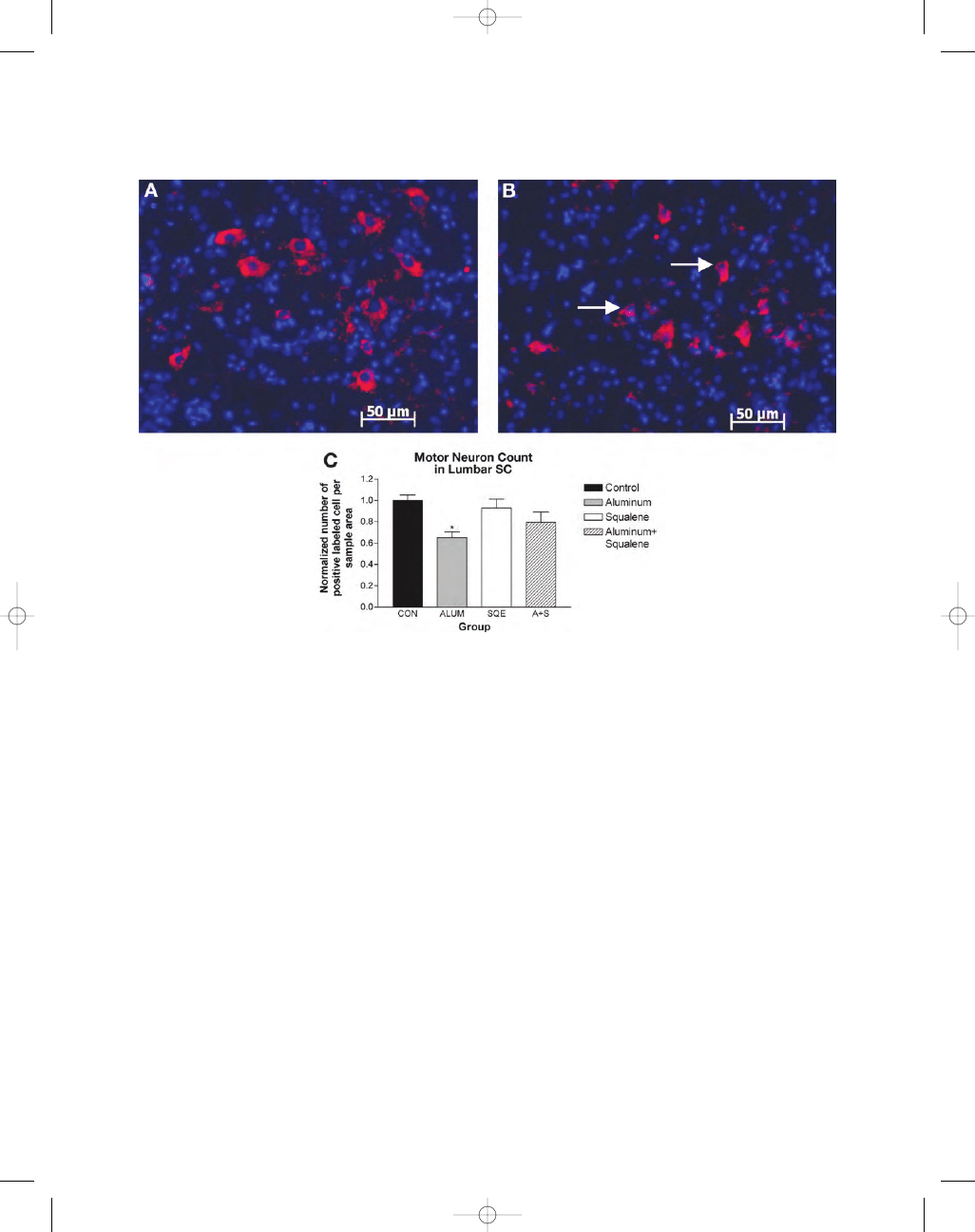
Only cells labeled with ChAT were included in
the motor neuron counts of lumbar spinal cord. Alu-
minum-injected mice showed a significant reduc-
tion in motor neurons (35%) compared with the
controls (Fig. 4A–C). The squalene and combined
group also showed a reduction in motor neuron
number that did not achieve statistical significance.
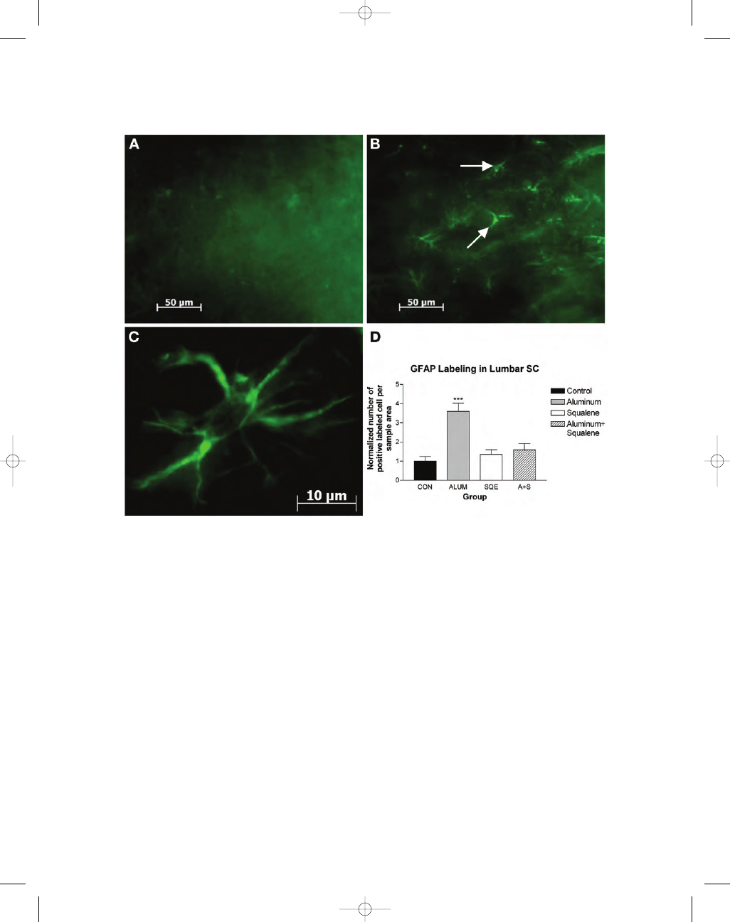
The aluminum-injected group showed a highly
significant increase in the expression of GFAP-
positive astrocytes (350%) greater than the con-
trols (Fig. 5A–D). Animals treated with squalene
or aluminum with squalene showed small
increases in the number of astrocytes present when
compared with the controls, but these differences
were not statistically significant.
Squalene-Antibodies Assay
Two out of ten control animals showed the
presence of squalene antibodies (SA) in the first
serum specimen taken at 4 wk (2 wk postsecond
injection). Alarger number of animals, 4/10, injected
with squalene possessed detectable levels of SA at
this time-point; however, this difference was not
statistically significant. Three out of the eleven ani-
mals injected with aluminum hydroxide and 1/10
injected with both adjuvants also showed increased
SA. The presence of SA was generally stable over
time in individual animals tested. However, one
animal that had been injected with both adjuvants
developed SA at a later time-point (24 wk).
Non-CNS Features
In addition to behavioral changes and CNS
pathology, various physiological changes were
observed. Hair loss at the injection site (0.5–1.0-cm
diameter region around the injections site) was
common to all adjuvant treated groups; 2/10 from
the aluminum hydroxide group, 4/10 from the
squalene group, and 3/10 mice from the combined
group. No control animals developed hair loss in
the injection area. Four of the ten mice injected with
both adjuvants developed an allergic skin reaction
(dermatitis; inflammation of the skin characterized
by itchiness and redness with scaling) showing in
a 0.5-cm diameter region around the injection site.
Discussion
Although, several animal studies using the
anthrax vaccine have been published (Ivins et al.,
1995; Fellows et al., 2001; Williamson et al., 2005),
none of these experiments examined neurological
outcomes or behavioral side-effects.
The present results indicate that anthrax vaccine
adjuvants mimicking a minimal AVA administration
regime (two injections) resulted in some neuro-
pathological outcomes postinjection (Nass, personal
communication). Aluminum hydroxide induced both
behavioral and motor deficits, and the increased pres-
ence of apoptotic neurons and in various regions of
Aluminum Adjuvant Linked to GWI Induces Motor Neuron Death in Mice
91
NeuroMolecular Medicine
Volume 9, 2007
Fig. 2. (Opposite page)
NeuN and activated caspase-3 fluorescent labeling in ventral horn of lumbar spinal
cord. Green
= NeuN; red = activated caspase-3; yellow = colocalization of NeuN and activated caspase-3; blue =
nuclear DAPI. (A,B) NeuN labeling in control and aluminum hydroxide injected mouse lumbar spinal cord sec-
tions, respectively. (C,D) Control and aluminum hydroxide mouse lumbar spinal cord sections labeled with cas-
pase-3. (E,F) Merge of NeuN and caspase. Magnification
×40 A–F. White arrow indicates neuron enlarged in (G,H).
Enlargement of neurons E,F at
×100 magnification. (I,J) Enlargement of another activated caspase-3 positive motor
neuron at
×100 magnification. J, Merged image of activated caspase-3 and NeuN. A–F; Scale bar = 50 µm. G,H;
Scale bar
= 20 µm. I,J, Scale bar = 10 µm.
M_15 Petrik 9/22/06 10:06 PM Page 91
92
Petrik et al.
NeuroMolecular Medicine
Volume 9, 2007
Fig. 3. (A) Cell counts for NeuN and activated caspase-3 labeling in ventral horn of lumbar spinal cord. NeuN
counts between groups (n
= 32, eight per group) show no significant differences indicating similar numbers of
neuronal cells labeled in all groups. Activated caspase-3 marker shows significantly increased positive capsase-3
labeling (255%) in mice injected with aluminum hydroxide compared with the controls. NeuN and activated
caspase-3 double labeling show significantly increased apoptotic neuronal cells (233%) in mice injected with
aluminum hydroxide compared with the control and squalene injected groups. (B) NeuN counts (n
= 20, five
per group) in the primary motor cortex show no significant difference between groups. Animals injected with
aluminum hydroxide show a significant increase in activated caspase-3 (192%) and double labeling (185%) in
primary motor cortex compared with the controls. Aluminum hydroxide-injected mice showed a significant
increase (165%) in double labeling when compared with the squalene-injected mice. (C) Cell counts (n
= 20,
five per group) performed in the red nucleus show a non significant increase in activated caspase-3 and double
labeling in both aluminum groups compared with the controls. (D) SNpc; there was no significant difference
in cell counts (n
= 20, five per group) of NeuN and activated caspase-3 labeling between groups in the sub-
stantia nigra region. (E) Hippocampal cell counts (n
= 20, five per group) performed on the polymorphic layer
of the dentate gyrus show increased activated caspase-3 and double labeling in the squalene group, whereas,
the combined group showed the greatest activated caspase-3 and double labeling. These results were not sta-
tistically significant. Histograms show means
± S.E.M *
, #
p < 0.05 vs control and squalene mice, **p < 0.01 vs
control mice using one-way ANOVA.
M_15 Petrik 9/22/06 10:06 PM Page 92
CNS with significant motor neuron loss in the lumbar
spinal cord. The presence of caspase-3 labeling in cells
not labeled with NeuN suggests that non-neural cells
also undergo apoptosis under these conditions.
These results are consistent with a potential role
for aluminum in motor neuron death in ALS. In
those CNS areas tested to date (spinal cord), reac-
tive astrocytes were present in significant numbers,
indicating an inflammatory response. Previous
studies have shown the increased presence of reac-
tive astrocytes in human ALS and animal models
of the disease (Nagy et al., 1994; O’Reilly et al., 1995;
Levine et al., 1999; Barbeito et al., 2004).
Aluminum Adjuvant Linked to GWI Induces Motor Neuron Death in Mice
93
NeuroMolecular Medicine
Volume 9, 2007
Fig. 4. Choline acetyltransferase (ChAT) fluorescent labeling in ventral horn of lumbar spinal cord. (A) Con-
trol section shows ChAT labeling of motor neurons (
×20 magnification). (B) Aluminum-injected animal shows
decreased ChAT labeling and abnormal morphology of motor neurons (white arrows) compared with the con-
trols (
×20 magnification). Scale bar = 50 µm. (C) Only cells positively labeled with ChAT were counted as motor
neurons (n
= 32, eight per group). Mice injected with aluminum hydroxide showed a statistically significant
decrease in motor neuron number (35%) compared with the controls. There was no significant difference in motor
neuron counts between all other groups compared with the controls. Data are means
± S.E.M ***p < 0.05 vs con-
trol mice using one-way ANOVA.
The squalene adjuvant alone produced a small
change in locomotion and anxiety testing, but the
differences in the cell counts of this group with
respect to controls were not significant in any CNS
region. The combination of both the adjuvants
showed a significant long-term memory deficit
with some indications of neuronal apoptosis in the
red nucleus and DG region of the hippocampus.
Thus, while squalene does not appear to have the
same overall impact as aluminum at sacrifice, the
change in cognitive function might suggest that
possible longer-term squalene effects should be
examined in future studies. Regarding to the SA
M_15 Petrik 9/22/06 10:06 PM Page 93
94
Petrik et al.
NeuroMolecular Medicine
Volume 9, 2007
assays, we were able to detect antibodies in 40% of
the mice injected with squalene. This outcome was
the highest incidence level of all treatment groups;
however, the other groups including the controls
showed some SA-positive mice. Previous studies
have suggested that naturally occurring antibodies
against squalene develop in mice, as well as humans,
during the aging process (Matyas et al., 2004).
BALB/c, B10.Br, and C57BL/6 mice showed SA in
approx 12% of animals, a number qualitatively sim-
ilar to the control and aluminum hydroxide injected
CD-1 mice. The relatively low incidence of SA in
squalene injected mice might reflect a transient anti-
body production. Future experiments with more
specific antibodies may resolve this issue.
Aluminum can access CNS following injections
with aluminum-adjuvanted vaccines (Wen and
Wisniewski, 1985; Redhead et al., 1992; Sahin et al.,
Fig. 5. GFAP-fluorescent labeling in ventral horn of lumbar spinal cord. (A) Control sections show little GFAP
labeling. (B) Sections from mice injected with aluminum hydroxide show increased GFAP labeling and greater
number of astrocytes (white arrows) compared with the controls (A,B
×40 magnification). Scale bar = 50 µm.
(C) Astrocyte from aluminum injected mouse observed under
×100 magnification. Scale bar = 10 µm. (D) Nor-
malized cell counts for GFAP-labeling of astrocytes in ventral horn of lumbar spinal cord (n
= 32, eight per
group). Squalene treated animals show a small increase in GFAP-labeled astrocytes when compared with the
controls. Animals treated with both aluminum hydroxide and squalene showed a larger increase in astrocyte
cell number whereas mice injected with aluminum showed the greatest increase in GFAP-labeled astrocytes
(350%). Data are means
± S.E.M ***p < 0.001 vs control mice using one-way ANOVA.
M_15 Petrik 9/22/06 10:06 PM Page 94

Aluminum Adjuvant Linked to GWI Induces Motor Neuron Death in Mice
95
NeuroMolecular Medicine
Volume 9, 2007
1994). Various studies have clearly demonstrated
that aluminum can be neurotoxic (Crapper et al.,
1973; Banks and Kastin, 1989; Joshi, 1990; Kawahara
et al., 2001). For example, aluminum-injected ani-
mals show severe anterograde degeneration of
cholinergic terminals in cortex and hippocampus
(Platt et al., 2001). Potential toxic mechanisms of
action include interference with cholinergic projec-
tions, blockage of synaptic transmission, defective
phosphorylation—dephosphorylation reactions,
altered rate of transmembrane diffusion and selec-
tive changes in saturable transport systems in the
blood–brain barrier (BBB), reduced glucose utiliza-
tion, and site-specific damage inflicted by free rad-
icals produced by altered iron metabolism.
Aluminum has also been proposed as a factor in neu-
rodegenerative diseases based on its demonstrated
neurotoxic potential and its association with degen-
erating neurons in specific CNS areas (Perl et al.,
1982; Perl and Pendlebury, 1986; Rao et al., 1998;
Savory and Garruto, 1998).
Squalene has been shown to induce antibodies
associated with lupus (Satoh et al., 2003) and to trig-
ger chronic T-cell-mediated rheumatoid arthritis
(Carlson et al., 2000). Its actions in the CNS have not
been extensively investigated, but some studies using
very high concentrations have demonstrated swelling
of astrocytic processes (Gajkowska et al., 1999).
In addition to direct toxic actions on the CNS,
aluminum, and squalene might act indirectly by
stimulation of a generalized immune response. In
fact, this is, what the adjuvants are placed in vac-
cines to do in the first place. Another possibility
is that of an imbalanced immune response. Rook
and Zumla (1997) hypothesize that multiple Th2
(T helper cell type-2)-inducing vaccinations,
stressful circumstances, and the method of vac-
cine administration (oral vs subcutaneous vs
intramuscularly) could lead to a shift from Th2 to
Th1 (T-helper cell type-1) immunity (Rook and
Zumla, 1997, 1998). Both aluminum hydroxide and
squalene have previously been shown to stimu-
late a Th2-cytokine response (Valensi et al., 1994;
Brewer et al., 1999). A recent study comparing
inbred and outbred mouse strains injected with
recombinant protective antigen (AVA) vaccine and
challenged with Bacillus anthracis, found that both
mouse strains displayed a predominantly Th2-
based immune response (Flick-Smith et al., 2005).
Such a Th1–Th2 shift could stimulate autoimmune
processes that target the neurons. Whereas a plau-
sible mechanism, a recent study of blood samples
from Gulf war veterans showed evidence for Th1
immune activation (Skowera et al., 2004).
Whereas significant behavioral and neuro-
pathological outcomes with aluminum hydroxide
and some additionally significant outcomes to the
combination of adjuvants, it is important to rec-
ognize that these were achieved under minimal
conditions was demonstrated. Table 1 shows a
summary of human ALS and GWI symptoms com-
pared with the symptoms observed in aluminum-
injected mice. The likelihood that a synergistic
effect exists between adjuvants and other vari-
ables such as stress, multiple vaccinations, and
environmental toxic exposure is another possibil-
ity that cannot be ruled out. A recent study exam-
ining some of these combinations showed that
stress, vaccination, and pyridostigmine bromide,
a carbamate anticholinesterase inhibitor, may syn-
ergistically act on multiples stress-activated
kinases in the brain to cause neurological impair-
ments in GWI (Wang et al., 2005). In addition,
genetic background might play a crucial role.
Regarding to this last point, gene–toxin interac-
tions remain a largely unexplored area in GWI and
neurological disease in general.
However, interactions of various stessors or adju-
vants does not have to be necessarily synergistic,
for example, in the present study the combination
of aluminum hydroxide and squalene seemed to
Table 1
Summary of Human ALS and GWI Symptoms
Compared With the Symptoms Observed
in Aluminum-Injected Mice
Comparison of human ALS and GWI symptomology
with symptoms observed in aluminum-injected mice
Aluminum-
Symptoms
ALS
a
GWI
b
injected mice
Muscular strength
and endurance loss
+
+
+
Enhanced anxiety
+
+
+
Memory impairment
+
+
+
Dermatitis
–
+
+
This table also outlines the similarities between
human ALS and Gulf War illness.
a
Bromberg (2002);
b
Haley et al. (1997).
M_15 Petrik 9/22/06 10:06 PM Page 95

96
Petrik et al.
NeuroMolecular Medicine
Volume 9, 2007
have less effect on motor behavior and anxiety than
either aluminum hydroxide or squalene alone. The
possibility of competing effects on immune
response cannot be over ruled and deserves fur-
ther investigation.
The current DOD immunization schedule
requires a higher number of injections (six) than
used in 1990–1991. The majority of those vaccinated
with the AVA vaccine to date have been service
personnel. As serious as this might be for the poten-
tial for adjuvant-associated complications in this
population, legislation now before US Congress
might mandate similar vaccination regimes for the
civilian population as well (e.g., the Biodefense and
Pandemic Vaccine and Drug Development Act of
2005). If a significant fraction of the military and
civilians vaccinated were to develop neurological
complications, then the impact on US society would
be profound.
In addition, the continued use of aluminum adju-
vants in various vaccines (i.e., Hepatitis A and B,
DPT, and so on) for the general public may have
even more widespread health implications. Until
vaccine safety can be comprehensively demon-
strated by controlled long-term studies that exam-
ine the impact on the nervous system in detail, many
of those already vaccinated as well as those cur-
rently receiving injections may be at risk in the
future. Whether the risk of protection from a dreaded
disease outweighs the risk of toxicity is a question
that demands urgent attention.
Animal Ethics Committee Approval
Protocols governing the use of animals were
approved by review committees of the University
of British Columbia and were in compliance with
guidelines published by the Canadian Council on
Animal Care and are in accordance with the inter-
national guidelines including the NIH Guide for the
Care and Use of Laboratory Animals, as well as the
EEC Council Directive.
Conflict of Interest Statement
None of the authors have received any grants
or funding from Bioport, Chiron, and Corixa, nor
any other pharmaceutical companies named in this
article.
Acknowledgments
This work was supported by grants from the Scot-
tish Rite Charitable Foundation of Canada and the
Natural Science and Engineering Research Council
of Canada (to CAS). We thank Dr. Jason Wilson (Uni-
versity of British Columbia, B.C., Canada), Dr. Meryl
Nass (Mount Desert Island Hospital, Maine, USA.),
Dr. Reyniel Cruz-Aguado (University of British
Columbia, B.C., Canada), and Lt. Col. John A.
Richardson (USAFR, ret.) for their invaluable com-
ments and advisory contributions to this project and
manuscript.
References
Abou-Donia M. B., Wilmarth K. R., Jensen K. F., Oehme
F. W., and Kurt T. L. (1996) Neurotoxicity resulting
from coexposure to pyridostigmine bromide, deet,
and permethrin: implications of Gulf War chemi-
cal exposures. J. Toxicol. Environ. Health 48, 35–56.
Asa P. B., Cao Y., and Garry R. F. (2000) Antibodies to
squalene in Gulf War syndrome. Exp. Mol. Pathol.
68,
55–64.
Asa P. B., Wilson R. B., and Garry R. F. (2002) Anti-
bodies to squalene in recipients of anthrax vac-
cine. Exp. Mol. Pathol. 73, 19–27.
Banks W. A. and Kastin A. J. (1989) Aluminum-induced
neurotoxicity: alterations in membrane function
at the blood–brain barrier. Neurosci. Biobehav. Rev.
13,
47–53.
Barbeito L. H., Pehar M., Cassina P., et al. (2004) A
role for astrocytes in motor neuron loss in amy-
otrophic lateral sclerosis. Brain Res. Brain Res. Rev.
47,
263–274.
Baylor N. W., Egan W., and Richman P. (2002) Alu-
minum salts in vaccines—US perspective. Vaccine
20(Suppl 3),
S18–S23.
Benisek Z., Suli J., Elias D., et al. (2004) Experimental
squalene adjuvant. II. Harmlessness and local reac-
togenity. Vaccine 22, 3470–3474.
Bilkei-Gorzo A. (1993) Neurotoxic effect of enteral
aluminium. Food Chem. Toxicol. 31, 357–361.
Brewer J. M., Conacher M., Hunter C. A., Mohrs M.,
Brombacher F., and Alexander J. (1999) Aluminium
hydroxide adjuvant initiates strong antigen-specific
Th2 responses in the absence of IL-4- or IL-13-medi-
ated signaling. J. Immunol. 163, 6448–6454.
Bromberg M. B. (2002) Diagnostic criteria and outcome
measurement of amyotrophic lateral sclerosis.
Adv. Neurol. 88, 53–62.
M_15 Petrik 9/22/06 10:06 PM Page 96

Aluminum Adjuvant Linked to GWI Induces Motor Neuron Death in Mice
97
NeuroMolecular Medicine
Volume 9, 2007
Carlson B. C., Jansson A. M., Larsson A., Bucht A., and
Lorentzen J. C. (2000) The endogenous adjuvant
squalene can induce a chronic T-cell-mediated
arthritis in rats. Am. J. Pathol. 156, 2057–2065.
Charatan F. (2002) US links motor neurone disease
with Gulf war service. BMJ 324, 65.
Crapper D. R., Krishnan S. S., and Dalton A. J. (1973)
Brain aluminum distribution in Alzheimer’s dis-
ease and experimental neurofibrillary degenera-
tion. Science 180, 511–513.
Crawley J. N. (2000) What’s Wrong With My Mouse?
Behavioral Phenotyping of Trangenic and Knock-
out Mice. 65–69.
Crawley J. N., Belknap J. K., Collins A., et al. (1997)
Behavioral phenotypes of inbred mouse strains:
implications and recommendations for molecular
studies. Psychopharmacology (Berl.) 132, 107–124.
DeFries, J. C., Hegmann, J. P., and Halcomb, R. A.
(1974) Response to 20 generations of selection for
open-field activity in mice. Behav. Biol. 11, 481–495.
Duan W. R., Garner D. S., Williams S. D., Funckes-
Shippy C. L., Spath I. S., and Blomme E. A. (2003)
Comparison of immunohistochemistry for acti-
vated caspase-3 and cleaved cytokeratin 18 with
the TUNEL method for quantification of apopto-
sis in histological sections of PC-3 subcutaneous
xenografts. J. Pathol. 199, 221–228.
Dyer O. (2004) Inquiry finds that Gulf war veterans
face extra burden of disease. BMJ 329, 1257.
Eisen A. and Weber M. (2001) The motor cortex and
amyotrophic lateral sclerosis. Muscle Nerve
24,
564–573.
Everts H. G. and Koolhaas J. M. (1999) Differential
modulation of lateral septal vasopressin receptor
blockade in spatial learning, social recognition,
and anxiety-related behaviors in rats. Behav. Brain
Res. 99, 7–16.
Fellows P. F., Linscott M. K., Ivins B. E., et al. (2001)
Efficacy of a human anthrax vaccine in guinea pigs,
rabbits, and rhesus macaques against challenge
by Bacillus anthracis isolates of diverse geograph-
ical origin. Vaccine 19, 3241–3247.
Ferguson E. and Cassaday H. J. (2001 and 2002) The-
oretical accounts of Gulf War Syndrome: from
environmental toxins to psychoneuroimmunol-
ogy and neurodegeneration. Behav. Neurol.
13,
133–147.
Flick-Smith H. C., Waters E. L., Walker N. J., et al.
(2005) Mouse model characterisation for anthrax
vaccine development: comparison of one inbred
and one outbred mouse strain. Microb. Pathog.
38,
33–40.
Fukuda K., Nisenbaum R., Stewart G., et al. (1998)
Chronic multisymptom illness affecting Air Force
veterans of the Gulf War. JAMA 280, 981–988.
Fulco C. E. , Liverman C. T., and Sox H. C. (2000) Gulf
War and Health: Volume 1. Depleted Uranium, Pyri-
dostigmine, Bromide, Sarin, and Vaccines. Institute
of Medicine. National Academy Press, pp. 89–168.
Gabutti G., Guido M., Durando P., et al. (2005) Safety
and immunogenicity of conventional subunit and
MF59-adjuvanted influenza vaccines in human
immunodeficiency virus-1-seropositive patients.
J. Int. Med. Res. 33, 406–416.
Gajkowska B., Smialek M., Ostrowski R. P., Piotrowski
P., and Frontczak-Baniewicz M. (1999) The exper-
imental squalene encephaloneuropathy in the rat.
Exp. Toxicol. Pathol. 51, 75–80.
Garruto R. M., Shankar S. K., Yanagihara R., Salazar
A. M., Amyx H. L., and Gajdusek D. C. (1989)
Low-calcium, high-aluminum diet-induced motor
neuron pathology in cynomolgus monkeys. Acta
Neuropathol. (Berl.) 78, 210–219.
Haley R. W. (2003) Excess incidence of ALS in young
Gulf War veterans. Neurology 61, 750–756.
Haley R. W., Kurt T. L., and Hom J. (1997) Is there a
Gulf War Syndrome? Searching for syndromes by
factor analysis of symptoms. JAMA. 277, 215–222.
Hom J., Haley R. W., and Kurt T. L. (1997) Neuropsy-
chological correlates of Gulf War syndrome. Arch
Clin. Neuropsychol. 12, 531–544.
Horner R. D., Kamins K. G., Feussner J. R., et al. (2003)
Occurrence of amyotrophic lateral sclerosis among
Gulf War veterans. Neurology 61, 742–749.
Hotopf M., David A., Hull L., Ismail K., Unwin C., and
Wessely S. (2000) Role of vaccinations as risk
factors for ill health in veterans of the Gulf war:
cross sectional study. BMJ 320, 1363–1367.
Hunter D., Zoutman D., Whitehead J., Hutchings J.,
and MacDonald K. (2004) Health effects of anthrax
vaccination in the Canadian forces. Mil Med. 169,
833–838.
Ivins B., Fellows P., Pitt L., et al. (1995) Experimental
anthrax vaccines: efficacy of adjuvants combined
with protective antigen against an aerosol Bacillus
anthracis spore challenge in guinea pigs. Vaccine
13,
1779–1784.
Jefferson T., Rudin M., and Di Pietrantonj C. (2004)
Adverse events after immunisation with alu-
minium-containing DTP vaccines: systematic
review of the evidence. Lancet Infect Dis. 4, 84–90.
Joshi J. G. (1990) Aluminum, a neurotoxin which
affects diverse metabolic reactions. Biofactors
2,
163–169.
M_15 Petrik 9/22/06 10:06 PM Page 97

98
Petrik et al.
NeuroMolecular Medicine
Volume 9, 2007
Kalra R., Singh S. P., Razani-Boroujerdi S., et al. (2002)
Subclinical doses of the nerve gas sarin impair T
cell responses through the autonomic nervous
system. Toxicol. Appl. Pharmacol. 184, 82–87.
Kang H. K., Mahan C. M., Lee K. Y., et al. (2002) Evi-
dence for a deployment-related Gulf War syndrome
by factor analysis. Arch Environ. Health 57, 61–68.
Kanra G., Viviani S., Yurdakok K., et al. (2003) Effect
of aluminum adjuvants on safety and immuno-
genicity of Haemophilus influenzae type b-CRM197
conjugate vaccine. Pediatr. Int. 45, 314–318.
Kawahara M., Kato M., and Kuroda Y. (2001) Effects
of aluminum on the neurotoxicity of primary cul-
tured neurons and on the aggregation of beta-
amyloid protein. Brain Res. Bull. 55, 211–217.
Kurland L. T. (1988) Amyotrophic lateral sclerosis and
Parkinson’s disease complex on Guam linked to an
environmental neurotoxin. Trends Neurosci. 11,51–54.
Kurt T. L. (1998) Epidemiological association in US
veterans between Gulf War illness and exposures
to anticholinesterases. Toxicol. Lett. 102–103,
523–536.
Lee V. M., Page C. D., Wu H. L., and Schlaepfer W. W.
(1984) Monoclonal antibodies to gel-excised glial fil-
ament protein and their reactivities with other inter-
mediate filament proteins. J. Neurochem. 42, 25–32.
Levine J. B., Kong J., Nadler M., and Xu Z. (1999) Astro-
cytes interact intimately with degenerating motor
neurons in mouse amyotrophic lateral sclerosis
(ALS). Glia 28, 215–224.
Maatkamp A., Vlug A., Haasdijk E., Troost D., French
P. J., and Jaarsma D. (2004) Decrease of Hsp25 pro-
tein expression precedes degeneration of
motoneurons in ALS-SOD1 mice. Eur. J. Neurosci.
20,
14–28.
Matyas G. R., Rao M., Pittman P. R., et al. (2004) Detec-
tion of antibodies to squalene: III. Naturally occur-
ring antibodies to squalene in humans and mice.
J. Immunol. Methods 286, 47–67.
Morris R. (1984) Developments of a water-maze pro-
cedure for studying spatial learning in the rat.
J. Neurosci. Methods 11, 47–60.
Mullen R. J., Buck C. R., and Smith A. M. (1992) NeuN,
a neuronal specific nuclear protein in vertebrates.
Development 116, 201–211.
Murakami N. (1999) Parkinsonism-dementia complex
on Guam—overview of clinical aspects. J. Neurol.
246(Suppl. 2),
II16–II18.
Nagy D., Kato T., and Kushner P. D. (1994) Reactive
astrocytes are widespread in the cortical gray
matter of amyotrophic lateral sclerosis. J. Neurosci.
Res. 38, 336–347.
Nass M. (1999) Anthrax vaccine. Model of a response
to the biologic warfare threat. Infect Dis. Clin. North
Am. 13, VIII187–VIII208.
Nass M. (2002) The Anthrax Vaccine Program: an analy-
sis of the CDC’s recommendations for vaccine use.
Am. J. Public Health 92, 715–721.
Nass M., Fisher B. L., and Robinson S. (2005) Com-
ments and Questions regarding FDA’s proposed
rule and order to licnese Anthrax Vaccine
Absorbed. FDA Anthrax vaccine docket submission.
Proposed rule and proposed order, 29 Fed. Reg.
78,281–78,293. December 29, 2004.
Nicolson G. L., Nasralla M. Y., Haier J., and Pomfret J.
(2002) High frequency of systemic mycoplasmal
infections in Gulf War veterans and civilians with
Amyotrophic Lateral Sclerosis (ALS). J. Clin.
Neurosci. 9, 525–529.
O’Reilly S. A., Roedica J., Nagy D., et al. (1995) Motor
neuron-astrocyte interactions and levels of Cu,
Zn superoxide dismutase in sporadic amy-
otrophic lateral sclerosis. Exp. Neurol. 131,
203–210.
Paxinos G. and Franklin K. B. J. (2001) The Mouse Brain
in Stereotoxic Coordinates 2nd ed. Academic Press.
Sydney.
Perl D. P., Gajdusek D. C., Garruto R. M., Yanagihara
R. T., and Gibbs C. J. (1982) Intraneuronal alu-
minum accumulation in amyotrophic lateral scle-
rosis and Parkinsonism-dementia of Guam.
Science. 217, 1053–1055.
Perl D. P. and Pendlebury W. W. (1986) Aluminum neu-
rotoxicity—potential role in the pathogenesis of
neurofibrillary tangle formation. Can. J. Neurol.
Sci. 13, 441–445.
Plaisier M. (2000) Letter dated March 20, 2000 from
Department of Health and Human Services to
former US member of Congress, Rep. Jack Met-
calf, admitting to squalene in anthrax vaccine
while denying that it was in the licencsed
formulation.
Platt B., Fiddler G., Riedel G., and Henderson Z. (2001)
Aluminium toxicity in the rat brain: histochemi-
cal and immunocytochemical evidence. Brain Res.
Bull. 55, 257–267.
Rao J. K., Katsetos C. D., Herman M. M., and Savory J.
(1998) Experimental aluminum ecephalomyelopa-
thy. Relationship to human neurodegenerative
disease. Clin. Lab. Med. 18, VIII687–VIII698.
M_15 Petrik 9/22/06 10:06 PM Page 98

Aluminum Adjuvant Linked to GWI Induces Motor Neuron Death in Mice
99
NeuroMolecular Medicine
Volume 9, 2007
Redhead K., Quinlan G. J., Das R. G., and Gutteridge
J. M. (1992) Aluminium-adjuvanted vaccines tran-
siently increase aluminium levels in murine brain
tissue. Pharmacol. Toxicol. 70, 278–280.
Rook G. A. and Zumla A. (1997) Gulf War syndrome:
is it due to a systemic shift in cytokine balance
towards a Th2 profile? Lancet 349, 1831–1833.
Rook G. A. and Zumla A. (1998) Is the Gulf War syn-
drome an immunologically mediated phenome-
non? Hosp. Med. 59, 10–11.
Sahin G., Varol I., Temizer A., Benli K., Demirdamar
R., and Duru S. (1994) Determination of aluminum
levels in the kidney, liver, and brain of mice treated
with aluminum hydroxide. Biol. Trace Elem. Res.
41,
129–135.
Salamon R., Verret C., Jutand M. A., et al. (2006) Health
consequences of the first Persian Gulf War on
French troops. Int J Epidemiol. 35, 479–487.
Samson K. (2002) VA study finds ALS spike in Gulf
War vets. Neurol. Today 2(1), 13–14.
Sartin J. S. (2000) Gulf War illnesses: causes and con-
troversies. Mayo Clin. Proc. 75, 811–819.
Sasaki S., Tsutsumi Y., Yamane K., Sakuma H., and
Maruyama S. (1992) Sporadic amyotrophic lateral
sclerosis with extensive neurological involvement.
Acta Neuropathol. (Berl.) 84, 211–215.
Satoh M., Kuroda Y., Yoshida H., et al. (2003) Induction
of lupus autoantibodies by adjuvants. J. Autoim-
mun. 21, 1–9.
Savory J. and Garruto R. M. (1998) Aluminum, tau pro-
tein, and Alzheimer’s disease: an important link?
Nutrition 14, 313–314.
Schumm W. R., Webb F. J., Jurich A. P., and Bollman S. R.
(2002a) Comments on the Institute of Medicine’s
2002 report on the safety of anthrax vaccine. Psychol.
Rep. 91, 187–191.
Schumm W. R., Reppert E. J., Jurich A. P., et al. (2002b)
Self-reported changes in subjective health and
anthrax vaccination as reported by over 900 Persian
Gulf War era veterans. Psychol. Rep. 90, 639–653.
Schumm W. R., Jurich A. P., Bollman S. R., Webb F. J.,
and Castelo C. S. (2005) The long term safety of
anthrax vaccine, pyridostigmine bromide (PB)
tablets, and other risk factors among Reserve
Component Veterans of the First Persian Gulf War.
Medical Veritas 2, 348–362.
Shaw C. A. and Wilson J. M. (2003) Analysis of neuro-
logical disease in four dimensions: insight from
ALS-PDC epidemiology and animal models.
Neurosci. Biobehav. Rev. 27, 493–505.
Shawky S. (2002) Depleted uranium: an overview of
its properties and health effects. East Mediterr.
Health J. 8, 432–439.
Sidman R. L., Angevine J. B. Jr., and Pierce E. T. (1971)
Atlas of the Mouse Brain and Spinal Cord.
Skowera A., Hotopf M., Sawicka E., et al. (2004) Cel-
lular immune activation in Gulf War veterans.
J. Clin. Immunol. 24, 66–73.
Steele L. (2000) Prevalence and patterns of Gulf War
illness in Kansas veterans: association of symp-
toms with characteristics of person, place, and
time of military service. Am. J. Epidemiol. 152,
992–1002.
Suli J., Benisek Z., Elias D., et al. (2004) Experimental
squalene adjuvant. I. Preparation and testing of
its effectiveness. Vaccine 22, 3464–3469.
Taylor D. N., Sanchez J. L., Smoak B. L., and DeFraites R.
(1997) Helicobacter pylori infection in Desert
Storm troops. Clin. Infect Dis. 25, 979–982.
Tohyama T., Lee V. M., Rorke L. B., and Trojanowski J.
Q. (1991) Molecular milestones that signal axonal
maturation and the commitment of human spinal
cord precursor cells to the neuronal or glial
phenotype in development. J. Comp. Neurol.
310,
285–299.
Tsuchiya K., Takahashi M., Shiotsu H., et al. (2002)
Sporadic amyotrophic lateral sclerosis with cir-
cumscribed temporal atrophy: a report of an
autopsy case without dementia and with ubiqui-
tinated intraneuronal inclusions. Neuropathology
22,
308–316.
Unwin C., Blatchley N., Coker W., et al. (1999) Health
of UK servicemen who served in Persian Gulf War.
Lancet 353, 169–178.
Valensi J. P., Carlson J. R., and Van Nest G. A. (1994)
Systemic cytokine profiles in BALB/c mice immu-
nized with trivalent influenza vaccine containing
MF59 oil emulsion and other advanced adjuvants.
J. Immunol. 153, 4029–4039.
Wagner-Recio M., Toews A. D., and Morell P. (1991)
Tellurium blocks cholesterol synthesis by inhibit-
ing squalene metabolism: preferential vulnera-
bility to this metabolic block leads to peripheral
nervous system demyelination. J. Neurochem.
57,
1891–1901.
Wang D., Perides G., and Liu Y. F. (2005) Vaccination
alone or in combination with pyridostigmine pro-
motes and prolongs activation of stress-activated
kinases induced by stress in the mouse brain. J.
Neurochem. 93, 1010–1020.
M_15 Petrik 9/22/06 10:06 PM Page 99

100
Petrik et al.
NeuroMolecular Medicine
Volume 9, 2007
Weisskopf M. G., O’Reilly E. J., McCullough M. L.,
et al. (2005) Prospective study of military service
and mortality from ALS. Neurology 64, 32–37.
Wen G. Y. and Wisniewski H. M. (1985) Histochemical
localization of aluminum in the rabbit CNS. Acta
Neuropathol (Berl.) 68, 175–184.
Wetts R. and Vaughn J. E. (1996) Differential vulnera-
bility of two subsets of spinal motor neurons in amy-
otrophic lateral sclerosis. Exp. Neurol. 141, 248–255.
Williamson E. D., Hodgson I., Walker N. J., et al.
(2005) Immunogenicity of recombinant protec-
tive antigen and efficacy against aerosol
challenge with anthrax. Infect Immun. 73,
5978–5987.
Wolf H. K., Buslei R., Schmidt-Kastner R., et al. (1996)
NeuN: a useful neuronal marker for diagnostic
histopathology. J. Histochem. Cytochem. 44,
1167–1171.
Wolfe J., Proctor S. P., Erickson D. J., and Hu H. (2002)
Risk factors for multisymptom illness in US Army
veterans of the Gulf War. J. Occup. Environ. Med.
44,
271–281.
M_15 Petrik 9/22/06 10:06 PM Page 100
Wyszukiwarka
Podobne podstrony:
Wojny w Zatoce Perskiej, Stosunki międzynarodowe
Analysis of the Persian Gulf War
GAO squalene Gulf War Illneasses
Mutacja związana z chorobą nerek
Telomery związane z chorobą zwyrodnieniową stawów, ortop, Ortopedia
Ways of War (koncepcje wojny)
Wpływ terapii z zastosowaniem okładów borowinowych na dolegliwości związane z chorobą zwyrdnieniową
Faces of War Oblicza Wojny poradnik do gry
sla i inne choroby neuronu ruchowego-konspekt wykladu, Lekarski, Neurologia
wojny grecko-perskie, notatki
Jabłońska, Wojny greckie-perskie, Wojny greckie-perskie
Wojny grecko perskie
Jabłońska, Wojny greckie-perskie II, Wojny greckie-perskie
WOJNY GRECKO PERSKIE
11 IB Wojny grecko perskie
Wojny grecko perskie
Lekcja 12 Wojny Grecko perskie
więcej podobnych podstron