CHAPTER 18
Vascular Tumours
Primary vascular tumours of bone are rare. Haemangiomas
occur as incidental findings in the skull or in the spine. The
roentgenographic features are almost always diagnostic. They
rarely cause clinical symptoms.
The terminology for malignant vascular tumours has been
controversial. Angiosarcoma is the most acceptable term for
malignant vascular tumours. They have a peculiar tendency to
involve multiple bones. Histological grading correlates well with
prognosis.
Epithelioid haemangioendothelioma is a distinct entity with
histological features identical to those of the soft tissue counter-
part and is associated with an favourable clinical course.
bb5_26.qxd 13.9.2006 13:49 Page 319
Haemangioma and related lesions
C.P. Adler
L. Wold
Definition
A benign vasoformative neoplasm or
developmental condition of endothelial
origin.
ICD-O code
9120/0
Synonyms
Capillary haemangioma, cavernous hae-
mangioma, venous haemangioma,
angioma, histocytoid haemangioma,
angiomatosis.
Epidemiology
Haemangiomas are relatively common
lesions; autopsy studies have identified
them in the vertebrae of approximately
10% of the adult population {18}.
However, clinically significant sympto-
matic tumours are very uncommon and
account for less than 1% of primary bone
tumours {539}. Haemangiomas occur at
any age, but most are diagnosed during
middle and late middle age with the
peak incidence in the 5th decade of life
{1875}. The male to female ratio is about
2:3 {18,539,1875,2153,2249}.
Sites of involvement
Vertebral bodies are the most common
site, followed by the craniofacial skeleton,
and then the long bones where they tend
to involve the metaphyses {18,539,2249}.
Clinical features / Imaging
The majority of haemangiomas, espe-
cially those arising in the spine, are inci-
dental radiographic findings. However,
large vertebral tumours may cause cord
compression, pain and neurological
symptoms. Symptomatic tumours occur-
ing elsewhere are painful and may cause
a pathologic fracture. Haemangiomas
present as a well demarcated lucent
mass that frequently contains coarse tra-
beculations or striations. In flat bones
like the calvarium, the tumour is expan-
sile and lytic and produces a sunburst
pattern of reactive bone formation.
Clinically, indolent lesions frequently
contain fat and sclerotic trabeculae on
CT and MRI. Symptomatic tumours usu-
ally show loss of fat and reveal a low sig-
nal on T1-weighted images and a high
signal on T2 {539,644,1280,1354,1875,
2287}.
Macroscopy
Haemangioma manifests as a soft well
demarcated dark red mass. It may also
have a honey-comb appearance with
intralesional sclerotic bone trabeculae
and scattered blood-filled cavities.
Histopathology
Haemangiomas have variable histologi-
cal features. Capillary and cavernous
haemangiomas are composed of thin-
walled blood-filled vessels lined by a sin-
gle layer of flat, cytologically banal
endothelial cells. The vessels permeate
the marrow and surround preexisting tra-
beculae. When capillary or cavernous
haemangiomas involve a large localized
B
A
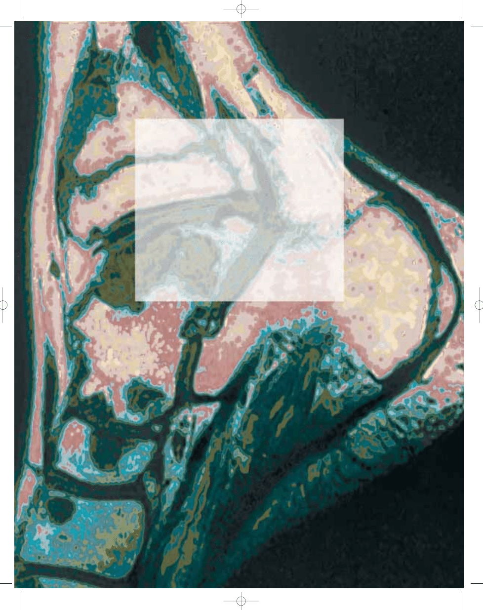
Fig. 18.01 Haemangioma of bone. A Plain radiographs show a lesion with multiple cystic defects within the
distal tibia. B CT cross-sectional appearance of a vertebral haemangioma where the coarse trabeculae
result in a “polka-dot” pattern.
Haemangioma:
cavernous,
capillary,
epithelioid,
histiocytoid,
sclerosing
Papillary vegetant endothelial proliferation
(Masson type)
Angiolymphoid hyperplasia with eosinophilia
(Kimura disease)
Angiomatosis:
non-aggressive: regional,
disseminated: cystic angiomatosis
aggressive: massive osteolysis
(Gorham-Stout syndrome)
Osseous glomus tumour (glomangioma)
Lymphangioma
Lymphangiomatosis
Table 18.01
Variants of haemangiomas.
320
Vascular tumours
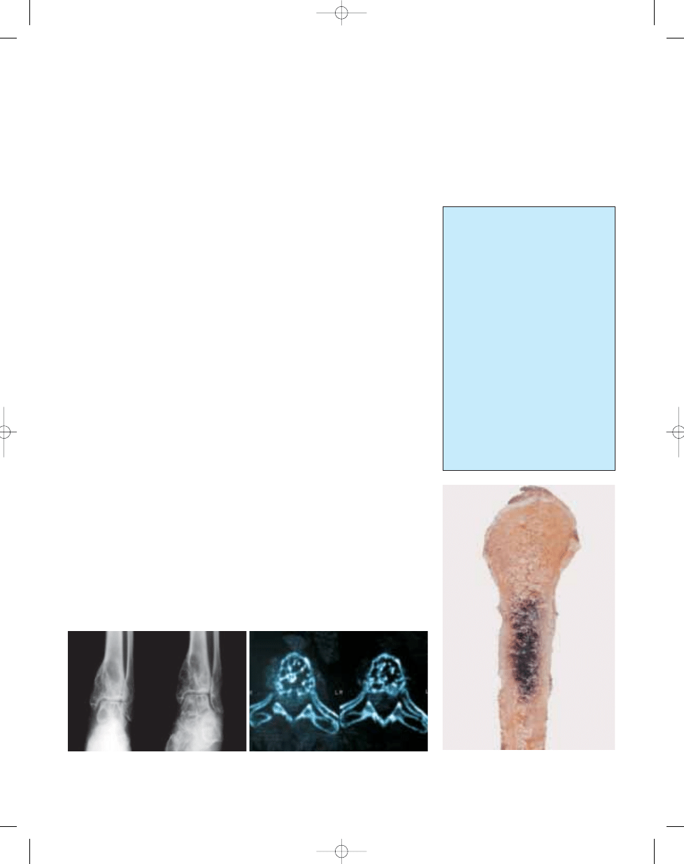
Fig. 18.02 Haemangioma of bone. Gross specimen of
a tumour of the proximal fibula with a focus of
brown-red appearance without marginal sclerosis.
bb5_26.qxd 13.9.2006 13:49 Page 320
321
Haemangioma and related lesions
region or are widespread throughout the
skeleton, it is known as angiomatosis.
Gorham disease may be associated with
a histological picture that resembles hae-
mangioma. Epithelioid haemangioma is
composed of large polyhedral neoplastic
endothelial cells that have vesicular
nuclei and abundant eosinophilic cyto-
plasm. Some tumour cells have round
clear cytoplasmic vacuoles that may
contain intact or fragments of red blood
cells. Vacuoles in neighbouring cells
often fuse forming vascular lumena. The
epithelioid cells may line well formed
vascular spaces or grow in solid cords or
sheets. The stroma consists of loose con-
nective tissue and may contain a mixed
inflammatory infiltrate including
eosinophils.
The vessels in lymphangioma are dilat-
ed, sinusoidal, filled with lymph fluid and
lined by a single layer of flat, banal
endothelial cells. The surrounding stroma
may contain lymphocytes.
Immunophenotype
The endothelial cells uniformly express
vimentin and many cells stain with anti-
bodies to F. VIII, CD31, and CD34.
Epithelial haemangiomas may also
express keratins and EMA. FLI1 has also
been observed in haemangiomas.
Ultrastructure
The endothelial cells contains Weibel-
Palade bodies. Cytoplasmic filaments are
abundant in epithelioid endothelial cells.
Prognostic factors
Haemangiomas have an excellent prog-
nosis and have a low rate of local recur-
rence. Progression to an angiosarcoma
is an extraordinarily rare event {528,611,
641,649,1628}.
Fig. 18.03 Haemangioma of bone. The radiated spicules
are demonstrated on this macerated specimen.
B
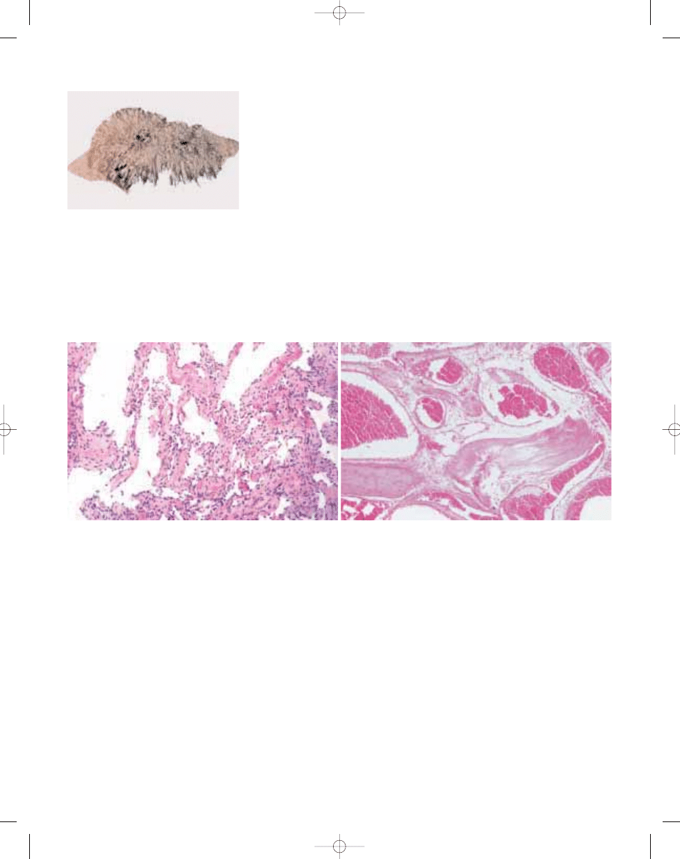
A
Fig. 18.04 Haemangioma of bone. A This bony haemangioma shows the morphology commonly associated with cavernous lesions which have been curetted. The
spaces often become collapsed, and blood is no longer present because of the processing.
B Histological pattern of a cavernous haemangioma showing broad thin-
walled blood vessels, lined by a single layer of flat endothelial cells and filled with blood, within the medullary cavity between the bone trabeculas.
bb5_26.qxd 13.9.2006 13:49 Page 321
Definition
Angiosarcomas of bone are composed
of tumour cells which show endothelial
differentiation.
ICD-O code
9120/3
Synonyms
Haemangiosarcoma, haemangioen-
dothelioma, haemangioendothelial sar-
coma, epithelioid angiosarcoma.
Epidemiology
Malignant vascular tumours of bone are
very rare and account for less than 1% of
malignant bone tumours. Age distribu-
tion shows a wide range with nearly
equal distribution from the second to the
eighth decade. Epithelioid haeman-
gioendothelioma tends to occur during
the second and third decades of life.
Males and females are affected approxi-
mately equally.
Sites of involvement
Malignant vascular tumours of bone
show a wide skeletal distribution. They
tend to affect the long tubular bones of
the extremity and the axial skeleton,
mainly the spine. These tumours reveal
the tendency to develop multicentric
lesions in bone. About a third of these
lesions are multifocal.
Clinical features / Imaging
Malignant vascular tumours most com-
monly present as painful lesions which
may be associated with a mass.
Angiosarcoma usually develops purely
lytic bone lesions. They are poorly mar-
ginated but can occassionally have a
sclerotic rim. A soft tissue mass is often
associated with less well differentiated
tumours. The radiological appearence of
epithelioid hemangioendothelioma is
also non-specific. They also present as
purely lytic lesions with varying degrees
of peripheral sclerosis. Although the
radiographic feature of malignant vascu-
lar tumours of bone are nonspecific,
clustering of multifocal lesions in a single
anatomic location suggests the diagno-
sis of a vascular neoplasm.
Aetiology
Angiosarcomas may arise at sites of
prior radiation {338,452,1716}. The aeti-
ology of the majority of malignant vascu-
lar tumours is unknown.
Macroscopy
Angiosarcomas are bloody and general-
ly firm in their consistency. Necrosis is
generally not observed. Epithelioid hae-
mangioendotheliomas tend to be firm
and tan-white. Both tumours can erode
the cortex and extend into the soft tis-
sue.
Histopathology
Tumour cells forming vascular spaces
constitute the general histological fea-
ture of angiosarcoma of bone.
Angiosarcoma of bone shows a wide
range of histology, ranging from well dif-
ferentiated cases mimicking haeman-
gioma to poorly differentiated tumours
which may be difficult to identify as a
vascular tumour. Histo-logically, reactive
bone formation can sometimes be
observed in angiosarcoma of bone. This
is more pronounced in the periphery, but
can also be found in the more central
portions of the lesion.
Poorly differentiated angiosarcomas are
composed of more atypical endothelial
cells. They exhibit very prominent nu clear
atypia and a considerably increased
number of mitoses with atypical mitotic
figures. Formation of intraluminal buds
can often be observed. Areas with
necroses may be present. Some tumours
may show epithelioid cytological features
and mimic the appearance of metastatic
carcinoma. Others show spindle cell cyto-
logical features and mimic other primary
bone sarcomas.
Epithelioid haemangioendothelioma is
composed of anastomosing cords, solid
nests, and strands of endothelial cells
that may sometimes form narrow vascu-
lar channels. The small capillary-sized
tumour vessels can mimic small reactive
vessels of granulation tissue. The epithe-
lioid cells tend to have eosinophilic cyto-
plasm which may show vacuolization
and sometimes signet ring-like appear-
ance. Of remarkable significance is the
A. Roessner
T. Boehling
Angiosarcoma
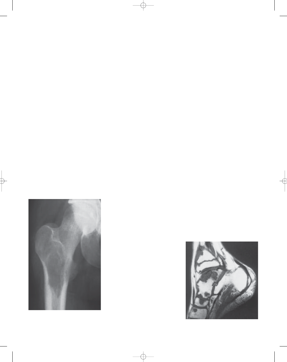
Fig. 18.05 Angiosarcoma. Plain X-ray of a tumour
involving the proximal femur, featuring a purely
lucent destructive process in the intertrochanteric
region. The radiological appearance is nonspecific.
Fig. 18.06 Angiosarcoma. T2 MRI of a multicentric
tumour involving multiple bones of the foot.
322
Vascular tumours
bb5_26.qxd 13.9.2006 13:49 Page 322
323
Angiosarcoma
myxoid and hyalinized appearance of
the connective tissues stroma. The
nuclei of the neoplastic cell show vary-
ing degrees of pleomorphism and
anaplasia.
Immunophenotype
The endothelial cells uniformly express
vimentin and many cells stain with anti-
bodies to Factor VIII, CD31, CD34, and
Ulex Europaeus. Epithelioid malignan-
cies may also express cytokeratins and
EMA {1134,2249}.
Ultrastructure
The endothelial cells contain Weibel-
Palade bodies, but are generally difficult
to find in poorly differentiated tumours.
Cytoplasmic filaments are abundant in
epithelioid neoplasms.
Genetics
Two epithelioid haemangioendothe-
liomas have shown an identical chromo-
somal translocation involving chromo-
somes 1 and 3 {1403}.
Prognostic factors
The histological degree of differentiation
is the most significant factor in the prog-
nosis of patients with malignant vascular
tumours of bone {300,2288}. Some stud-
ies have also suggested that multifocal
tumours show a survival advantage. This
survival advantage may in part be relat-
ed to the multifocal tumours showing
better differentation {1134,2142}.
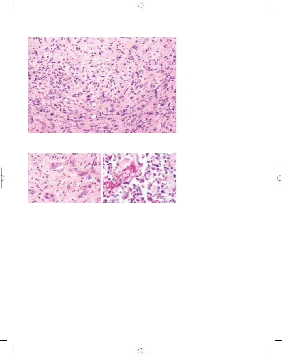
Fig. 18.07 Epithelioid haemangioendothelioma. The tumour cells are arranged in a cording fashion in a myxoid
stroma. Note the occasional cytoplasmic vacuoles.
B
A
Fig. 18.08 A Epithelioid angiosarcoma. The tumour cells form anastomosing channels, have large nuclei and
prominent nucleoli. B High grade angiosarcoma showing atypical cells with poorly formed papillae present
within spaces.
bb5_26.qxd 13.9.2006 13:49 Page 323
Wyszukiwarka
Podobne podstrony:
bb5 chap3
bb5 chap8
bb5 chap1
BB5 BOX
bb5 chap16
bb5 chap15
bb5 contents
bb5 chap12
bb5 chap4
bb5 references
bb5 chap6
bb5 chap17
bb5 chap20
bb5 chap5
Lista wszystkich dostępnych polskich Product Code dla telefonów platformy BB5
bb5 chap21
bb5 source
bb5 chap13
więcej podobnych podstron