CHAPTER 14
Ewing Sarcoma / Primitive
Neuroectodermal Tumour
Ever since its first description by Ewing as a "diffuse endothe-
lioma", controversy has persisted about its histogenesis. The
term primitive neuroectodermal tumour describes a small cell
malignancy which is considered by some to be similar to, but
distinct from, Ewing tumour. Recent immunoperoxidase and
cytogenic studies indicate that primitive neuroectodermal
tumour and Ewing sarcoma are the same entity and should be
considered to be of neuroectodermal derivation. The prognosis
of patients with Ewing tumour has improved dramatically since
the introduction of radiation and chemotherapy.
bb5_22.qxd 13.9.2006 13:21 Page 297
Ewing sarcoma / Primitive
neuroectodermal tumour (PNET)
S. Ushigome
R. Machinami
P.H. Sorensen
Definition
Ewing sarcoma and PNET are defined as
round cell sarcomas that show varying
degrees of neuroectodermal differentia-
tion. The term Ewing sarcoma has been
used for those tumours that lack evi-
dence of neuroectodermal differentiation
as assessed by light microscopy,
immunohistochemistry, and electron
microscopy, whereas, the term PNET has
been employed for tumours that demon-
trate neuroectodermal features as evalu-
ated by one or more of these modalities.
ICD-O codes
Ewing sarcoma
9260/3
PNET
9364/3
Askin tumour
9365/3
Synonyms
Ewing tumour, peripheral neuroepithe-
lioma, peripheral neuroblastoma, Askin
tumour.
Epidemiology
Ewing sarcoma / PNET is relatively
uncommon accounting for 6-8% of pri-
mary malignant bone tumours and is less
common than myeloma, osteosarcoma
and chondrosarcoma. It is the second
most common sarcoma in bone and soft
tissue in children. Ewing sarcoma / PNET
shows a predilection for males with the
ratio of 1.4 to 1. Nearly 80% of patients
are younger than 20 years, and the peak
age incidence is during the second
decade of life. Patients older than 30 are
extremely uncommon. Ewing sarcoma /
PNET rarely arises in Blacks.
Sites of involvement
Ewing sarcoma / PNET tends to arise in
the diaphysis or metaphyseal-diaphy-
seal portion of long bones. The pelvis
and ribs are also common locations. The
skull, vertebra, scapula, and short tubu-
lar bones of hands and feet are rarely
involved.
Clinical features / Imaging
Pain and a mass in the involved area are
the most common clinical symptoms.
Fever (remittent, about 38°C), anaemia,
leukocytosis and increase in sedimenta-
tion rate are often seen. Pathological
fracture is an uncommon complication.
Radiographically, an ill defined osteolytic
lesion involving the diaphysis of a long
tubular bone or flat bone is the most
common feature. Permeative or moth-
eaten bone destruction often associated
with "onion-skin" like multilayered
periosteal reaction is characteristic. The
cortex overlying the tumour is irregularly
thinned or thickened. A large, ill-defined
soft tissue mass is a frequent association
in Ewing tumour. Expansile bone
destruction with soap-bubble appear-
ance might be seen.
MRI and CT study help demonstrate the
extent of the tumour in the bone and soft
tissue.
Macroscopy
The tumour in bone and soft tissue is
tan–grey and often necrotic and haemor-
rhagic. Necrotic yellowish and semi-fluid
tissue obtained from intramedullary or
subperiosteal lesion at open biopsy
might grossly be erroneously interpreted
as pus by surgeons. Some soft tissue
tumours may be associated with a large
peripheral nerve.
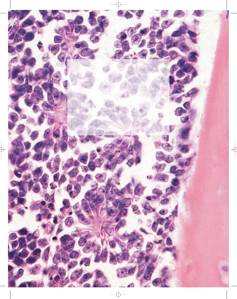
Histopathology
The morphology of the tumour is vari-
able. Most cases are composed of uni-
form small round cells with round nuclei
containing fine chromatin, scanty clear
or eosinophilic cytoplasm, and indistinct
cytoplasmic membranes, whereas in
others, the tumour cells are larger, have
prominent nucleoli, and irregular con-
tours {1540}. The cytoplasm of the
tumour cells frequently contains PAS
positive glycogen. In soft tissue tumours,
the tumour cells rarely have a spindle
cell morphology. In some cases
Homer–Wright rosettes are present
{2161}. Necrosis is common with viable
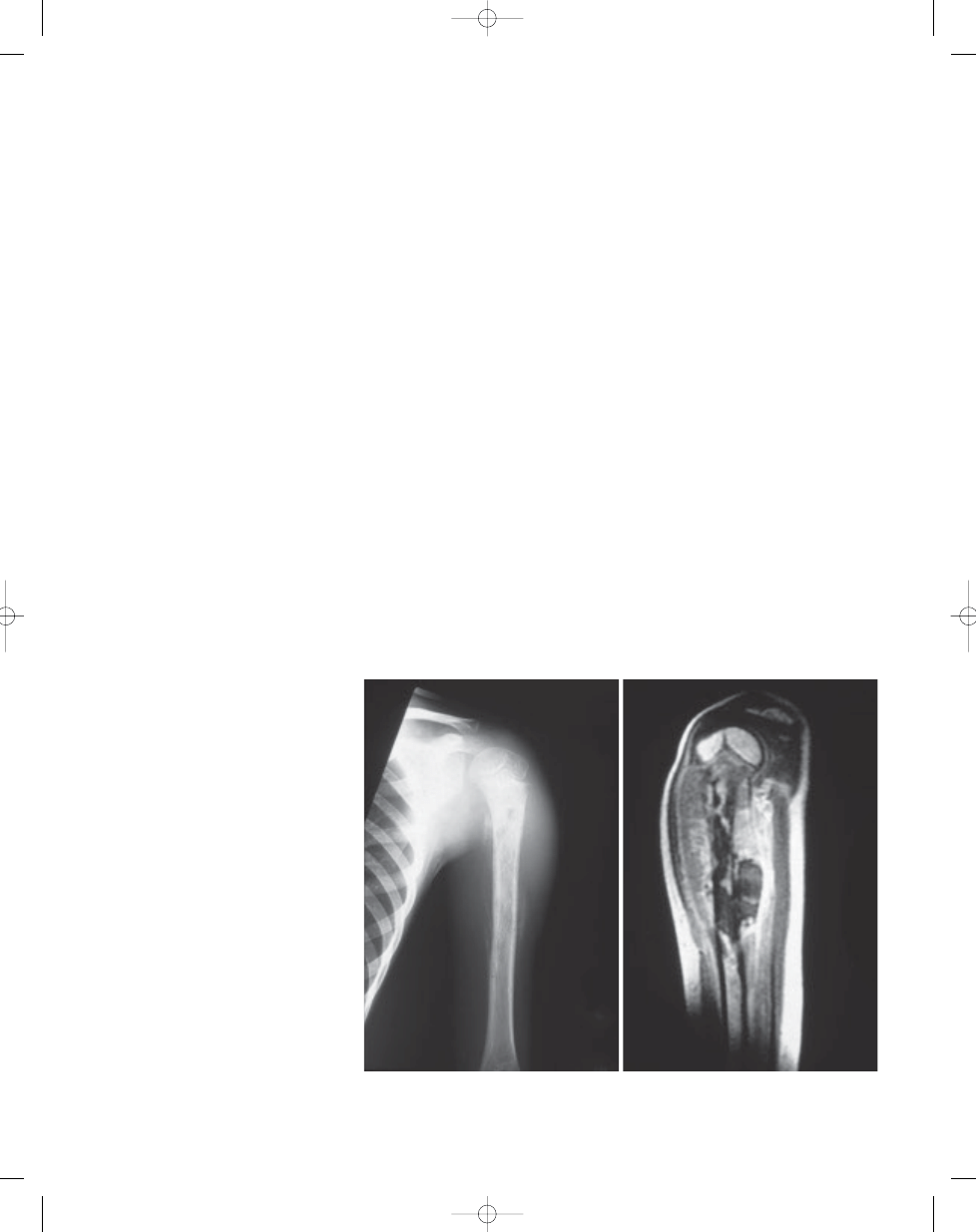
Fig. 14.01 Ewing sarcoma of the left humerus in a 6-year-old boy. A Periosteal new bone formation show-
ing "onion-skin" appearance. B Axial T1-weighted MRI of the same lesion. Both intraosseous and
extraosseous tumours are more clearly demonstrated than on plain X-ray.
B
A
298
Ewing sarcoma / Primitive neuroectodermal tumours
bb5_22.qxd 13.9.2006 13:21 Page 298
cells frequently perivascular in distribu-
tion.
Immunophenotype
CD99 is expressed in almost all cases in
a characteristic membranous fashion,
though it is not specific. Vimentin stains
most tumour cells and neural markers
such as neuron specific enolase (NSE),
are frequently expressed. Ewing sarco-
ma / PNET has also been shown to stain
with keratin in some cases.
Ultrastructure
Ewing sarcoma / PNET is composed of
primitive round to oval tumour cells often
with glycogen aggregates in the cyto-
plasm. Fine cytoplasmic processes are
often observed. Primitive intercellular
junctions are often seen. Neurosecretory
granules (100-150 nm) and microtubules
may be present.
Genetics
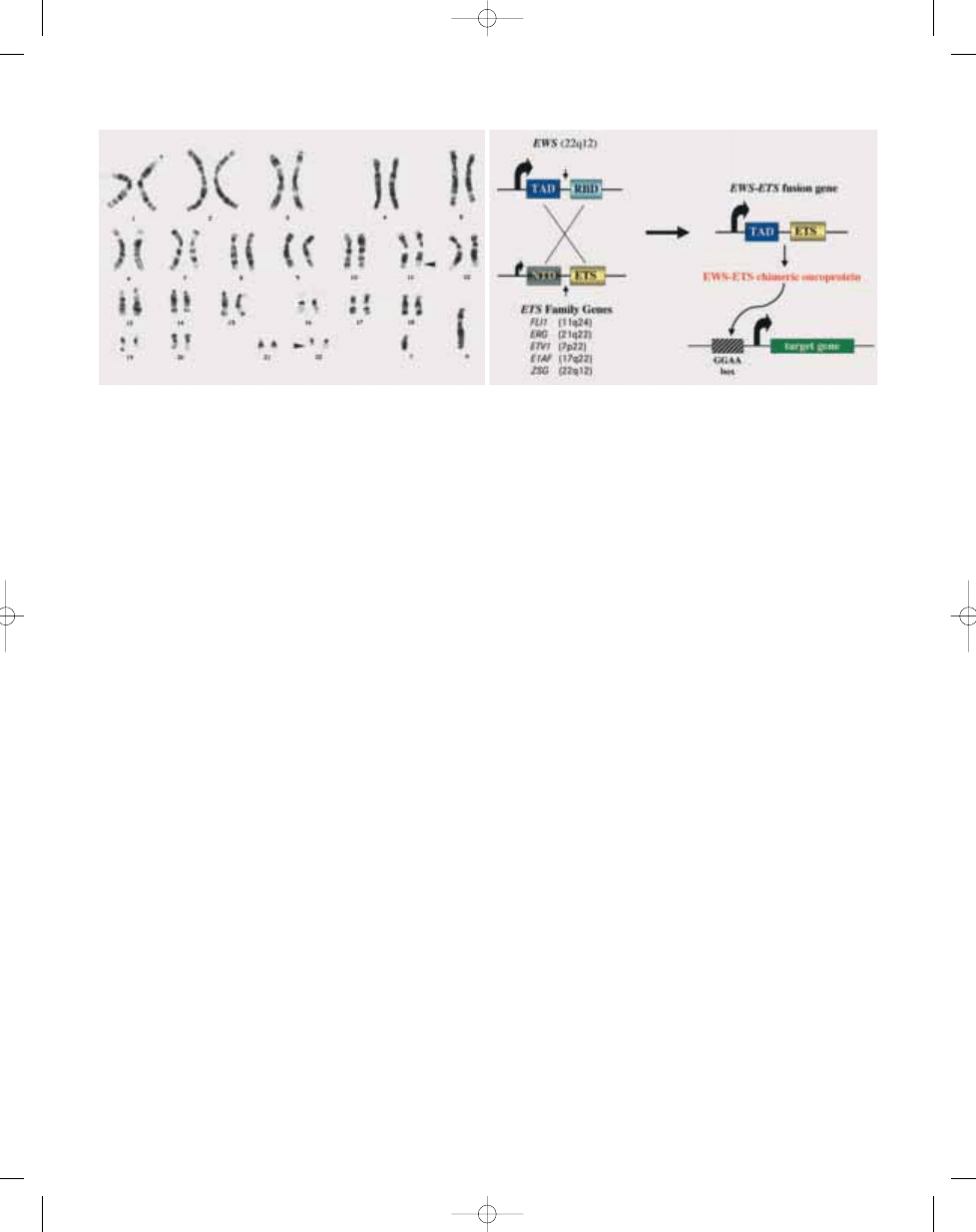
The Ewing family of tumours (EFT) is
characterized by a recurrent t(11;22)
(q24;q12) chromosomal translocation,
detectable in approximately 85% of the
cases {96,2146,2257}. Secondary chro-
mosomal aberrations, notably gains of
chromosome arm 1q and chromosomes
8 and 12 occur in more than half of the
cases. Molecular cloning of the t(11;22)
breakpoints revealed an in-frame fusion
between the 5’ end of the
EWS gene from
chromosome band 22q12 with the 3’ por-
tion of the 11q24
FLI1 gene, a member of
the ETS family of transcription factors
{497,1360}. It was subsequently found
that another 10-15% of cases have a
variant t(21;22)(q22;q12) translocation
fusing
EWS to a closely related ETS
gene,
ERG from chromosome band
21q22 {790,1995,2351}. In 1% or less of
EFT cases, t(7;22), t(17;22), and t(2;22)
translocations and inv(22) have been
described that give rise to fusions
between
EWS and the ETS genes ETV1,
E1AF, FEV, and ZSG, respectively
{1038,1060,1693,2159}. Therefore, virtu-
ally all EFTs appear to express some
form of
EWS/ETS gene fusion {496}.
Chimeric transcripts analysed to date all
encode the N-terminal transcriptional
activation domain of EWS fused to the C-
terminal DNA binding domain of the ETS
partner (reviewed in {89}). EWS/FLI1 has
potent oncogenic activity {1360}, and
many studies have suggested that it and
other EWS/ETS chimeric proteins func-
tion as aberrant transcription factors
binding to ETS target genes {111,1242,
1361,1598}. In this regard, a number of
up-regulated genes have been identified
in EWS/FLI1 expressing cells {88, 248,
1359, 2110}. One target is suggested by
the observation that EWS/ETS proteins
down-regulate expression of the TGF-
β
type II receptor (TGFBR2), a putative
tumour suppressor {865,1003}. TGF-
β
signalling induces apoptosis in many cell
types, and, therefore, repression of
TGFBR2 may provide EFT cells with a
mechanism to avoid programmed cell
death. Inactivation of the INK4a locus
encoding the CDKN2A cell cycle
inhibitor is the second most common
genetic alteration in EFTs {1162}. The sig-
nificance of this finding is underscored
by the recent observation that loss of
CDKN2A stabilises the EWS/FLI1 onco-
B
A
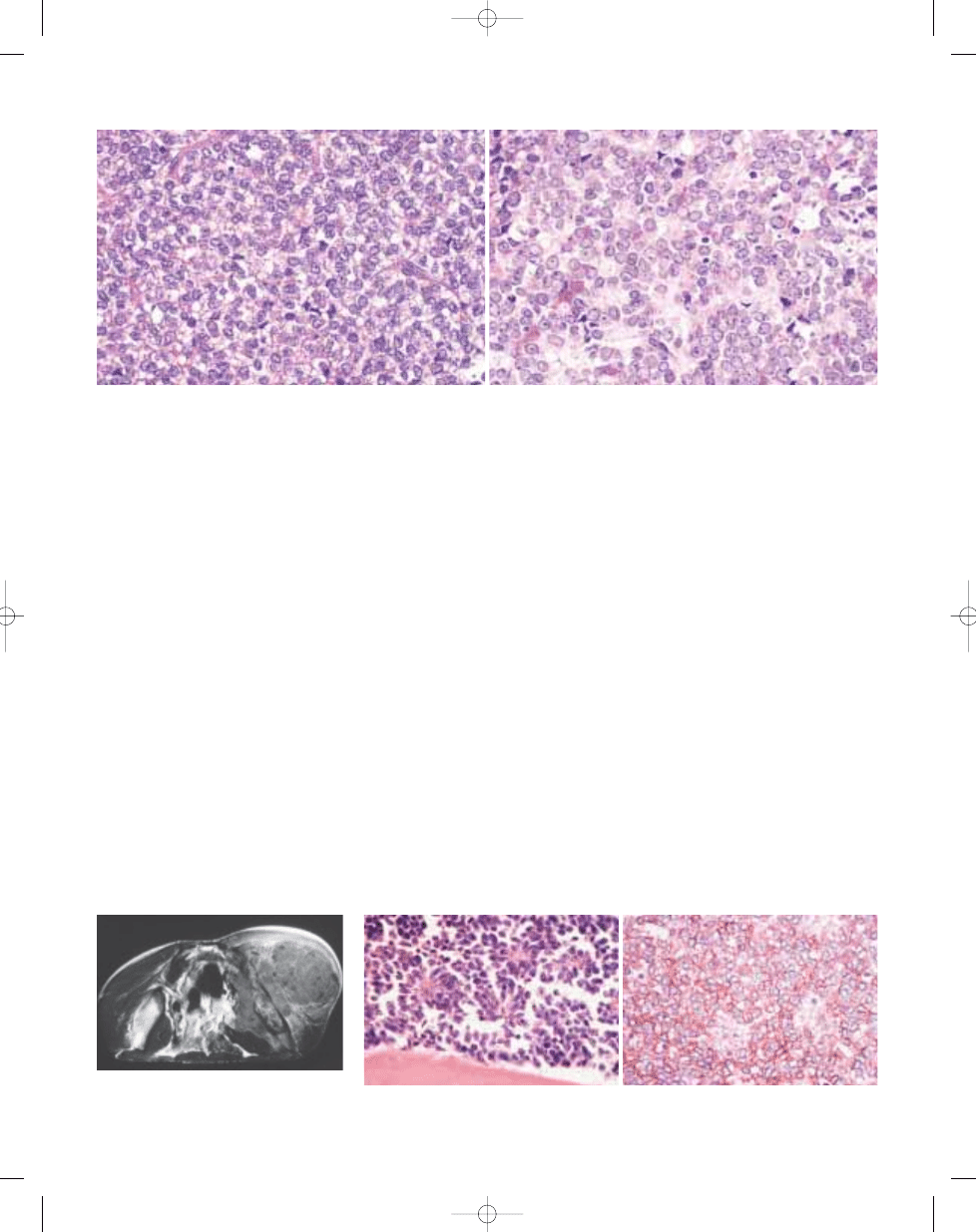
Fig. 14.04 Ewing sarcoma / PNET. A Rosette-like structures are occasionally found. B Immunohistochemi-cal
expression of CD99 showing characteristic reactivity on the cell membranes.
B
A
Fig. 14.02 Ewing sarcoma. A Uniform round cells with uniform round nuclei. B Histology of Ewing tumour / PNET predominantly composed of rosettes of small round cells
and luminal fibrillary processes.
299
Ewing sarcoma / Primitive neuroectodermal tumour (PNET)
Fig. 14.03 Ewing sarcoma. MRI (T1 image) of pelvic
tumour showing a huge soft tissue mass outside and
inside the iliac wing.
bb5_22.qxd 13.9.2006 13:21 Page 299
protein {501}, and that
CDKN2A muta-
tions may be associated with poor out-
come in EFTs {2228}.
Genetic diagnostic approaches include
chromosome banding analysis, inter-
phase fluorescence in situ hybridisation,
RT-PCR assays, and Southern blotting. It
is advisable to have available more than
one diagnostic modality, to be able to
confirm unexpected or discrepant results
{126,549,1181,1204,1380,1694,1996}.
Detection of fusion transcripts in periph-
eral blood or bone marrow is a sensitive
marker of minimal residual disease {462,
2252,2348}, although the clinical signifi-
cance of such a finding remains to be
determined {94,1380}
Prognostic factors
The prognosis in Ewing sarcoma / PNET
has improved in the modern era of treat-
ment and current survival rate is estimat-
ed to be 41%. Important prognostic fea-
tures include the stage, anatomic loca-
tion and the size of the tumour. Tumours,
that are metastatic at the time of diagno-
sis, arise in the pelvis, and are large tend
to do poorly. In addition to its diagnostic
utility,
EWS/ETS fusion status also pro-
vides prognostic information. Further
diversity of these rearrangements is con-
ferred by different combinations of
exons from
EWS and its partner genes
giving rise to variably sized chimeric
proteins {2351}. Among loco-regional
tumours with
EWS/FLI1 gene fusions, the
most common so-called type 1 gene
fusion (in which
EWS exon 7 is fused to
FLI1 exon 6) has been reported to be
associated with a better prognosis than
cases with larger, less common, fusion
types {460}.
B
A
Fig. 14.05 Ewing sarcoma. A Karyotype showing the most common rearrangement, a translocation t(11;22)(q24;q12). Arrowheads indicate breakpoints. B Schematic dia-
gram of
EWS-ETS gene fusions in Ewing family of tumours.
300
Ewing sarcoma / Primitive neuroectodermal tumours
bb5_22.qxd 13.9.2006 13:21 Page 300
Wyszukiwarka
Podobne podstrony:
bb5 chap3
bb5 chap8
bb5 chap1
BB5 BOX
bb5 chap16
bb5 chap15
bb5 contents
bb5 chap12
bb5 chap4
bb5 references
bb5 chap6
bb5 chap17
chap14 2
bb5 chap20
bb5 chap5
Lista wszystkich dostępnych polskich Product Code dla telefonów platformy BB5
bb5 chap21
bb5 source
więcej podobnych podstron