
Properties of Guaiacol Peroxidase Activities Isolated from
Corn Root Plasma Membranes
1
Angela Mika and Sabine Lu¨thje*
Universita¨t Hamburg, Institut fu¨r Allgemeine Botanik, Ohnhorststrasse 18, D–22609 Hamburg, Germany
Although several investigations have demonstrated a plasma membrane (PM)-bound peroxidase activity in plants, this
study is the first, to our knowledge, to purify and characterize the enzymes responsible. Proteins were extracted from highly
enriched and thoroughly washed PMs. Washing and solubilization procedures indicated that the enzymes were tightly
bound to the membrane. At least two distinct peroxidase activities could be separated by cation exchange chromatography
(pmPOX1 and pmPOX2). Prosthetic groups were identified in fractions with peroxidase activity by absorption spectra, and
the corresponding protein bands were identified by heme staining. The activities of the peroxidase enzymes responded
different to various substrates and effectors and had different thermal stabilities and pH and temperature optima. Because
the enzymes were localized at the PM and were not effected by p-chloromercuribenzoate, they were probably class III
peroxidases. Additional size exclusion chromatography of pmPOX1 revealed a single activity peak with a molecular mass
of 70 kD for the native enzyme, whereas pmPOX2 had two activity peaks (155 and 40 kD). Further analysis of these fractions
by a modified sodium dodecyl sulfate-polyacrylamide gel electrophoresis in combination with heme staining confirmed the
estimated molecular masses of the size exclusion chromatography.
Peroxidases (EC 1.11.1.7, etc.) belong to a large
family of enzymes that are ubiquitous in fungi,
plants, and vertebrates. These proteins usually con-
tain a ferriprotoporphyrin IX prosthetic group and
oxidize several substrates in the presence of hydro-
gen peroxide (H
2
O
2
; Penel et al., 1992; Vianello et al.,
1997). In higher plants, the number of isoenzymes
may be extremely high, up to 40 genes corresponding
to isoperoxidases for each plant, and several other
isoforms can be generated by posttranscriptional and
posttranslational modifications (Welinder et al., 1996;
De Marco et al., 1999).
Although many soluble intracellular and extracel-
lular peroxidases have been characterized in detail
(for refs., see Gaspar et al., 1982; Hiraga et al., 2001;
Shigeoka et al., 2002), less is known about membrane-
bound enzymes, in particular the peroxidases of
plant plasma membranes (PMs). Evidence for a
PM-bound peroxidase activity in higher plants has
been demonstrated frequently. Lin (1982) reported an
increased oxygen consumption by intact corn (Zea
mays) root protoplasts in the presence of extracellular
NADH. Pantoja and Willmer (1988) obtained similar
results using guard cell protoplasts from Commelina
communis in the presence of NAD(P) H. PMs iso-
lated from several species and plant parts showed
NAD(P) H oxidase activities, which were compara-
ble with a peroxidase (Møller and Be´rczi, 1986;
Askerlund et al., 1987; Vianello et al., 1990, 1997; De
Marco et al., 1995; Zancani et al., 1995; Sagi and
Fluhr, 2001). Because the application of detergents did
not significantly affect the activity observed and be-
cause activity could be detected with intact proto-
plasts, peroxidase activity has been suggested to be
located at the apoplastic surface of the PM.
The NADH oxidation by PM from cauliflower
(Brassica oleracea) could be stimulated by phenolic
substances or inhibited by typical effectors of peroxi-
dases like catalase, superoxide dismutase, cyanide, or
azide (Askerlund et al., 1987). In PM-enriched frac-
tions of Arabidopsis and Chinese cabbage (Brassica
campestris L. subsp. pekinensis) seedlings, oxidation of
Trp was reported in the presence of H
2
O
2
(Ludwig-
Mu¨ller et al., 1990; Ludwig-Mu¨ller and Hilgenberg,
1992). PM isolated from soybean (Glycine max) roots
showed a peroxidase activity in the presence of sub-
strates like o-dianisidine, guaiacol, and ascorbate (Vi-
anello et al., 1997). The oxidation of ascorbate could
be strongly stimulated by phenolic acids, like caffeic
and ferulic acid. Guaiacol or o-dianisidine oxidation
rates were increased by CaCl
2
and inhibited by po-
tassium cyanide and azide. When proteins solubi-
lized by SDS from non-washed PM were separated
by SDS-PAGE, two bands (38 and 45 kD) could be
detected by heme staining. Peroxidase activity of
these bands was not demonstrated, and only one, less
intensive band remained after partial washing of the
membrane vesicles (Vianello et al., 1997).
In addition to these experiments, antibodies spe-
cific for apoplastic peroxidases were used to detect
PM-bound peroxidases by immunogold labeling and
electron microscopy in situ (Hu et al., 1989; Penel and
Castillo, 1991; Crevecoeur et al., 1997). However,
1
This work was supported by the Deutsche Forschungsgemein-
schaft (grant no. DFG Lu 668/1–2) and by the University of Ham-
burg (PhD student’s grant no. HmbNFG to A.M.).
* Corresponding author; e-mail s.luthje@botanik.uni-hamburg.
de; fax 49 – 40 – 82282–254.
Article, publication date, and citation information can be found
at www.plantphysiol.org/cgi/doi/10.1104/pp.103.020396.
Plant Physiology, July 2003, Vol. 132, pp. 1489–1498, www.plantphysiol.org © 2003 American Society of Plant Biologists
1489
Askerlund et al. (1987) demonstrated that the pres-
ence of peroxidases in PM preparations depends
largely on the final PM washing procedure, which
decreases the level of peroxidases significantly. A
PM-bound peroxidase has not yet been isolated and
characterized from highly purified and properly
washed PM (Be´rczi and Møller, 2000).
In the present work, we demonstrate the occur-
rence of at least two distinct peroxidase activities
(pmPOX1 and 2) in corn root PM. A purification
protocol for the isolation of these enzymes was de-
veloped, and the properties of the partially purified
proteins were investigated by comparing them with
soluble peroxidase activities.
RESULTS AND DISCUSSION
Binding to the PM
To check if peroxidase activities were loosely
bound to the PM or entrapped inside the vesicles,
different washing procedures were carried out. Inde-
pendent of the salt concentrations used a maximum
of 40% of the activity could be washed off in the
presence of 1 mm EDTA and 0.01% (w/v) Triton
X-100, i.e. 79%
⫾ 7.2% (n ⫽ 2) of the activity re-
mained in the PM at 150 mm KCl and 60%
⫾ 1.9%
(n
⫽ 4) at 500 mm KCl, respectively. Using 1 mm
EGTA instead of EDTA did not change this result. A
combination of 150 mm KCl, 1 mm EDTA, 0.01%
(w/v) Triton X-100, and 0.1% (w/v) CHAPS (i.e. a
detergent:protein ratio of 6:1 [w:w]) removed 62%
⫾
0.4% (n
⫽ 2) of the peroxidase activity from the PM.
Due to the fact that neither physiological or high
salt concentrations in the presence of detergent and
EDTA or EGTA nor high detergent concentrations
were able to remove the activity completely from the
PM, we conclude that these enzymes are probably
tightly bound to the PM. Salts should have minimal
effects on the micellar size of Triton X-100, whereas
effects on the zwitterionic detergent CHAPS cannot
be excluded. Thus, the presence of higher salt con-
centrations could change the critical micellar concen-
tration of CHAPS, thereby increasing the proportion
of washed off peroxidase activity as a result of partial
solubilization.
However, because peroxidase activity remains in
the low detergent phase after Triton X-114 solubili-
zation and temperature-induced phase separation
(data not shown), the peroxidases were probably not
strongly hydrophobic. Independently of the detergent
to protein ratio used, none of the detergents tested
(Triton X-114, Triton X-100, CHAPS, or octylglucopy-
ranoside) could solubilize the activity completely from
the PM. The mechanism of the binding to the PM is
unknown, but sequence analysis of intracellular per-
oxidases indicated that transmembrane domains may
exist in plant peroxidases (Bunkelmann and Trelease,
1996; Jespersen et al., 1997; Nito et al., 2001).
In Arabidopsis, three genes encoding membrane-
bound ascorbate peroxidases were found (Jespersen
et al., 1997). One of the corresponding proteins is
probably bound to microbodies by a C-terminal
transmembrane domain like membrane-bound intra-
cellular peroxidases of other plant species (Bunkel-
mann and Trelease, 1996; Nito et al., 2001). However,
sequence analysis of these peroxidases revealed that
they are class I peroxidases, which implies they can-
not occur in the PM.
Purification
Solubilization by CHAPS yields about 30%
⫾ 1%
(n
⫽ 2) of the activity and increased up to 73% ⫾ 4%
(n
⫽ 5) in the presence of the dipole aminocaproic
acid. Two activity peaks (pmPOX1 and pmPOX2)
could be separated by cation exchange chromatogra-
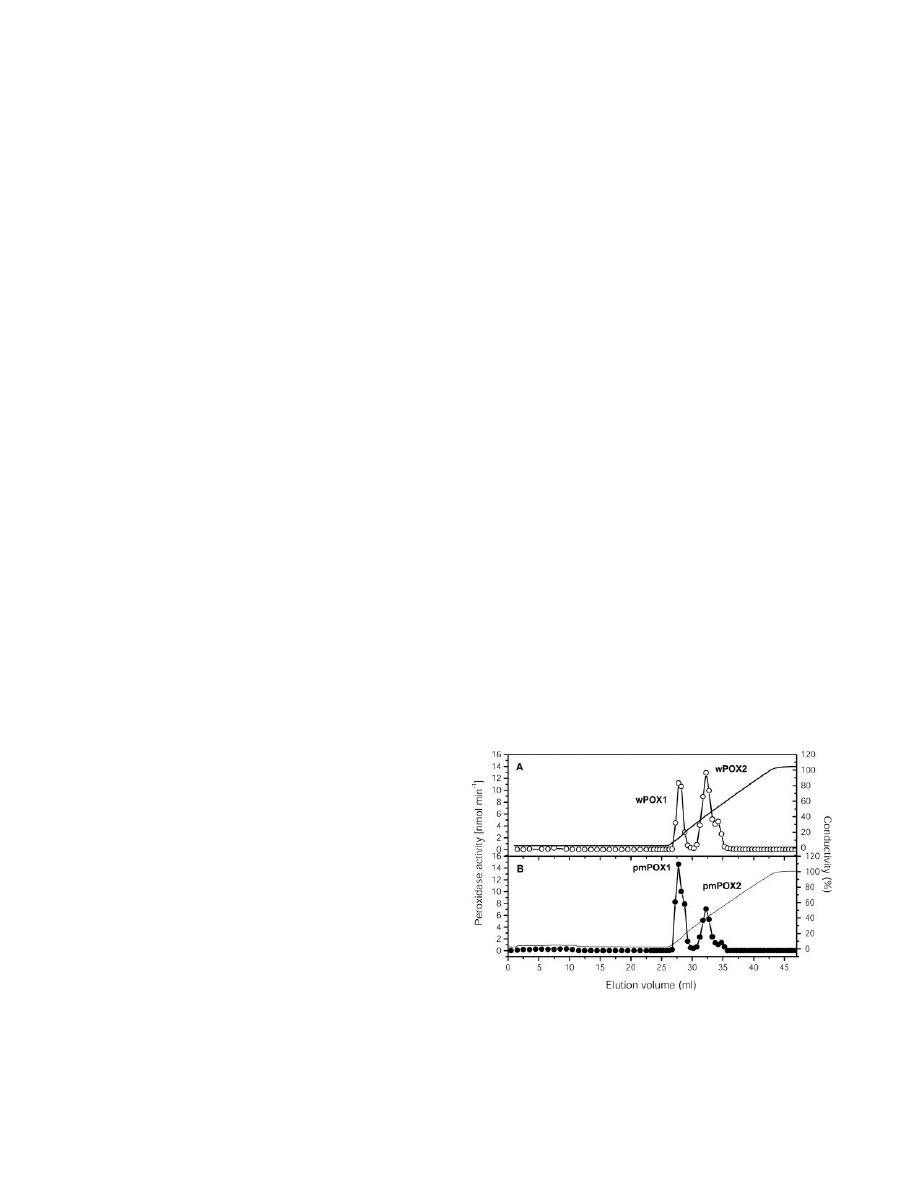
phy (Fig. 1). Peroxidase activities were eluted at 115
and 395 mm KCl. The total activity was divided into
59%
⫾ 3% and 41% ⫾ 2% (n ⫽ 4) for pmPOX1 and
pmPOX2, respectively. Starting from washed PM
(specific activity 401
⫾ 52 nmol min
⫺1
mg protein
⫺1
;
n
⫽ 5), a 24.0- and 8.8-fold purification for peak
fractions of pmPOX1 and pmPOX2 with an overall
yield of 31.4% was achieved. To compare the prop-
erties of pmPOX with soluble peroxidases, activities
of the washing fluid of the PM (wPOX) were concen-
trated and separated by the same protocol (Fig. 1). The
elution profile obtained was similar to that from the
PM-bound POX. The total activity was divided into
30% and 70% for wPOX1 and wPOX2, respectively.
Relative Molecular Mass
As shown in Figure 2, pmPOX1 displayed a single
peak after size exclusion chromatography. By modi-
Figure 1. Elution profiles of POX after cation exchange chromatog-
raphy. Enzyme activities isolated from corn root PM were separated
on a Uno S1 column. Bound proteins were eluted by a KCl gradient
from 0 to 1
M
. The flow rate was 1 mL min
⫺1
, and fractions of 1.0 and
0.5 mL were collected. A, Separation of POX activities from washing
fluid of PM (wPOX; E). B, Elution profile of PM-bound peroxidase
activities (pmPOX; F). Enzyme activities were measured in the pres-
ence of 8.26 m
M
guaiacol and 8.8 m
M
H
2
O
2
.
Mika and Lu¨thje
1490
Plant Physiol. Vol. 132, 2003
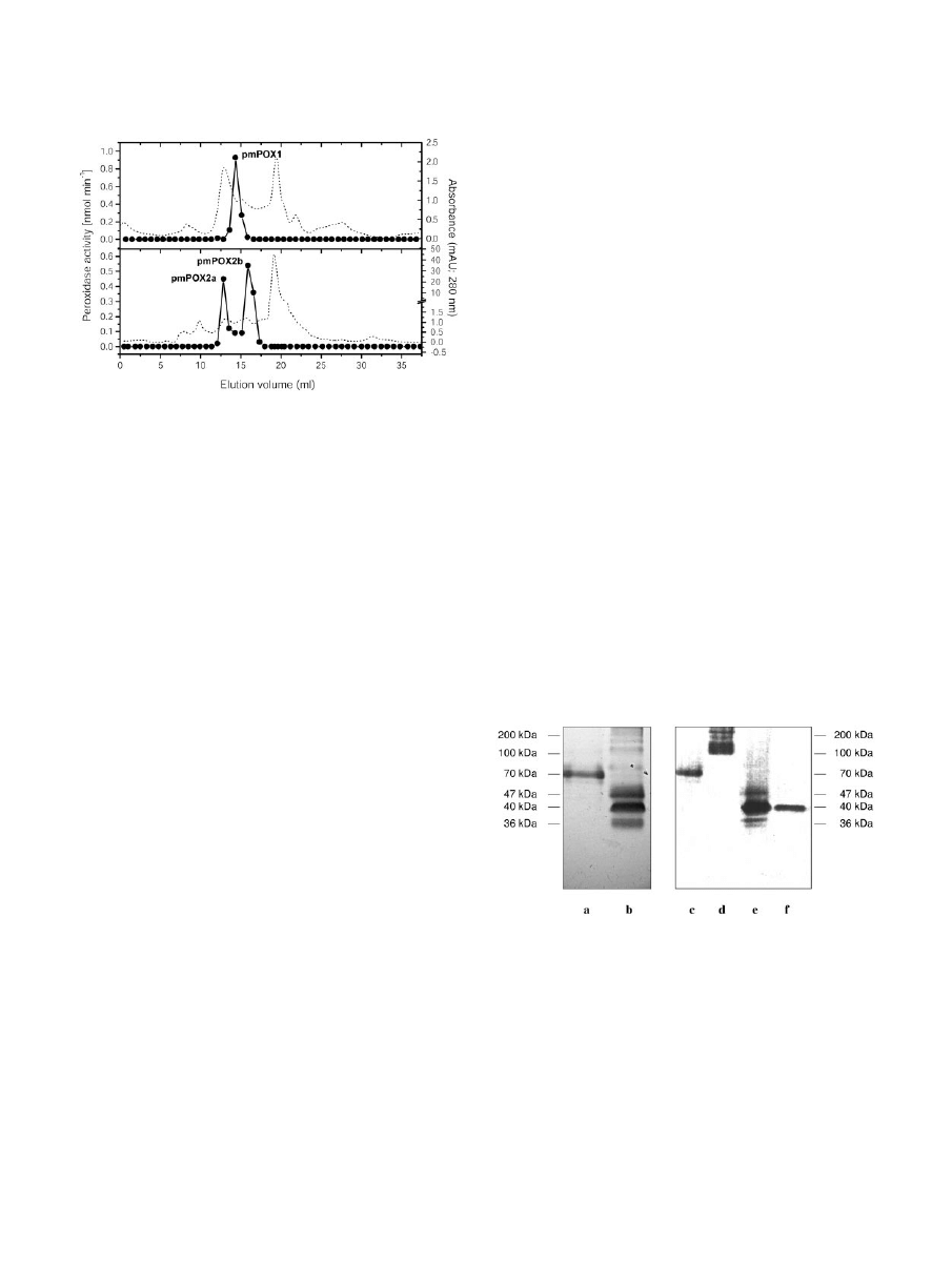
fied SDS-PAGE and heme staining, a protein band
with an apparent molecular mass of 70 kD could be
identified (Fig. 3). However, pmPOX2 was clearly
separated into two peaks after size exclusion chro-
matography (pmPOX2a and pmPOX2b; Fig. 2). In
comparison with peak fractions eluted during the
cation
exchange
chromatography,
analysis
of
pmPOX2b showed a significant increase in intensity
of a 40-kD band after heme staining (Fig. 3). pmPOX2a
exhibited a protein band between 100 and 170 kD.
Due to the modifications of the SDS-PAGE, these are
molecular masses of whole enzymes, i.e. oligomers
were not separated into subunits.
Molecular masses were also calculated by elution
volumes of the size exclusion purification step in
comparison with marker proteins. The native en-
zymes revealed apparent molecular masses of 70,
155, and 38 kD for pmPOX1, pmPOX2a, and
pmPOX2b, respectively, confirming results obtained
by gel electrophoresis and suggesting the presence of
three distinct peroxidases at the plant PM. On the
other hand, the separation of pmPOX2 into two per-
oxidase peaks by size exclusion chromatography
could be due to proteins that were not fully solubi-
lized and remained as aggregates (i.e. protein deter-
gent or protein aggregates). However, the data ob-
tained by SDS-PAGE excluded this hypothesis.
Known class III peroxidases revealed molecular
masses in a range of 28 to 60 kD (Hiraga et al., 2001)
and a 70- or a 155-kD protein have not been described
for soluble peroxidases from higher plants.
In PM isolated from soybean roots, 38- and 45-kD
bands were identified by SDS-PAGE and heme stain-
ing (Vianello et al., 1997), masses comparable with
that found for pmPOX2b (Fig. 3). However, both
bands decreased in intensity after partial washing of
the PM vesicles with NaCl, and several apoplastic
peroxidases with these molecular masses were iden-
tified in different plant species (Hendriks et al., 1991;
Melo et al., 1996; De Marco et al., 1999; Blee et al.,
2001). The molecular masses of pmPOX1 and
pmPOX2a were different compared with the protein
bands identified in soybean PM. However, this could
be due to the different material.
Prosthetic Groups
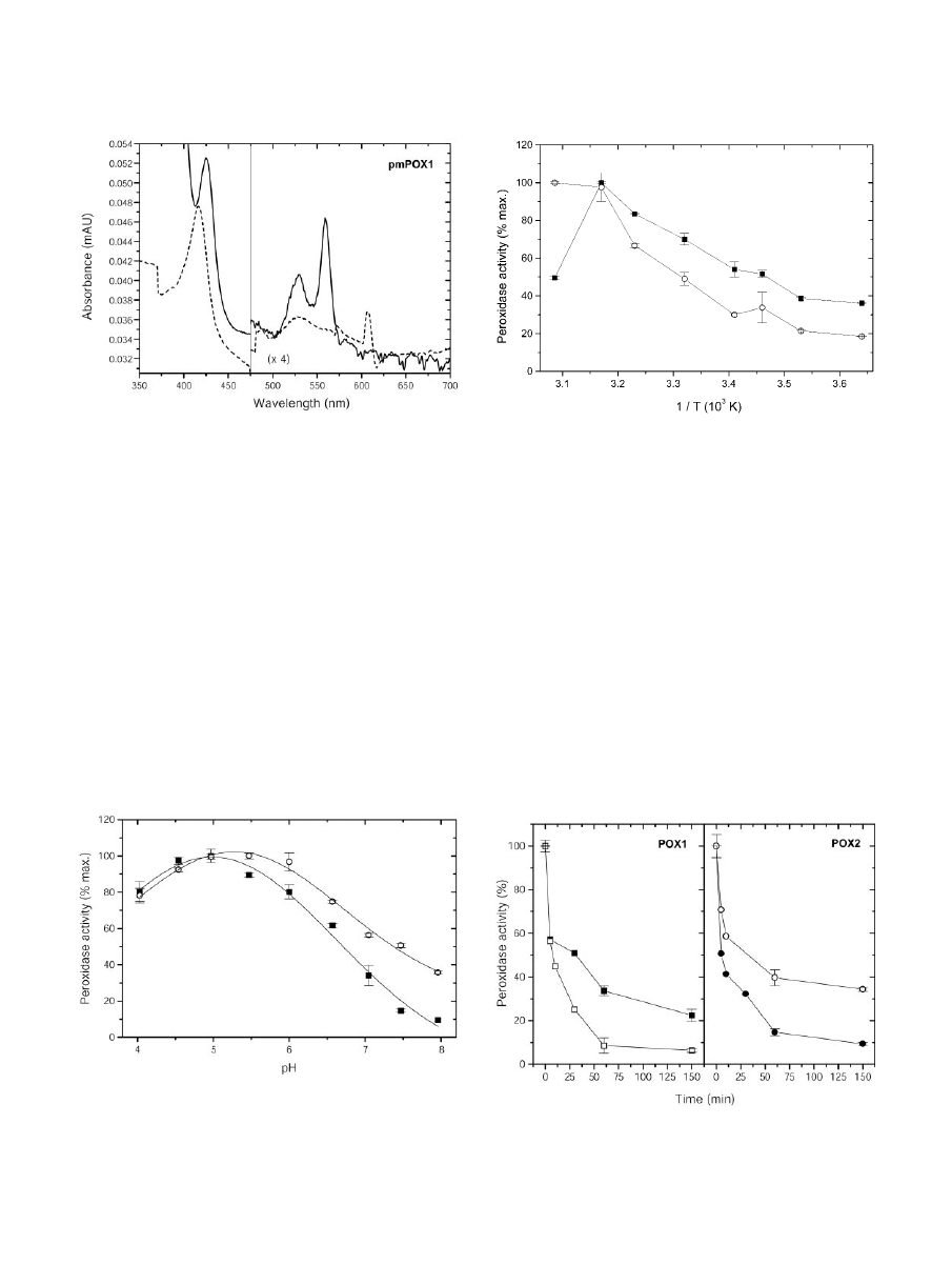
UV/visible absorption spectra of pmPOX1 and
pmPOX2 were almost identical and typical for heme-
containing proteins (e.g. Converso and Fernandez,
1995; Kvaratskhelia et al., 1997). Both pmPOXs exhib-
ited a Soret peak at 416 nm (Fig. 4). In addition to
these, the oxidized enzymes showed
␣- and -
absorption bands at 607 and 528 nm, respectively.
The Soret peak and the
␣-band shifted to 425 and
559 nm when the proteins were reduced by sodium
dithionite. The A
416
/A
280
values, which are a crite-
rion of purity and heme content, were 0.5 and 0.2 for
pmPOX1 and pmPOX2, suggesting that the enzymes
were not purified to homogeneity, which was also
shown by SDS-PAGE. Thus, the
␣-absorption band at
607 nm cannot be definitely ascribed to the heme
group of the peroxidase. The spectra of both pmPOXs
more closely resemble those of guaiacol rather than
ascorbate peroxidases (Chen and Asada, 1989; Con-
verso and Fernandez, 1995; Kvaratskhelia et al.,
1997). Peroxidase staining of the isolated proteins
suggests a relatively strong binding of the heme
Figure 2. Elution profiles of PM-bound POX after size exclusion
chromatography. Peak fractions collected from several Uno S runs
were combined, concentrated, and applied onto a Superdex 200
column. Proteins were separated by a flow rate of 0.5 mL min
⫺1
. The
fraction size was automatically adjusted between 0.75 and 0.5 mL
depending on increase or decrease in A
280
(dotted line) Enzyme
activities were measured as described in Figure 1. PM-bound perox-
idase activities could be separated into three peaks.
Figure 3. Heme staining of pmPOX fractions after modified SDS-
PAGE. Electrophoresis was performed using a low concentrated SDS-
PAGE, i.e. 0.1% (w/v) SDS in all solutions and gels without dithio-
threitol or mercaptoethanol. Thus, the oligomers were not separated
into their subunits. Heme-containing protein bands were visualized
by their reaction with the peroxidase substrates tetramethylbenzidine
(TMB) and H
2
O
2
as described in “Materials and Methods.” Left,
pmPOX1 (a) and pmPOX2 (b) are shown after cation exchange
chromatography. Further purification of these fractions by size ex-
clusion is presented on the right: pmPOX1 (c), pmPOX2a (d), and
pmPOX2b (e). In addition, f shows pmPOX2b treated with 25 m
M
dithiothreitol. Bars indicate the corresponding molecular masses.
After size exclusion chromatography, the PM-bound enzymes had
apparent molecular masses of 70 and 40 kD for pmPOX1 and
pmPOX2b, whereas pmPOX2a exhibited a broad protein band be-
tween 100 and 170 kD.
Plasma Membrane-Bound Peroxidases
Plant Physiol. Vol. 132, 2003
1491
groups to the enzymes. Only pmPOX2b could be
detected by heme staining after treatment with dithio-
threitol and revealed the same molecular mass as
without reducing compounds (Fig. 3). Thus,
pmPOX2b was identified as a monomer. pmPOX1
and pmPOX2a did not reveal any visible band after
the same treatment (data not shown). Conforma-
tional changes due to the cleavage of disulfide
bridges within the molecules possibly resulted in a
release of the heme groups.
pH Optimum and Kinetic Studies
The properties of POX, which were separated by
cation exchange chromatography, were further char-
acterized. As shown in Figure 5, the highest activity
with guaiacol as a substrate was observed between
pH 4.5 and 5.5 for pmPOX1, whereas pmPOX2 ex-
hibited a pH optimum in the range of 5.0 to 6.0. With
guaiacol as substrate, acidic pH optima have often
been reported for the apoplastic peroxidases of sev-
eral plant species (Hendriks et al., 1991; Melo et al.,
1996; Nair and Showalter, 1996). Variations in pH
optima could represent efficient regulatory means in
vivo to shift optimal conditions from one isoenzyme
to another and thereby favor different processes (De
Marco et al., 1999).
Figure 4. Absorption spectra of partially purified pmPOX1. Samples
(1.1 mg protein mL
⫺1
) containing the native enzyme (dashed line)
were measured in 50 m
M
sodium phosphate buffer (pH 7.0) with
buffer as reference. Ferric enzymes were reduced by the addition of
approximately 1.5 m
M
dithionite (straight line). The spectra were
measured at 50 nm min
⫺1
. n
⫽ 2 independent preparations showing
identical results. The spectra indicate the presence of heme groups as
the prosthetic group.
Figure 5. Dependence of the guaiacol peroxidase activity of partially
purified pmPOX on pH. The rates of guaiacol oxidation were deter-
mined under the standard assay conditions except that 25 m
M
so-
dium acetate (pH 4.0–5.0), MES (pH 5.5–6.5), or HEPES (pH 7.0–8.0)
buffers were used. Data presented are average values
⫾
SD
of n
⫽ 3
experiments. f, pmPOX1; E, pmPOX2.
Figure 6. Dependence of the guaiacol peroxidase activity of purified
pmPOX on temperature (Arrhenius plot). The rates of guaiacol oxi-
dation were determined under the standard assay conditions except
for temperature. Data presented are average values
⫾
SD
of n
⫽ 3
experiments. f, pmPOX1; E, pmPOX2.
Figure 7. Thermal stability of guaiacol peroxidase activities. Soluble
and PM-bound POX were incubated at 50°C at different time slices.
Data presented are average values
⫾
SD
of n
⫽ 2 experiments. f,
pmPOX1; F, pmPOX2;
䡺, wPOX1; E, wPOX2.
Mika and Lu¨thje
1492
Plant Physiol. Vol. 132, 2003

The K
m
s of both PM-bound peroxidase activities
for guaiacol were comparable (12.2 mm for pmPOX1
and 14.3 mm for pmPOX2, calculated by Eadie-
Hofstee plots). K
m
values in a millimolar range are
typical for peroxidases with artificial substrates like
guaiacol. For instance, soluble peroxidases from kiwi-
fruit (Actinidia deliciosa) and tomato (Lycopersicon
esculentum) fruits had K
m
values of 7.4 and 10 mm,
respectively (Soda et al., 1991).
Temperature Optima and Thermal Stability
At low temperatures the enzyme activity of
pmPOX2 was about 2-fold lower compared with
pmPOX1 (Fig. 6). The activity of both protein frac-
tions increased with higher temperatures. Although
the activity of pmPOX2 more or less continuously
increased in the range of 2°C to 51°C, pmPOX1
showed a maximum of activity at 43°C and decreased
dramatically afterward.
In a second set of experiments, the thermal stability
of soluble and PM-bound peroxidases was investi-
gated (Fig. 7). All enzymes lost between 40% and 50%
of their activities within 5 min at 50°C. During an
incubation time of 3 h, the guaiacol peroxidase activ-
ities decreased exponentially to values between 5.7%
and 34.3%. After 3 h, pmPOX1 showed twice the
activity of pmPOX2. Most peroxidases from plants
and animals seemed to have high temperature op-
tima and show high thermal stabilities (Bakardjieva
et al., 1996; Madhavan and Naidu, 2000). Apoplastic,
cytosolic, and soluble peroxidases of several plant
tissues showed temperature optima between 30°C
and 60°C, the most between 50°C and 60°C (Soda et
al., 1991; Bakardjieva et al., 1996; Nair and Showalter,
1996; Bernards et al., 1999; Loukili et al., 1999). Due to
the fact that pmPOX1 had a lower temperature opti-
mum than pmPOX2, the latter enzyme seemed to be
more stable (Fig. 6). However, for longer treatments
of higher temperatures, pmPOX1 revealed a higher
thermal stability (Fig. 7).
Effector Studies
As shown in Table I, classical peroxidase inhibitors
like potassium cyanide or sodium azide caused a
complete loss of the peroxidase activities or de-
creased the rates more than 90%. These results were
consistent with the presence of heme groups as pros-
thetic groups.
The localization of the enzymes at the plant PM
suggests that they may be part of the secretory path-
way. According to Welinder et al. (1996), pCMB, a
sulfhydryl inhibitor, is often used to distinguish be-
tween class I and class III peroxidases. As shown in
Table I, this inhibitor did not effect the PM-bound
activities of pmPOX1 or pmPOX2, indicating that SH
groups did not participate in the active center or
maintenance of the conformation of the isoenzymes.
Thus, the PM-bound peroxidases were probably
class III peroxidases. In contrast to the pmPOX,
wPOX1 and wPOX2 were slightly inhibited in the
presence of 1 mm pCMB.
Both PM-bound peroxidase activities were de-
creased by distinct concentrations of the lectins con-
canavalin A (Con A) and wheat germ agglutinin
(WGA; Table II), whereas the Ulex europaeus agglu-
tinin (UEA1) was without significant effect (data not
shown). Inhibition of wPOX1 and wPOX2 was weak
and occurred only at higher concentrations of Con A
and WGA (Table II). The effects of lectins indicate
glycosylation of the enzymes. These results are con-
sistent with the finding of Vianello et al. (1997) that
treatment of soybean roots with tunicamycin, an in-
hibitor of glycoprotein synthesis, reduced the guaia-
col peroxidase activity of unwashed PM vesicles by
40%. Due to the possible glycosylation, which was
also indicated by diffuse protein bands in SDS gels
(Fig. 3), the real molecular masses of all identified
proteins may be different from the calculated values.
However, the structures of the proteins have to be
further elucidated.
Table I. Guaiacol-dependent activity in the absence or presence of typical peroxidase effectors
Peroxidase activity was measured with the partially purified enzymes after cation exchange chromatography in the presence of 8.26 m
M
guaiacol and 8.8 m
M
H
2
O
2
at pH 7.0 as described in “Materials and Methods.” Data are given as mean
⫾
SD
(n).
Substance
Concentration
Peroxidase Activity
pmPOX1
pmPOX2
wPOX1
wPOX2
mol min
⫺1
mg protein
⫺1
Control
5.2
⫾ 0.1 (3)
1.6
⫾ 0.1 (3)
12.0
⫾ 0.3 (3)
16.8
⫾ 0.1 (3)
(% of control)
Control
100.0
⫾ 1.5 (3)
100.0
⫾ 4.9 (3)
100.0
⫾ 2.3 (3)
100.0
⫾ 0.3 (3)
KCN
1 m
M
n.d.
a
(3)
n.d. (3)
1.2
⫾ 1.6 (3)
0.6
⫾ 0.8 (3)
Azide
1 m
M
10.6
⫾ 0.4 (3)
b
2.9
⫾ 0.6 (3)
b
0.2
⫾ 0.3 (2)
b
7.4
⫾ 0.9 (2)
b
p-Chloromercuribenzoate (pCMB)
50
M
112.5
⫾ 7.1 (3)
111.1
⫾ 5.7 (3)
99.7
⫾ 9.6 (3)
96.4
⫾ 4.6 (3)
200
M
105.7
⫾ 2.3 (3)
102.2
⫾ 5.0 (3)
101.4
⫾ 2.0 (3)
102.1
⫾ 8.3 (3)
1 m
M
110.2
⫾ 4.3 (3)
106.9
⫾ 0.6 (3)
54.5
⫾ 10.9 (3)
77.2
⫾ 9.1 (3)
a
n.d., Not detectable.
b
pH 5.0.
Plasma Membrane-Bound Peroxidases
Plant Physiol. Vol. 132, 2003
1493

Ca
2
⫹
reduced the activity of pmPOX2 and wPOX2.
Mn
2
⫹
had no effect on pmPOX1 or pmPOX2
(Table III). In contrast to the PM-bound enzymes,
many peroxidases exhibit increased activities after
treatment with Ca
2
⫹
or Mn
2
⫹
(Gaspar et al., 1982;
Van Huystee et al., 1996; Greppin et al., 1999). Cal-
cium probably maintains the conformation of the
proteins, whereas Mn
2
⫹
could be involved in regu-
latory processes (Van Huystee et al., 1996). However,
Loukili et al. (1999) characterized plant peroxidases
that were not influenced by these ions. Furthermore,
Mn
2
⫹
was not detectable in PM from corn roots
(Lu¨thje et al., 1995). On the other hand, unwashed
PM from soybean roots showed a 42% increase of
activity in the presence of CaCl
2
(Vianello et al.,
1997). Possibly, this increase was caused by soluble
peroxidases that were loosely attached to the PM or
due to the different plant material.
DMSO had no effect at 0.5% (v/v), the final con-
centration of DMSO used in experiments with phe-
nolic compounds as effectors (Table III). PM showed
90.4%
⫾ 0.3% (n ⫽ 3) peroxidase activity in the
presence of 2% (w/v) DMSO. pmPOX1 was not ef-
fected by this concentration, whereas pmPOX2 and
the washed off peroxidase activities were inhibited.
Detergents like Triton X-100 and Triton X-114 in-
duced a decrease or increase of the enzyme activities.
The activities of cell wall-bound and apoplastic
peroxidases have often been reported to be stimu-
lated by phenolic substances, like ferulic acid and
coumaric acid (Ma¨der and Fu¨ssl, 1982; Lobarzewski
et al., 1996; De Marco et al., 1999). As shown in
Table IV, activities of the partially purified peroxi-
dases increased to 220% and 400% of the control in
the presence of ferulic acid as a substrate, whereas
coumaric acid had no effect on pmPOX2 and wPOX2,
and pmPOX1 and wPOX1 were slightly decreased.
The phenolic compound propyl gallate inhibited the
guaiacol-dependent peroxidase activity of all frac-
tions. In the presence of IAA, the activity of pmPOX2
decreased slightly, whereas all other activities were
not effected. The inhibitory effects of propyl gallate
and IAA suggest a competition between the sub-
strates, which was further indicated by their substrate
specificity.
Substrate Specificity
Artificial electron donors were used by pmPOX1 in
the following order: o-dianisidine
⬎ guaiacol ⬎ TMB
⬎⬎
2,2
⬘-azino-bis(3-ethylbenz-thiazoline-6-sulfonic
acid) (ABTS; Table V). In contrast to pmPOX1,
pmPOX2 showed a higher affinity for TMB than for
guaiacol. Both pmPOXs oxidized natural substrates
like phenolic acids and alcohols in the following
order: coniferyl alcohol
⬎ ferulic acid ⬎ coumaric
acid. Hydroxycinnamyl alcohol species are used by
apoplastic peroxidases to participate in lignin poly-
merization, whereas hydroxycinnamic acids could be
incorporated into suberin (for refs., see Hiraga et al.,
2001).
In vitro IAA oxidation by peroxidases has been
reported several times (Converso and Fernandez,
1995; Gazaryan and Lagrimini, 1996). This plant hor-
Table II. The effects of lectins on guaiacol-dependent peroxidase activity of POX
Guaiacol-dependent peroxidase activity was measured at pH 5.0 after cation exchange chromatography. Fractions were measured in absence
or presence of different lectins. Pre-incubation was for 3 min. Data are given as mean
⫾
SD
(n). For control rates, see Table I.
Substance
Concentration
pmPOX1
pmPOX2
wPOX1
wPOX2
g mL
⫺1
% of control
Control
100.0
⫾ 1.5 (3)
100.0
⫾ 4.9 (3)
100.0
⫾ 2.3 (3)
100.0
⫾ 0.3 (3)
Con A
1
79.8
⫾ 3.3 (4)
79.8
⫾ 2.0 (4)
97.9
⫾ 5.2 (3)
98.4
⫾ 3.6 (3)
5
n.m.
a
78.1
⫾ 2.8 (3)
111.5
⫾ 1.9 (3)
90.5
⫾ 4.1 (3)
WGA
1
78.2
⫾ 2.4 (3)
85.6
⫾ 1.9 (4)
106.1
⫾ 9.0 (3)
90.4
⫾ 1.9 (3)
5
81.9
⫾ 4.9 (3)
n.m.
88.8
⫾ 2.6 (3)
83.6
⫾ 3.2 (3)
a
n.m., Not measured.
Table III. Guaiacol-dependent activity in presence of salts, solvents, or detergents
Guaiacol-dependent peroxidase activity was measured at pH 5.0 after cation exchange chromatography. Data are given as mean
⫾
SD
(n).
Substance
Concentration
pmPOX1
pmPOX2
wPOX1
wPOX2
% of control
Control
100.0
⫾ 1.5 (3)
100.0
⫾ 4.9 (3)
100.0
⫾ 2.3 (3)
100.0
⫾ 0.3 (3)
CaCl
2
500
M
108.2
⫾ 7.4 (4)
85.1
⫾ 1.7 (3)
95.0
⫾ 0.4 (2)
88.5
⫾ 4.8 (2)
MnCl
2
100
M
105.3
⫾ 1.2 (3)
107.4
⫾ 3.4 (3)
94.4
⫾ 5.4 (3)
89.1
⫾ 1.3 (3)
500
M
102.3
⫾ 3.6 (3)
101.6
⫾ 2.4 (3)
99.0
⫾ 3.5 (3)
86.7
⫾ 0.5 (3)
Dimethyl sulfoxide (DMSO)
2% (v/v)
104.9
⫾ 8.6 (3)
77.0
⫾ 2.3 (3)
75.6
⫾ 4.9 (2)
78.0
⫾ 1.2 (2)
Triton X-100
0.02% (w/v)
98.2
⫾ 5.1 (3)
81.3
⫾ 6.7 (2)
n.m.
n.m.
Triton X-114
0.02% (w/v)
119.5
⫾ 3.5 (2)
119.5
⫾ 5.0 (2)
n.m.
n.m.
Mika and Lu¨thje
1494
Plant Physiol. Vol. 132, 2003

mone was used by pmPOX1, pmPOX2, and wPOX1,
whereas the auxin was not oxidized by wPOX2
(Table V).
The highest peroxidase activities were reached
with coniferyl alcohol as substrate for both pmPOX.
Because the accumulation of the enzymes was differ-
ent, the specific activities of the soluble POX were
apparently higher than the specific activities of the
pmPOX.
The washed off peroxidase activities could not only
be distinguished from the PM-bound POX by their
different substrate specificities for phenolic com-
pounds and IAA but also by their ability to oxidize
ascorbate (Table V). Only wPOX2 revealed an ascor-
bate peroxidase activity, suggesting that intracellular
or extracellular soluble peroxidases were attached to
the PM during the isolation procedure and removed
by washing of the membranes. Also, both pmPOXs
did not show any ascorbate peroxidase activity in
presence of twice the amounts of enzyme into the
assay (data not shown). However, the ability to oxi-
dize ascorbate may have been lost during the purifi-
cation process, as has been described for several
ascorbate peroxidases extracted in the absence of
ascorbate (Chen and Asada, 1989; Amako et al.,
1994). Other cytosolic ascorbate peroxidases are re-
sistant to depletion of ascorbate (Mittler and Zilins-
kas, 1991; Koshiba, 1993). Vianello et al. (1997)
reported ascorbate peroxidase activities at non-
washed plant PM isolated in the absence of ascorbate
from soybean roots, confirming the presented results.
In general, pmPOX1 and pmPOX2 showed more
properties corresponding to apoplastic than to cyto-
solic peroxidases. On the other hand, a localization
on the outside or inside of the plant PM cannot be
concluded by these properties.
CONCLUDING REMARKS
The results of the present work demonstrate the
presence of at least two distinct PM-bound peroxi-
dase activities in corn roots. Although peroxidases
are usually difficult to distinguish due to their simi-
lar characteristics (De Marco et al., 1999), both
pmPOXs showed definitely distinct properties in de-
pendence on substrate concentration, pH optima,
temperature, and effectors. The biochemical charac-
teristics of both activities are typical for class III
peroxidases. Thus, it is the first time, to our knowl-
edge, that enzymes of this class have been found with
such high molecular masses in plants.
Until now, the physiological function of PM-bound
POX is not clear, and several distinct functions have
been postulated (Møller and Be´rczi, 1986; Askerlund
et al., 1987; Pantoja and Willmer, 1988; Ludwig-
Mu¨ller et al., 1990; Ludwig-Mu¨ller and Hilgenberg,
Table IV. Guaiacol-dependent peroxidase activity in presence of different substrates
Guaiacol-dependent peroxidase activity of the partially purified enzymes was measured at pH 5.0. Substrates were added to the assay at
concentrations as indicated. Data are given as mean
⫾
SD
(n).
Substance
Concentration
pmPOX1
pmPOX2
wPOX1
wPOX2
M
% of control
Control
100.0
⫾ 1.5 (3)
100.0
⫾ 4.9 (3)
100.0
⫾ 2.3 (3)
100.0
⫾ 0.3 (3)
Coumaric acid
100
86.7
⫾ 2.4 (2)
111.0
⫾ 0.1 (2)
86.9
⫾ 2.8 (2)
101.9
⫾ 2.1 (2)
Ferulic acid
100
221.5
⫾ 8.2 (3)
402.0
⫾ 7.1 (2)
372.7
⫾ 3.9 (2)
264.1
⫾ 5.8 (2)
Propyl gallate
500
2.4
⫾ 0.8 (3)
5.0
⫾ 0.1 (3)
7.2
⫾ 0.2 (2)
5.7
⫾ 0.1 (2)
Indole-3-acetic acid (IAA)
10
98.5
⫾ 0.5 (3)
83.2
⫾ 0.2 (3)
102.7
⫾ 4.8 (3)
99.1
⫾ 6.0 (3)
Table V. Substrate specifity of soluble and pmPOX
Enzyme activities were measured in presence of 8.8 m
M
H
2
O
2
and given concentrations of common peroxidase substrates at pH 5.0. Data are
given as mean
⫾
SD
(n). Guaiacol oxidation rates are shown in Table I.
Substrate
Concentration
Relative Activity
pmPOX1
pmPOX2
wPOX1
wPOX2
m
M
%
Guaiacol
8.26
100.0
⫾ 1.5 (3)
100.0
⫾ 4.9 (3)
100.0
⫾ 2.3 (3)
100.0
⫾ 0.3 (3)
ABTS
0.36
1.8
⫾ 0.1 (3)
10.9
⫾ 0.3 (3)
1.0
⫾ 0.4 (3)
7.6
⫾ 0.4 (5)
o-Dianisidine
0.127
133.1
⫾ 6.1 (4)
206.9
⫾ 10.7 (3)
124.9
⫾ 9.9 (3)
231.0
⫾ 4.6 (3)
TMB
0.083
86.0
⫾ 4.4 (3)
179.6
⫾ 8.5 (3)
85.6
⫾ 3.5 (3)
194.6
⫾ 9.4 (4)
Ascorbate
0.5
n.d. (4)
a
n.d. (4)
a
n.d. (3)
a
20.6
⫾ 0.9 (3)
a
Coniferyl alcohol
0.1
225.9
⫾ 4.6 (3)
288.3
⫾ 9.2 (3)
235.3
⫾ 16.4 (3)
177.9
⫾ 2.3 (3)
Coumaric acid
0.1
9.5
⫾ 0.2 (3)
4.3
⫾ 0.1 (3)
n.d. (3)
38.8
⫾ 1.6 (3)
Ferulic acid
0.1
71.1
⫾ 2.4 (3)
30.1
⫾ 1.5 (3)
121.0
⫾ 16.4 (3)
59.2
⫾ 3.3 (3)
IAA
0.2
60.3
⫾ 3.4 (4)
56.1
⫾ 4.3 (4)
14.2
⫾ 0.8 (3)
n.d. (3)
a
pH 7.0
Plasma Membrane-Bound Peroxidases
Plant Physiol. Vol. 132, 2003
1495

1992; De Marco et al., 1995; Zancani et al., 1995;
Vianello et al., 1997). Due to the differences observed
for pmPOX1 and pmPOX2, it is possible that these
enzymes have distinct functions. Most of the possible
functions like detoxifying or production of reactive
oxygen species as signal mediators or antimicrobial
agents at the interface cell wall/PM could be part of
defense mechanisms against pathogen infection
(Hiraga et al., 2001). A flavonoid-peroxidase reaction
as a mechanism for H
2
O
2
scavenging was demon-
strated by Yamasaki et al. (1997). Thus, protection
and membrane repair mechanisms of the PM may
also be possible.
However, the location (cytoplasmic or apoplastic
side of the PM), the binding properties to the PM,
and the physiological function of PM-bound POX
activities have to be further elucidated.
MATERIALS AND METHODS
PMs
PM have been prepared from 5-d-old corn (Zea mays L. cv Jet, Saaten-
union, Hannover, Germany) roots by phase partitioning as described earlier
(Lu¨thje et al., 1998). The final pellet was stored at
⫺80°C until use.
Solubilization of Membrane Proteins
Isolated PM were washed according to Be´rczi and Møller (1998) with
minor modifications. PM were incubated in 25 mm sodium acetate-HCl
(pH 4.0), 500 mm KCl, 1 mm EDTA, and 0.01% (w/v) Triton X-100 for 30 min
at 4°C under continuous stirring to remove peripheral and adsorbed soluble
proteins. Washed membranes were pelleted at 105,000g for 45 min at 4°C,
resuspended in acetate buffer (25 mm sodium acetate-HCl [pH 4.0] and
1 mm EDTA), and solubilized with CHAPS at a detergent:protein ratio of
30:1 (w/v) in the presence of 0.5 mm aminocaproic acid. After incubation for
1 h at 4°C, solubilized proteins were separated by ultracentrifugation (1 h at
105,000g and 4°C).
Protein Purification
Proteins were purified by a combination of cation exchange chromatog-
raphy and size exclusion using an HPLC-System (AKTA, Amersham Phar-
macia Biotech, Freiburg, Germany) with a 10-mL super-loop. All of the
following purification steps were performed at 4°C. Solubilized enzymes
were applied on an Uno S1 column (HR 5/5, Bio-Rad, Munich) equilibrated
with 25 mm sodium acetate-HCl (pH 4.0), 1 mm EDTA, 1% (w/v) glycerol,
and 1 mm CHAPS. After loading, the matrix was washed with 10 column
volumes of sodium acetate buffer, and bound proteins were eluted by a
continuous KCl gradient (0–1 m KCl in sodium acetate buffer, flow rate,
1 mL min
⫺1
; total volume, 13 column volumes), followed by 2 column
volumes of 1 m KCl. Fractions of 1 and 0.5 mL were collected for the flow
through and gradient, respectively. Peak fractions of several Uno S runs
were combined and concentrated using Centricon YM-10 concentrators
(Millipore, Bedford, MA). Concentrated fractions (500
L) or calibration
proteins (thyroglobulin [669 kD], ferritin [440 kD], catalase [232 kD], aldo-
lase [158 kD], bovine serum albumin [68 kD], horseradish peroxidase
[44 kD], and ribonuclease A [13.7 kD], Amersham Pharmacia Biotech) were
applied on a Superdex 200 column (HR 10/30, Amersham Pharmacia Bio-
tech) equilibrated with 4 column volumes of phosphate buffer (50 mm
Na
3
PO
4
[pH 7.0], 150 mm NaCl, 1 mm CHAPS, and 1 mm EDTA). Proteins
were eluted by 1.5 column volumes of buffer. The flow rate was 0.5
mL min
⫺1
. The fraction size was automatically adjusted between 0.75 and
0.5 mL depending on the absorption (
⫽ 280 nm). Estimates of the molec-
ular masses of native pmPOX were calculated using a semilogarithmic plot
of the molecular mass values for the calibration proteins against the elution
volumes.
SDS-PAGE
Successive steps of purification were monitored by SDS-PAGE, which
were performed with 11% (w/v) polyacrylamide slab gels according to
Laemmli (1970). Protein bands were visualized by the method of Merril et al.
(1984) using a silver staining kit (Bio-Rad).
PAGE for heme staining was performed at room temperature by modified
SDS-PAGE. The final concentration of SDS was 0.1% (w/v) in all solutions
and gels (Trost et al., 2000). Concentrated samples (0.9–5.0
g of protein)
were diluted in loading buffer to final concentrations of 62.5 mm Tris-HCl,
0.1% (w/v) SDS, 10% (w/v) glycerol, and 0.002% (w/v) bromo-phenol blue
without reducing compounds, and loaded onto the gels within 30 min
without heating. Horseradish peroxidase as a positive control and each
sample were loaded twice once on each one-half of the gel. The gels were cut
in one-half after the run. One-half of the gel was used for silver staining,
whereas the other one-half was stained in the presence of 6.3 mm TMB and
30 mm H
2
O
2
(Thomas et al., 1976). Because the running characteristics of
monomers inside the gels were not effected by the lower SDS concentration,
molecular mass standards (Broad Range, Bio-Rad) were used according to
Laemmli (1970).
Enzyme Assays
Peroxidase activities were measured as oxidation of guaiacol (8.26 mm,
⑀ ⫽ 26.6 mm
⫺1
cm
⫺1
) in the presence of 8.8 mm H
2
O
2
within 2 min. The
assay (1 mL) contained 25 mm sodium acetate-HCl (pH 5.0) and 25
L of
fraction. PM vesicles (50
g of protein) were measured in 25 mm sodium
acetate (pH 5.0), 1 mm EDTA, and 0.01% (w/v) Triton X-100. The reaction
was started by addition of guaiacol and followed spectrophotometrically
(DU 7500, Beckmann, Munich) as the increase of absorption at 470 nm. Rates
were corrected by chemical control experiments. The oxidation rates of
other substrates were measured as increases or decreases in absorption
using the same reaction mixture and assay conditions but with guaiacol
replaced by ABTS (A
405
;
⑀ ⫽ 36.8 mm
⫺1
cm
⫺1
), coniferyl alcohol (A
265
;
⑀ ⫽
7.5 mm
⫺1
cm
⫺1
), coumaric acid (A
310
;
⑀ ⫽ 16.6 mm
⫺1
cm
⫺1
), o-dianisidine
(A
460
;
⑀ ⫽ 30.0 mm
⫺1
cm
⫺1
), ferulic acid (A
310
;
⑀ ⫽ 16.6 mm
⫺1
cm
⫺1
), IAA
(A
261
;
⑀ ⫽ 3.2 mm
⫺1
cm
⫺1
), or tetramethyl-benzidine (A
652
;
⑀ ⫽ 39.0 mm
⫺1
cm
⫺1
). The peroxidase activity with ascorbate as the reducing substrate was
determined in a reaction mixture containing 50 mm potassium phosphate
(pH 7.0), 0.5 mm ascorbate, and 8.8 mm H
2
O
2
. Oxidation of ascorbate was
followed by the decrease in A
290
(
⑀ ⫽ 2.8 mm
⫺1
cm
⫺1
) within 1 min. The pH
dependence of peroxidase activities was ascertained in 25 mm sodium
acetate (pH 4.0–5.0), MES (pH 5.5–6.5), and HEPES (pH 7.0–8.0), respec-
tively. Phenolic compounds were dissolved in 50% (v/v) DMSO, resulting
in a final concentration of 0.5% (v/v) DMSO per assay. Absorption spectra
were recorded in quartz cuvettes (1 cm) on a UV/Vis spectrophotometer
(Uvikon 943, Kontron Instruments, Milano, Italy) with a scan speed of 50 nm
min
⫺1
. Data presented were calculated with Microcal Origin (version 5.0,
Additive GmbH, Friedrichsdorf/TS, Germany).
ACKNOWLEDGMENTS
The authors appreciate support by Michael Bo¨ttger (University of
Hamburg, Germany), helpful discussions with Alajos Be´rczi (Academy of
Sciences, Szeged, Hungary), and critical reading of the manuscript by
Richard Becket (University of Natal, Scottsville, South Africa).
Received January 13, 2003; returned for revision January 28, 2003; accepted
March 3, 2003.
LITERATURE CITED
Amako K, Chen GX, Asada K
(1994) Separate assays specific for ascorbate
peroxidase and guaiacol peroxidase and for the chloroplastic and cyto-
solic isozymes of ascorbate peroxidase in plants. Plant Cell Physiol 35:
497–504
Askerlund P, Larsson C, Widell S, Møller IM
(1987) NAD(P) H oxidase and
peroxidase activities in purified plasma membranes from cauliflower
inflorescences. Physiol Plant 71: 9–19
Mika and Lu¨thje
1496
Plant Physiol. Vol. 132, 2003

Bakardjieva NT, Cristova NV, Cristov K
(1996) Reaction of peroxidase
from different plant species to increased temperatures and the effect of
calcium and zinc ions. In C Obinger, U Burner, R Ebermann, C Penel, H
Greppin, eds, Proceedings of the IV International Symposium on Plant
Peroxidases: Biochemistry and Physiology. University of Vienna, Aus-
tria, and University of Geneva, Switzerland, pp 345–351
Be´rczi A, Møller IM
(1998) Characterization and solubilization of residual
redox activity in salt-washed and detergent-treated plasma membrane
vesicles from spinach leaves. Protoplasma 205: 59–65
Be´rczi A, Møller IM
(2000) Redox enzymes in the plant plasma membrane
and their possible roles. Plant Cell Environ 23: 1287–1302
Bernards MA, Fleming WD, Llewellyn DB, Priefer R, Yang X, Sabatino A,
Plourde GL
(1999) Biochemical characterization of the suberization-
associated anionic peroxidase of potato. Plant Physiol 121: 135–146
Blee KA, Jupe SC, Richard G, Zimmerlin A, Davies DR, Bolwell GP
(2001)
Molecular identification and expression of the peroxidase responsible for
the oxidative burst in French bean (Phaseolus vulgaris L.) and related
members of the gene family. Plant Mol Biol 47: 607–620
Bunkelmann J, Trelease RN
(1996) Ascorbate peroxidase: a prominent
membrane protein in oilseed glyoxysomes. Plant Physiol 110: 589–598
Chen GX, Asada K
(1989) Ascorbate peroxidase in tea leaves: occurrence of
two isozymes and the differences in their enzymatic and molecular
properties. Plant Cell Physiol 30: 987–998
Converso DA, Fernandez ME
(1995) Peroxidase isozymes from wheat germ:
purification and properties. Phytochemistry 40: 1341–1345
Crevecoeur M, Pinedo M, Greppin H, Penel C
(1997) Peroxidase activity in
shoot apical meristem from spinach. Acta Histochem 99: 177–186
De Marco A, Guzzardi P, Jamet E´
(1999) Isolation of tobacco isoperoxi-
dases accumulated in cell-suspension culture medium and character-
ization of activities related to cell wall metabolism. Plant Physiol 120:
371–382
De Marco A, Pinton R, Fischer-Schliebs E, Varanini Z
(1995) Possible
interaction between peroxidase and NAD(P) H-dependent nitrate re-
ductase activities of plasma membranes of corn roots. J Exp Bot 46:
1677–1683
Gaspar T, Penel C, Thorpe T, Greppin H, editors
(1982) Peroxidases
1970–1980: A Survey of their Biochemical and Physiological Roles in
Higher Plants. University of Geneva Press, Switzerland
Gazaryan IG, Lagrimini LM
(1996) Tobacco anionic peroxidase overex-
pressed in transgenic plants: aerobic oxidation of indole-3-acetic acid.
Phytochemistry 42: 1271–1278
Greppin H, Wiater RG, Ginalska G, Lobarzewski J
(1999) The cabbage
peroxidase isoforms changes influenced by Ca
2
⫹
and Mg
2
⫹
ions. Plant
Peroxidase Newslett 13: 129–135
Hendriks T, Wijsman HJ, Van Loon LC
(1991) Petunia peroxidase a:
isolation, purification and characteristics. Eur J Biochem 199: 139–146
Hiraga S, Sasaki K, Ito H, Ohashi Y, Matsui H
(2001) A large family of class
III plant peroxidases. Plant Cell Physiol 42: 462–468
Hu C, Smith R, Van Huystee R
(1989) Biosynthesis and localization of
peanut peroxidases: a comparison of the cationic and the anionic
isozymes. Plant Physiol 135: 391–397
Jespersen HM, Kjærsgård IVH, Østergaard L, Welinder KG
(1997) From
sequence analysis of three novel ascorbate peroxidases from Arabidopsis
thaliana to structure, function and evolution of seven types of ascorbate
peroxidase. Biochem J 326: 305–310
Koshiba T
(1993) Cytosolic ascorbate peroxidase in seedlings and leaves of
maize (Zea mays). Plant Cell Physiol 34: 713–721
Kvaratskhelia M, Winkel C, Thorneley RNF
(1997) Purification and char-
acterization of a novel class III peroxidase isoenzyme from tea leaves.
Plant Physiol 114: 1237–1245
Laemmli UK
(1970) Cleavage of structural proteins during the assembly of
the head of bacteriophage T4. Nature 227: 680–685
Lin W
(1982) Responses of corn root protoplasts to exogenous reduced
nicotinamide adenine dinucleotide: oxygen consumption, ion uptake and
membrane potential. Proc Natl Acad Sci USA 79: 3773–3776
Lobarzewski J, Brzyska M, Greppin H
(1996) The fungal peroxidase kinet-
ics with some phenolics hydrogen donors in relation to lignin degrada-
tion. In C Obinger, U Burner, R Ebermann, C Penel, H Greppin, eds,
Proceedings of the IV International Symposium on Plant Peroxidases:
Biochemistry and Physiology. University of Vienna, Austria, and Uni-
versity of Geneva, Switzerland, pp 153–156
Loukili A, Limam F, Ayadi A, Boyer N, Ouelhazi L
(1999) Purification and
characterization of a neutral peroxidase induced by rubbing tomato
internodes. Physiol Plant 105: 24–31
Ludwig-Mu¨ller J, Hilgenberg W
(1992) Tryptophan oxidizing enzyme and
basic peroxidase isoenzymes in Arabidopsis thaliana (L.) Heynh.: are they
identical? Plant Cell Physiol 33: 1115–1125
Ludwig-Mu¨ller J, Rausch T, Lang S, Hilgenberg W
(1990) Plasma mem-
brane bound high pI peroxidase isoenzymes convert tryptophan to
indole-3-acetaldoxime. Phytochemistry 29: 1397–1400
Lu¨thje S, Niecke M, Bo¨ttger M
(1995) Iron and copper in plasma mem-
branes of maize (Zea mays L.) roots investigated by proton induced X-ray
emission. Protoplasma 184: 145–150
Lu¨thje S, Van Gestelen P, Co´rdoba-Pedregosa MC, Gonza´les-Reyes JA,
Asard H, Villalba JM, Bo¨ttger M
(1998) Quinones in plant plasma
membranes: a missing link? Protoplasma 205: 43–51
Ma¨der M, Fu¨ssl R
(1982) Role of peroxidase in lignification of tobacco cells:
II. Regulation by phenolic compounds. Plant Physiol 70: 1132–1134
Madhavan ND, Naidu KA
(2000) Purification and partial characterization
of peroxidase from human term placenta of non-smokers: metabolism of
benzo(a) pyrene-7,8-dihydrodiol. Placenta 21: 501–509
Melo NS, Calvete JJ, Thole HH, Welinder KG, To¨pfer-Petersen E, Fe-
vereiro PS
(1996) Cationic peroxidases from Vaccinium myrtillus cell
suspension cultures. Preliminary sequence results. In C Obinger, U
Burner, R Ebermann, C Penel, H Greppin, eds, Proceedings of the IV
International Symposium on Plant Peroxidases: Biochemistry and Phys-
iology. University of Vienna, Austria, and University of Geneva, Swit-
zerland, pp 217–221
Merril CR, Goldman D, Van Keuren ML
(1984) Gel protein stains: silver
stain. Methods Enzymol 104: 441–447
Mittler R, Zilinskas A
(1991) Purification and characterization of pea cyto-
solic ascorbate peroxidase. Plant Physiol 97: 962–968
Møller IM, Be´rczi A
(1986) Salicylhydroxamic acid-stimulated NADH oxi-
dation by purified plasmalemma vesicles from wheat roots. Physiol Plant
68:
67–74
Nair AR, Showalter AM
(1996) Purification and characterization of a
wound-inducible cell wall cationic peroxidase from carrot roots. Biochem
Biophys Res Commun 226: 254–260
Nito K, Yamaguchi K, Kondo M, Hayashi M, Nishimura M
(2001) Pump-
kin peroxisomal ascorbate peroxidase is localized on peroxisomal mem-
branes and unknown membranous structures. Plant Cell Physiol 42:
20–27
Pantoja O, Willmer CM
(1988) Redox activity and peroxidase activity
associated with the plasma membrane of guard-cell protoplasts. Planta
174:
44–50
Penel C, Castillo FJ
(1991) Peroxidases of plant plasma membranes, apo-
plastic ascorbate, and relation of redox activities to plant pathology. In FL
Crane, DJ Morre´, H Loew, eds, Oxidoreduction at the Plasma Membrane,
Vol II. CRC Press, Boca Raton, FL, pp 121–147
Penel C, Gaspar T, Greppin H
(1992) Plant Peroxidases: 1980–1990: Topics
and Detailed Literature on Molecular, Biochemical, and Physiological
Aspects. University of Geneva, Switzerland
Sagi M, Fluhr R
(2001) Superoxide production by plant homologues of the
gp91(phox) NADPH oxidase: modulation of activity by calcium and by
tobacco mosaic virus infection. Plant Physiol 126: 1281–1290
Shigeoka S, Ishikawa T, Tamoi M, Miyagawa Y, Takeda T, Yabuta Y,
Yoshimura K
(2002) Regulation and function of ascorbate peroxidase
isoenzymes. J Exp Bot 53: 1305–1319
Soda I, Hasegawa T, Suzuki T, Ogura N
(1991) Purification and some
properties of peroxidase from kiwifruit. Agric Biol Chem 55: 1677–1678
Thomas PE, Ryan D, Levin W
(1976) An improved staining procedure for
the detection of the peroxidase activity of cytochrome P-450 on sodium
dodecyl sulfate polyacrylamide gels. Anal Biochem 75: 168–176
Trost P, Berczi A, Sparla F, Sponza G, Marzadori B, Asard H, Pupillo P
(2000) Purification of cytochrome b-561 from bean hypocotyls plasma
membrane: evidence for the presence of two heme centers. Biochim
Biophys Acta 1468: 1–5
Van Huystee RB, Rodriguez Maran˜o´n MJ, Wan L
(1996) Peanut peroxi-
dase, a trimetal glycoprotein. In C Obinger, U Burner, R Ebermann, C
Penel, H Greppin, eds, Proceedings of the IV International Symposium
on Plant Peroxidases: Biochemistry and Physiology. University of Vi-
enna, Austria, and University of Geneva, Switzerland, pp 42–44
Plasma Membrane-Bound Peroxidases
Plant Physiol. Vol. 132, 2003
1497

Vianello A, Zancani M, Macri F
(1990) Hydrogen peroxide formation and
iron ion oxidoreduction linked to NADH oxidation in radish plasma-
lemma vesicles. Biochim Biophys Acta 1023: 19–24
Vianello A, Zancani M, Nagy G, Macri F
(1997) Guaiacol peroxidase
associated to soybean root plasma membranes oxidizes ascorbate. J Plant
Physiol 150: 573–577
Welinder KG, Jespersen HM, Kjærsgård IVH, Ostergaard L, Abelskov
AK, Hansen LN, Rasmussen SK
(1996) What can we learn from Arabi-
dopsis peroxidases? In C Obinger, U Burner, R Ebermann, C Penel, H
Greppin, eds, Proceedings of the IV International Symposium on Plant
Peroxidases: Biochemistry and Physiology. University of Vienna, Aus-
tria, and University of Geneva, Switzerland, pp 173–178
Yamasaki H, Sakihama Y, Ikehara N
(1997) Flavonoid-peroxidase reaction
as a detoxification mechanism of plant cells against H
2
O
2
. Plant Physiol
115:
1405–1412
Zancani M, Nagy G, Vianello A, Macri F
(1995) Copper-inhibited NADH-
dependent peroxidase activity of purified soya bean plasma membranes.
Phytochemistry 40: 367–371
Mika and Lu¨thje
1498
Plant Physiol. Vol. 132, 2003
Wyszukiwarka
Podobne podstrony:
Charakterystyka peroksydaz
charakterystyka kuchni słowackiej
Najbardziej charakterystyczne odchylenia od stanu prawidłowego w badaniu
Charakterystyka rozwoju motorycznego
Kryteria charakteryzujące czystość uszlachetnionego pierza gęsiego i kaczego
Charakterystyka programu
charakterystyka kuchni ukraińskiej
Zarządzanie Kryzysowe charakterystyka powiatu czluchowskiego
charakterystyka II gr kationów
5 CHARAKTERYSTYKA INSTYTUCJI I ORGANIZACJI SPOLECZNYCH
Uwarunkowania i charakterystyczne cechy klimatu w Polsce
7 Sposób montażu charakterystycznych elementów
2 Charakterystyka wychowania jako procesu pedagogicznegoid 19780 ppt
Karta charakterystyki1
charakterystyka dochodow samorzadu terytorialnego (cz2
więcej podobnych podstron