KT Method
Rhomboid Major
Movie for broad-band (300kbps)
KINESIO TAPING
®
English
| Japanese | Korean
My Account
Bulge in an Artery
Functional Anatomy Testing Tape Specification
KT Method
Confirmation Testing
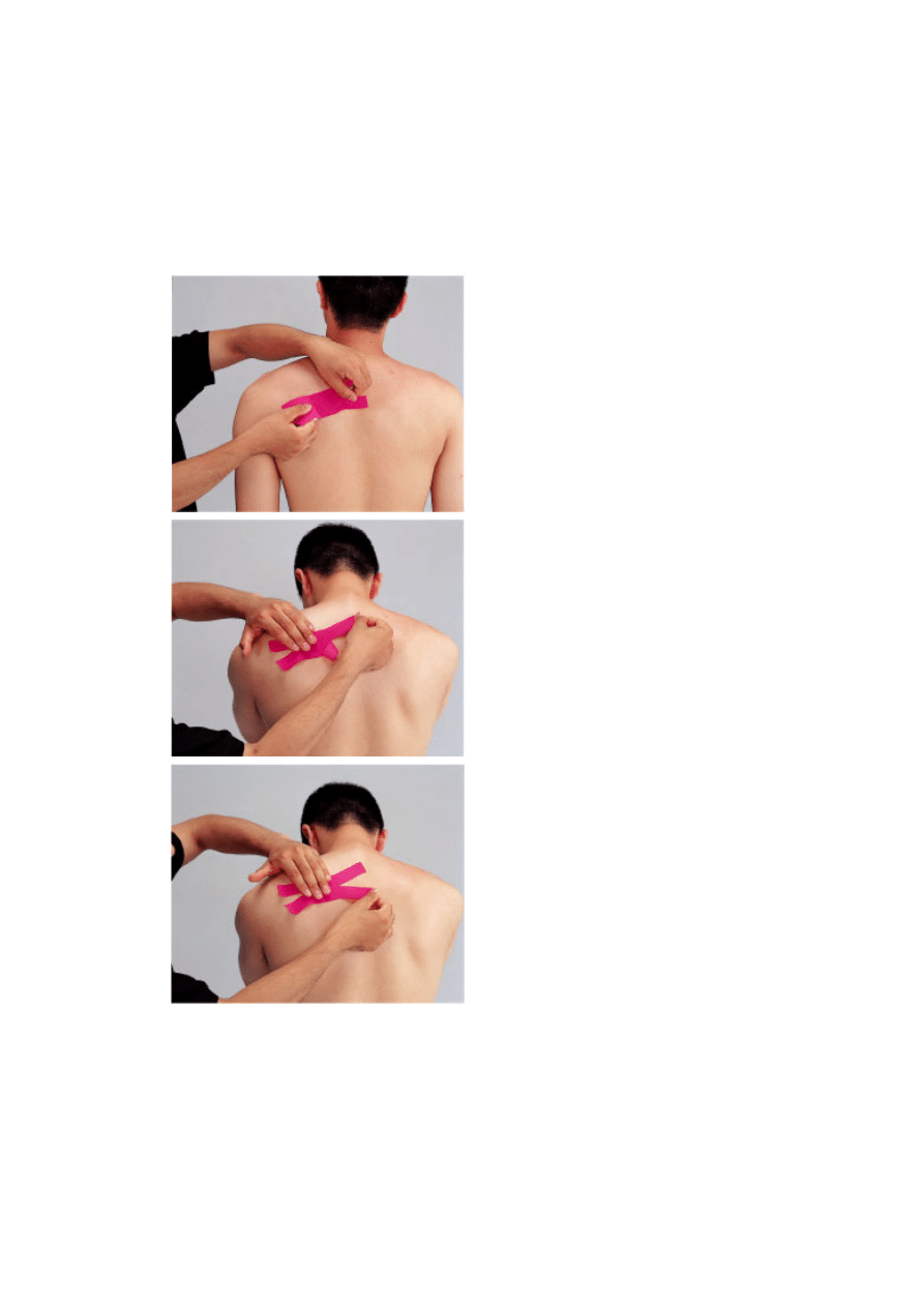
Patient position: seated
Apply the middle of the Kinesio Tex
strip over the muscle belly following
the fiber direction. The muscle is
located along the midpoint of the
medial vertebral border of the scapula
one to two fingerbreadths below the
spine of the scapula.
Peel the Kinesio Tex from the paper
liner and place the tails of the tape
temporarily on the skin. Do not
activate the glue by rubbing.
Cross the arms in front of the body,
raise the elbows, flex the head and
neck, and round the upper back to
place the tissues over the rhomboid
muscle under tension.
With the hand over the center of the
“X” shaped Kinesio Tex, pull the skin
laterally as the tails of the tape are
applied to increase tissue tension.
Apply the upper medial tail of the “X”
to spinous process of the 2nd thoracic
vertebra.
With the hand over the center of the
“X” shaped Kinesio Tex, pull the skin
laterally as the tails of the tape are
applied to increase tissue tension.
Apply the lower medial tail of the “X”
to the spinous process of the 5th
thoracic vertebra.
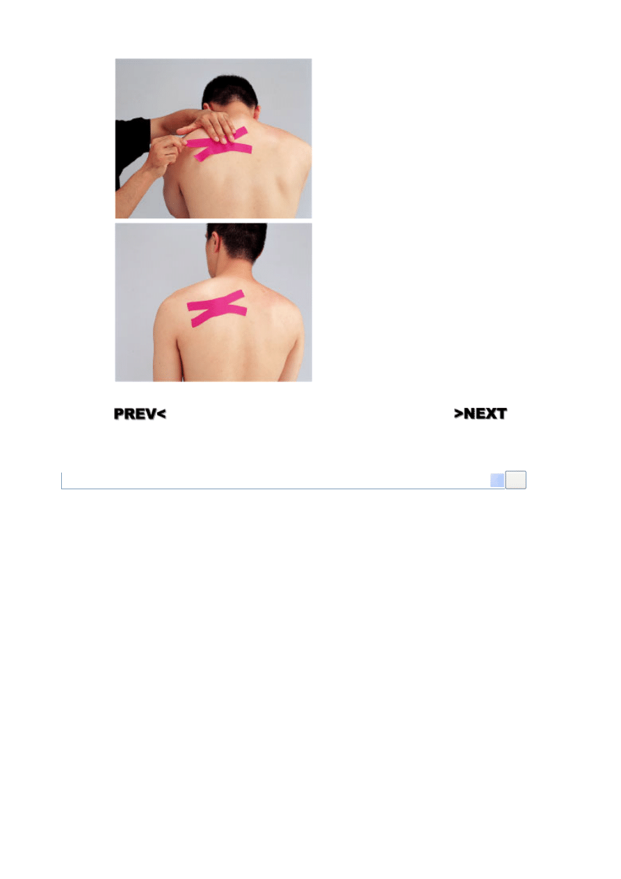
With the hand over the center of the
“X” shaped Kinesio Tex, pull the skin
medially as the tails of the tape are
applied to increase tissue tension.
Apply the two tails to the vertebral
border of the scapula, following the
fiber direction of the rhomboid major.
Strona 1 z 2
2007-12-14
http://www.kinesiotex.com/portal/systems/KinesioWebsite/jsp/ktmethod_enGB_6.kin
Copyright © 2005-2007 Kinesio Taping Association. All Rights Reserved
Completed Kinesio Tex application:
rhomboid major
0006-SH-XX Rhomboid Major
Go
Strona 2 z 2
2007-12-14
http://www.kinesiotex.com/portal/systems/KinesioWebsite/jsp/ktmethod_enGB_6.kin
Wyszukiwarka
Podobne podstrony:
Psoas Major KT method
Rhomboid Major and Minor Muscles KT method
Pectoralis Major (both Portions) KT method
Pectoralis Major (Sternocostal Portions) KT method
Pectoralis Major (Clavicular Portions) KT method
Opponens Pollicis KT method
Fibularis (Peroneus) Longus KT method
Flexor Hallucis Longus KT method
Popliteus KT method
Posterior Diaphragm KT method
Palmaris Longus KT method
Oblique Abdominis Externus KT method
Deltoid Anterior KT method
Iliocostalis Thoracis (Superficial Erector Spinae) KT method
Dorsal Muscles Cross KT method
Extensor Carpi Ulnaris KT method
Lower Abdomen KT method
Pharynx KT method
Middle Trapezus KT method
więcej podobnych podstron