
Biomaterials 23 (2002) 3749–3756
In vitro behavior of silicate glass coatings on Ti6Al4V
E. Saiz
a
, M. Goldman
a
, J.M. Gomez-Vega
a
, A.P. Tomsia
a,
*, G.W. Marshall
b
,
S.J. Marshall
b
a
Lawrence Berkeley National Laboratory, Materials Sciences Division, MS 62-203 1 Cyclotron Road, Berkeley, CA 94720, USA
b
University of California, Department of Restorative Dentistry, San Francisco, CA, USA
Received 8 October 2001; accepted 14 March 2002
Abstract
The in vitro response in simulated body fluid (SBF) of silicate glass coatings on Ti6Al4Vwas evaluated. Glasses belonging to the
SiO
2
–CaO–MgO–Na
2
O–K
2
O–P
2
O
5
system were used to prepare 50–70 mm thick coatings on Ti6Al4V, employing a simple
enameling technique. Glasses with silica content higher than 55 wt% can be used to prepare coatings that do not crack or delaminate
and exhibit good adhesion to the alloy. It has been found that coatings with silica content lower than 60 wt% are more susceptible to
corrosion and precipitate carbonated hydroxyapatite on their surface during in vitro tests. However, these coatings have a higher
thermal expansion than the metal and are under tension. After 2 months in SBF cracks grow in the coating that reach the glass/
metal interface and initiate delamination. Glasses with silica content higher than 60 wt% are more resistant to corrosion and have
lower thermal expansion. These coatings do not crack but they do not precipitate apatite even after 2 months in SBF. r 2002
Elsevier Science Ltd. All rights reserved.
Keywords: Coatings; Glass; Ti6Al4V; Hydroxyapatite; In vitro
1. Introduction
Titanium and Co–Cr alloys are the most popular
choices for fabrication of orthopedic implants where
high strength is required. These alloys exhibit good
mechanical properties but are bioinert and attach to the
bone through form fit or frictional connections. The
weak bone to implant adhesion can result in implant
loosening and failure. Coating the metallic implants
with bioactive layers allows biological interaction
between the bone and the implant and can consequently
improve adhesion. Furthermore, the coatings could
protect the implants from corrosion, limiting the release
of metallic ions into the body [1–4].
In previous work, we reported the development of
bioactive coatings using a new family of glasses in the
SiO
2
–Na
2
O–K
2
O–CaO–MgO–P
2
O
5
system [5–7]. The
glasses are based on Hench’s Bioglass
s
and have silica
contents ranging from 44 to 70 wt%. Coatings on Ti and
Ti6Al4Vwere successfully fabricated using glasses
whose silica content was >55%. At lower silica
contents, the coatings cracked due to the high stresses
that result from the large difference in thermal expan-
sion between the glasses and the alloys.
In vitro tests in cell-free solutions with ionic
concentrations similar to those of body fluids allow
analysis of the chemical and microstructural evolution
of the coatings under conditions that simulate their
biological interactions with the body. Because the
solution-precipitation processes that occur on the coat-
ing surfaces have a determinant role on their bone
bonding mechanisms, studies in simulated body fluid
(SBF) provide fundamental data to predict and under-
stand the in vivo behavior and long-term stability [8,9].
The purpose of the present work is to systematically
evaluate the in vitro behavior of the silicate glass
coatings on Ti6Al4V. The study focuses on the ability
of the coatings to precipitate apatite (the mineral
component of the bone) and the effects of long-term
immersion in SBF on the coating adhesion to the metal.
2. Experimental
The starting glasses were prepared using a conven-
tional procedure. The appropriate reagents (SiO
2
*Corresponding author. Tel.: +1-510-486-4918; fax: +1-510-486-
6086.
E-mail address:
aptomsia@lbl.gov (A.P. Tomsia).
0142-9612/02/$ - see front matter r 2002 Elsevier Science Ltd. All rights reserved.
PII: S 0 1 4 2 - 9 6 1 2 ( 0 2 ) 0 0 1 0 9 - 6

(99.5%, purity),
1
CaCO
3
(99.9%),
2
MgO (98.6%),
2
K
2
CO
3
(99%),
3
NaHCO
3
(99.5%)
2
and
NaPO
3
(99.7%)
3
) were mixed in ethanol using a high-speed
stirrer. The mixture was first dried at 801C for 12 h and
then fired in air at temperatures ranging between 14001C
and 15001C for 4 h in a Pt crucible. The melt was
cast into a graphite mold to obtain glass plates
(
B50 50 5 mm
3
) that were subsequently milled in a
planetary agate mill. In order to prepare the coatings, a
suspension of the glass powder (particle size
o20 mm) in
ethanol was deposited on Ti6Al4Vplates (99.0% purity,
15 10 1 mm
3
), which had been previously polished
with diamond (1 mm particle size) and cleaned in
acetone and ethanol. Afterwards, the coatings were
dried in air at 751C overnight and fired at temperatures
ranging between 8001C and 8201C for 30 s in order to
make the glass flow and adhere to the metal [5–7]. The
compositions of the coatings are shown in Table 1 (in
the glass designation the number after 6P corresponds to
its silica content in wt%). The coatings exhibited good
adhesion to the alloy (they did not delaminate during
interfacial indentation tests) and their final thickness
ranged between 50 and 70 mm [5,7].
The in vitro response of the coatings was studied by
immersing the specimens (10 10 mm
2
area) in 20 ml of
SBF (Table 2) at 36.51C for fixed periods of time up to 2
months. The solution was buffered at the physiological
pH 7.25 at 36.51C with 50 mm trishydroxymethyl
aminomethane [(CH
2
OH)
3
CNH
2
] and 45 mm hydro-
chloric acid (HCl). In order to analyze the effect of the
amount of SBF, the same tests were repeated using
200 ml of SBF. The role of the calcium ions in the
solution was analyzed by conducting in vitro tests in a
solution having identical composition of SBF and
buffered the same way, but without Ca
2+
(Table 2).
After the required times, the samples were removed
from the liquid, rinsed in distilled water and dried with
an air gun. Inductively coupled plasma (ICP) analysis
was performed on the remaining SBF to monitor the
concentrations of Ca, P, Si, and Mg. After drying, the
coating surfaces and polished cross sections (up to 1 mm
diamond) were analyzed by X-ray diffraction (XRD),
atomic force microscopy (AFM), X-ray photoemesion
(XPS), scanning electron microscopy with associated
calibrated energy dispersive analysis (SEM-EDS) and
Fourier
Transform
Infrared
Spectromicroscopy
(FTIRSM) in the Advanced Light Source (ALS) at
Lawrence Berkeley National Laboratory. The contact
AFM analysis was carried out using a Park M5
instrument (Park Scientific Instruments) using the
constant force mode and Ultralever silicon tips from
Park. The XPS analysis was performed on a Physical
Electronics PHI 5400 ESCA using an Mg anode as an
X-ray source. The SEM analysis was performed on a
DS130C microscope (Topcon) and the EDS analysis
was done using a DX-4 system (EDAX). Prior to the
EDS analysis the samples were coated with a thin
carbon layer and the system was calibrated using glasses
and calcium phosphates of known composition. The
accelerating voltage was 15 kV, resulting in a spot size of
B1 mm. The FTIRSM uses the synchrotron beam at the
ALS as an external light source in a Nicolet Magna 760
bench with Nic-Plan IR Microscope, which allows
focusing the beam in very small diameters with little
loss of signal. With a 32 objective, the full-width
half-maximum spot size is
B10 mm; this spot size
becomes diffraction limited at longer wavelengths.
The spectra were taken in the reflectance mode using a
KBr beam splitter and a mercury cadmium telluride
detector.
Crack growth in the coatings, when immersed in SBF,
was analyzed by following the evolution of well-defined
cracks generated by Vickers indentations (1.2 kg load)
on the coating surfaces. Crack growth was followed by
immersing the indented samples in SBF or dehydrated
mineral oil at 36.51C and measuring the crack
lengths after selected times up to 8 days using optical
microscopy. A qualitative test of adhesion between
the coatings and the metals after immersion in
SBF
was
performed
using
Vickers
indentations
at the glass/metal interfaces on polished cross sections
with
loads
ranging
from
0.05 to
1.2 kg in
air
and SBF, and analyzing the relative crack resistance
[5].
Table 1
Compositions and thermal expansion of the glasses used in the
preparation of the coatings analyzed in this work (in wt %)
SiO
2
Na
2
O
K
2
O
CaO
MgO
P
2
O
5
a (1C
1
)
Bioglass
s
45.0
24.5
24.5
6.0
15.1
6P57
56.5
11.0
3.0
15.0
8.5
6.0
10.8
6P61
61.1
10.3
2.8
12.6
7.2
6.0
10.2
6P68
67.7
8.3
2.2
10.1
5.7
6.0
8.8
Table 2
Ion concentrations of the solutions used in this research and of human
plasma.
Ion concentration (mM)
Na
+
K
+
Ca
2+
Mg
2+
Cl
HCO
3
HPO
2
4
SO
2
4
SBF
142.0 5.0 2.5
1.5
147.8
4.2
1.0
0.5
Ca-free solution 142.0 5.0 0
1.5
147.8
4.2
1.0
0.5
Human plasma 142.0 5.0 2.5
1.5
103.0 27.0
1.0
0.5
1
Cerac, USA.
2
JT Baker, USA.
3
Allied Chemical, USA.
E. Saiz et al. / Biomaterials 23 (2002) 3749–3756
3750
3. Results
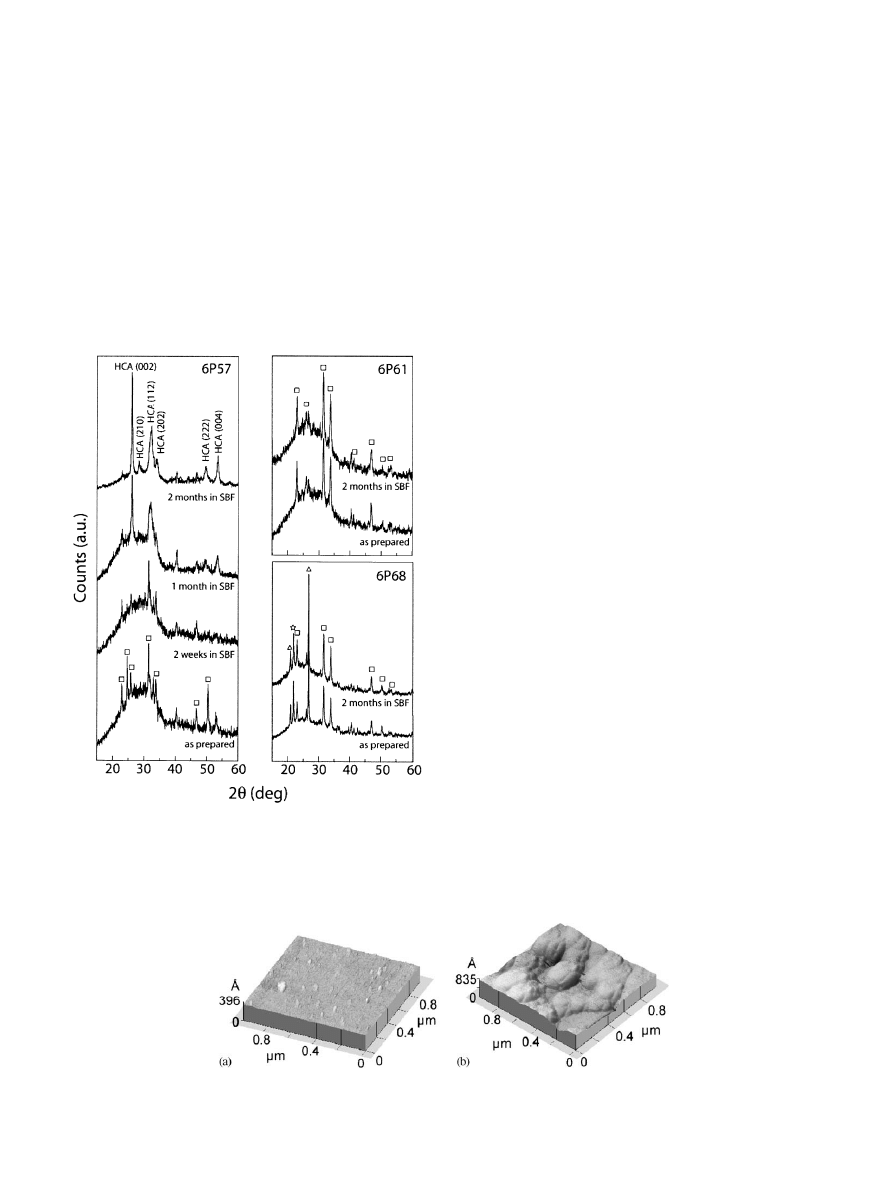
Fig. 1 shows the X-ray diffraction patterns of the
coating surfaces after different times in SBF. The peaks
that appear in the XRD of the starting coatings are due
to partial crystallization of the glass during the
fabrication procedure (Fig. 1). In all the cases the
volume of crystalline phases is below 5 vol% [5]. No
appreciable changes occur in the patterns of 6P61 and
6P68 coatings after 2 months in SBF (Fig. 1). After 1
month in SBF the apatite peaks are clearly visible in the
X-ray analysis of the 6P57 coatings. The relative
intensity of the peaks suggests that the apatite crystals
growth preferentially with the c-axis perpendicular to
the coating.
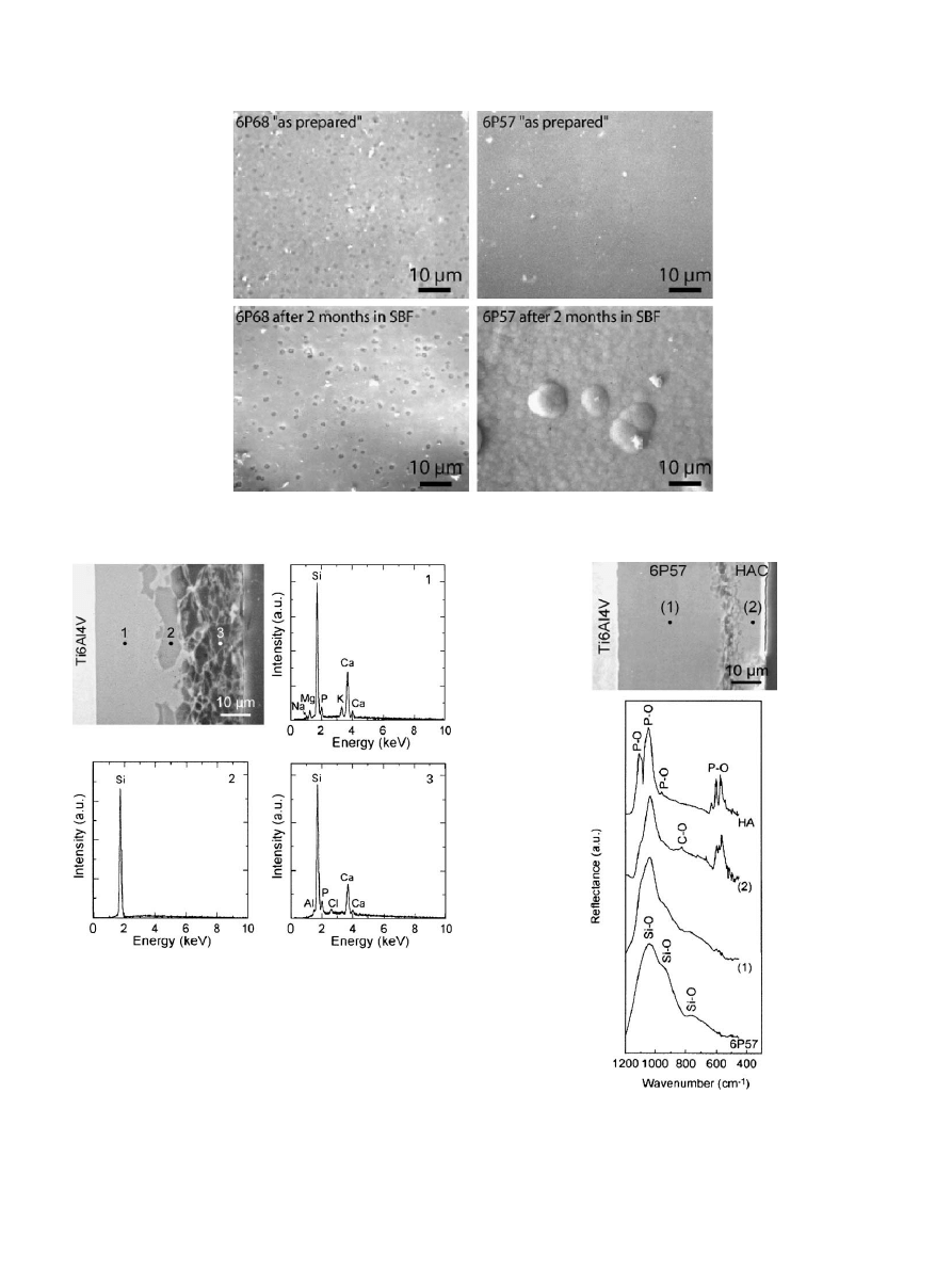
The 6P57 coatings corrode very fast in SBF (Figs. 2
and 3). After 1 day in SBF morphological changes can
be clearly observed on the surface of the coatings using
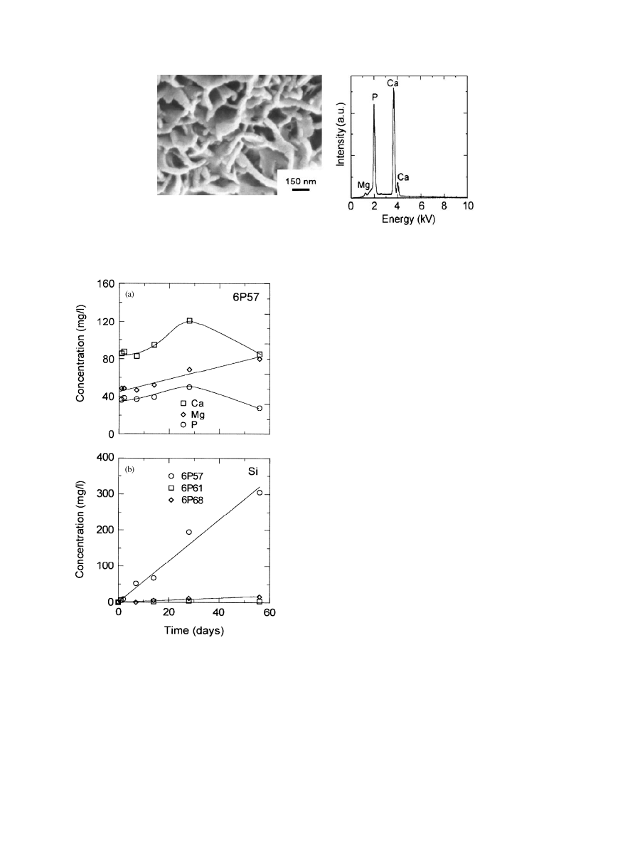
AFM (Fig. 2). Cross sectional analyses of the coatings
soaked for 2 weeks show three separate regions (Fig. 4):
a layer of the remaining glass in contact with the metal;
a surface layer rich in silica with small amounts of Ca, P
and Al; and a Si-rich layer between them. After 1 month
in SBF the surface layer disappears and is substituted by
a
B5 mm thick layer of HCA that grows on the Si-rich
surface (Fig. 5). The apatite crystals were detected by
XRD (Fig. 1) and they completely cover the coating
surface (Fig. 6). This behavior was similar for the
samples immersed in 20 or 200 ml of SBF. The apatite
crystals have flake-like morphology with sizes in
the range of 50–100 nm. In the analysis of the apatite
precipitates using FTIRSM (Fig. 5) the P–O peak at
B510 m
1
and the C–O shoulder at
B950 cm
1
(that
corresponds to the C–O vibration mode in CO
2
3
) were
consistent with carbonated hydroxyapatite (HCA,
Ca
10
(PO
4
)
3
(CO
3
)
3
(OH)
2
) [8,10]. The calibrated EDS
analysis indicates that the apatite incorporates 1–
5 wt% MgO in its structure. No other elements could
be detected in the precipitated apatite by EDS or XPS.
The precipitation of HCA was accompanied by a
decrease in the Ca and P concentrations in SBF
(Fig. 7). The apatite layer grew to a thickness of
B20 mm in the samples soaked for 2 months (Fig. 8).
The composition and structure of the glass layer that
remained attached to the metal were the same as the
original 6P57 coating (Figs. 5 and 8). Leaching of Si into
the solution continued at a constant linear rate even
after the precipitation of HCA. No apatite precipitated
on the coatings immersed in the solution without
calcium.
The coatings prepared using 6P61 and 6P68 glasses
are much more resistant to corrosion in SBF (Figs. 3
and 8). No corrosion or variation in the coating
composition could be observed in the coatings’ cross
Fig. 1. XRD diffraction patterns of 6P57, 6P61 and 6P68 coatings
after immersion in SBF for different times. The main crystalline phases
are: 2.4CaO 0.6Na
2
OP
2
O
5
(&); SiO
2
, quartz (n); SiO
2
, cristobalite
($); Ca
10
(PO
4
)
3
(CO
3
)
3
(OH)
2
carbonated hydroxyapatite,(HCA).
Fig. 2. AFM scans of the surface of a 6P57 after firing (a) and after 1 day in SBF (b), showing the morphological change during the early stages of
glass dissolution.
E. Saiz et al. / Biomaterials 23 (2002) 3749–3756
3751
sections using SEM and EDS elemental line profiles
even after soaking for 2 months in SBF. For 6P61
coatings, the leaching of silica into SBF is more than ten
times slower than for 6P57. After 2 months no
measurable increase in the Si content was observed in
the solution containing 6P67 coatings.
Fig. 3. SEM micrographs of the surface of 6P57 and 6P68 coatings after soaking in SBF. The surface of 6P68 coatings remains unaltered even after 2
months whereas apatite has precipitated in 6P57.
Fig. 4. SEM and associated EDS analysis of the cross section of a
6P57 coating after 2 weeks in SBF. The Cl peak on the surface layer is
due to infiltration of the mounting resin.
Fig. 5. Cross section of a 6P57 coating after 1 month in SBF.
FTIRSM of selected points is also presented. A layer of the original
glass remains attached to the metal. The P–O and C–O bands on the
FTIRSM analysis of the top layer are consistent with hydroxycarbo-
nate apatite.
E. Saiz et al. / Biomaterials 23 (2002) 3749–3756
3752
The corrosion of the 6P57 coatings is not homo-
geneous. In some areas the coating has corroded all the
way down to the glass/metal interface after 2 weeks in
SBF (Fig. 9). This resulted in the formation of cracks
that after 2 months reached the glass/metal interface and
initiated delamination (Fig. 10). No cracks were ob-
served on 6P61 and 6P67 coatings. The indentation
cracks placed on the 6P57 coatings grew during
immersion
in
SBF
with
decreasing
velocities
of
10
10
m/s or lower, up to the lengths of
B200 mm
(Fig. 11). No appreciable crack growth occurred on the
other coatings or on the 6P57 coatings placed in
dehydrated oil.
During the indentation tests performed at the glass/
metal interface after immersion in SBF the cracks do not
propagate along the interface but rather tend to be
driven into the glass as seen with the original coatings
before the in vitro tests [5,6].
4. Discussion
The in vitro response of the coatings depends strongly
on their composition, in particular on their silica
content. The observed behavior is similar to that
reported for bulk glasses in the SiO
2
–Na
2
O–CaO–P
2
O
5
system (which includes Bioglass
s
), and other related
compositions [11–18]. Low silica compositions have a
more open network structure that facilitates ion
exchange with the solution, resulting in faster glass
corrosion and precipitation of apatite. In the coatings
studied here, the critical silica content was around
60 wt%. A layer of carbonated apatite grew in vitro on
the surface of the coatings with lower silica content
(6P57) (Fig. 5) whereas coatings with higher silica
content were more resistant to corrosion and did not
form apatite. The HCA layer grew with time with the
crystalline c-axis preferentially oriented perpendicular to
the coating surface as observed for bulk bioactive
glasses [19].
The analysis of the coating behavior in SBF is
consistent with a mechanism of apatite formation
similar to that described by Hench for Bioglass
s
[13].
The steps involved are: the exchange of Na
+
and K
+
Fig. 7. Evolution of Ca–P and Si concentrations in SBF. The decrease
of the Ca and P concentrations in the solution containing 6P57
coatings coincides with the precipitation of HCA. The leaching of silica
continues at a linear constant rate in 6P57 coatings even after 2 months
in SBF. There is not a measurable increase in the Si concentration of
the solution containing 6P68 coatings.
Fig. 6. SEM image and associated EDS analysis of the apatite crystals precipitated on 6P57 after 2 months in SBF. The crystals contain 1–5 wt%
MgO.
E. Saiz et al. / Biomaterials 23 (2002) 3749–3756
3753
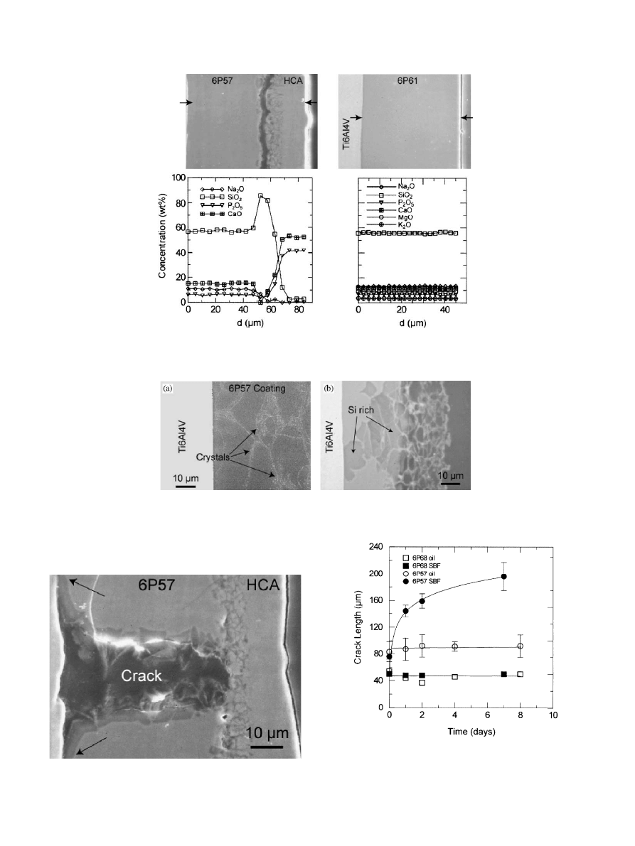
Fig. 8. Elemental cross section analysis (SEM-EDS) of 6P57 and 6P61 coatings after 2 months in SBF. A
B20 mm thick apatite layer is visible on the
6P57 coating surface growing on a Si-rich region. No appreciable changes can be observed on the 6P61 coatings.
Fig. 9. (a) SEM image of a 6P57 coating, after etching for 5 s with 5 vol% HFl the crystallization in the glass is visible; (b) inhomogeneous corrosion
in a 6P57 coating after 2 weeks in SBF.
Fig. 11. Growth of indentation cracks on 6P57 and 6P68 coatings in
SBF and dehydrated mineral oil. On the 6P57 coatings in SBF the
crack grows with decreasing velocity as it moves away from the
indentation stress field.
Fig. 10. Crack formed in the 6P57 coating after 2 months in SBF. The
crack has reached the interface and initiated delamination.
E. Saiz et al. / Biomaterials 23 (2002) 3749–3756
3754

from the glass with H
+
or H
3
O
+
from solution,
accompanied by the loss of soluble silica into the
solution and the formation of silanols on the glass
surface; condensation and repolymerization of a SiO
2
-
rich layer on the surface; migration of Ca
2+
and PO
3
4
through the silica-rich layer forming a CaO–P
2
O
5
-rich
film that incorporates calcium and phosphates from
solution; finally, the crystallization of the amorphous
calcium phosphate film to form an apatite layer. De Aza
et al. [20] have pointed out that the increase in pH on
the glass surface due to the ionic exchange between
the labile cations Na
+
, K
+
, Ca
+
, etc., is necessary for
the partial dissolution of the silica-rich layer and the
subsequent apatite precipitation. The observed dissolu-
tion of silica into SBF (Fig. 7) during the in vitro tests of
6P57 coatings is consistent with the observation of De
Aza et al. [20] that the high local pH attained at the
bioactive glass/SBF interface promotes the partial
dissolution of the silica hydrogel layer that forms on
the glass surface.
In coatings made from 6P57 glasses that were
immersed for 2 weeks in SBF the formation of a silica-
rich layer,
B10 mm thick, containing also Ca and P can
be observed (Fig. 4). This agrees with the sequence
proposed by Hench in which a silica-rich layer forms on
the surface, through which the Ca and P species migrate.
The presence of Al traces in this layer also was observed
on the related bulk glasses and can be due to the
presence of alumina impurities in the starting oxides
used in the glass preparation. Even though alumina
additions at levels higher than 7.5 wt% have been
reported to reduce the bone bonding ability of bioactive
glasses [21] the low levels found here (the Al
2
O
3
could
not be detected on the starting coatings using EDS) did
not inhibit apatite precipitation. This is in agreement
with the studies of the effect of Al
2
O
3
in bioactive glasses
published by Ohura et al. [22] and Andersson et al. [23].
The initial increase in the Ca, P, and Mg concentra-
tions observed in the SBF containing 6P57 coatings
(Fig. 7) is due to the ionic exchange with the glass. After
1 month the precipitation of apatite on the glass surface
resulted in the observed decrease of the Ca and P
concentrations. It should be noted that the apatite layer
is porous (Fig. 3) and does not protect the coating from
corrosion in SBF, confirmed by the constant leaching of
Si (Fig. 7). The overall process of leaching and HCA
formation was slower in the coatings than in the
Bioglass
s
due to their higher silica content that resulted
in a more compact glass structure. However, the
formation of apatite was independent of the amount
of SBF used in the test (from 20 to 200 ml), as expected
if the apatite precipitates after a process similar to that
described by Hench, and not because of saturation of
the solution with Ca and P from the glass. Regina et al.
[8] have pointed out that the presence of calcium and
phosphorus in solution accelerates the precipitation of
hydroxyapatite. Our results suggest that the high silica
glasses used in the coatings require Ca in the solution for
the formation of HCA, due to their slower ionic
exchange rate.
MgO and K
2
O were added to the glasses in order to
adjust their thermal expansions and softening points
such that enameling could be carried out at tempera-
tures below the a
-b transformation of Ti in the alloy
(955–10101C), without generating large thermal stresses.
[5] The work of Brink et al. [12] suggests that small
amounts of K
2
O and MgO do not affect bioactivity of
glasses, however, there have been reports that Mg
inhibits the precipitation of hydroxyapatite [8,9,24]. Our
results indicated that coatings containing as much as
8.5 wt% of MgO and 56.5 wt% of SiO
2
precipitated
apatite during the in vitro tests. The precipitated apatite
incorporates 1–5 wt% MgO into its structure. The lack
of MgO on the surface layer of the coatings immersed
for 2 weeks in SBF suggests that the magnesium is
coming from the solution. However, there has not been
a report of magnesium present on the apatite precipi-
tated on the surface of MgO-free glasses.
The small amount of crystallization that occurs
during the firing of the coatings does not compromise
the ability of the glass to form HCA. Crystallization and
inhomogenities in the initial coating can be the cause of
the non-uniform leaching of glass elements into the
solution. As a result, the amorphous SiO
2
-rich layer that
forms at intermediate times is not continuous and can
reach the glass/metal interface in some areas (Figs. 4 and
9).
A simple elastic analysis for 50 mm coatings on
Ti6Al4Vpredicts tensile stresses of
B50 and 25 MPa,
for 6P57 and 6P61, respectively, and compressive
stresses of
B–25 MPa for 6P68 [5].
In 6P57 coatings, these stresses could be high enough
to drive the slow growth of cracks in SBF. Crack
formation can be detrimental for the long-term stability
of the coating, since the cracks eventually reach the
interface and initiate delamination. The precipitation of
a hydroxyapatite layer on the coating surface does not
shield the crack from the solution and it continues
growing (Fig. 10). Because of the lower stresses and
higher resistance to corrosion, cracks did not grow in
the 6P61 and 6P68 coatings. The results of the
indentation tests qualitatively agree with these observa-
tions. In 6P68 coatings the compressive residual stresses
effectively arrest the growth of indentation cracks.
However, in the 6P57 coatings in SBF the crack growth
is driven by the combined stresses from the thermal
expansion mismatch and the indentation field. As the
cracks grow away from the indentation stress field, the
overall stresses decrease and the cracks decelerate.
Typically, the indentation stress field decreases signifi-
cantly at distances greater than the indent size
(
B40 mm). Accordingly, the results (Fig. 11) indicate
E. Saiz et al. / Biomaterials 23 (2002) 3749–3756
3755

an upper limit for the crack velocity in SBF under the
thermal stresses of
B10
10
m/s. After 7–8 days the
corrosion of the 6P57 surface hampered the measure-
ment of the crack length. The cracks did not grow on the
6P57 coatings in dehydrated mineral oil, emphasizing
the role of SBF on the corrosion of the glass.
As expected, immersion in SBF did not affect the
adhesion of the interfaces that were not in contact with
the solution. During the indentations at the glass/metal
interfaces after the in vitro tests, the cracks did not
propagate along the interface, but rather tended to be
driven into the glass as seen in the original coatings [5,6].
5. Conclusions
The in vitro behavior of the silicate glass coatings
analyzed in this work is similar to that of bulk glasses.
Coatings with silica content lower than 60 wt% pre-
cipitated apatite during in vitro testing. The mechanism
of apatite formation is similar to that described by
Hench for Bioglass
s
. However, due to their lower silica
content, the thermal expansion of these coatings is
higher than that of Ti and the tensile thermal stresses
generated during processing drove the slow growth of
cracks in SBF. The cracks eventually reached the
interface and initiated delamination.
Higher silica coatings did not form apatite but were
more resistant to corrosion and slow crack growth. At
the moment, graded coatings that have glasses with high
silica content in contact with the metal and a low silica
glass on their surface are under development in order to
improve their long term stability while maintaining a
good biological response.
Acknowledgements
This work was supported by the NIH/NIDCR grant
1R01DE11289. Jose M. Gomez-Vega wishes to thank
the Spanish Ministry of Education (MEC) for financial
support. The Advanced Light Source is supported by
the Director, Office of Science, Office of Basic Energy
Sciences, Materials Sciences Division, of the US
Department of Energy under Contract No. DE-AC03-
76SF00098 at Lawrence Berkeley National Laboratory.
References
[1] Hench LL, Andersoon O. Bioactive glass coatings. In: Hench LL,
Wilson J, editors. An introduction to bioceramics. New Jersey:
World Scientific, 1993. p. 239–60.
[2] Sousa SR, Barbosa MA. Electrochemistry of AISI-316L stainless
steel in calcium phosphate and protein solutions. J Mater Sci
1991;2:19–26.
[3] Sousa SR, Barbosa MA. The effect of hydroxyapatite thickness
on metal ion release from stainless steel substrates. J Mater Sci
1995;6:818–23.
[4] Sousa SR, Barbosa MA. Effect of hydroxyapatite thickness on
metal
ion
release
from
Ti6Al4Vsubstrates.
Biomaterials
1996;17:397–404.
[5] Gomez-Vega JM, Saiz E, Tomsia AP. Glass-based coatings for
titanium implant alloys. J Biomed Mater Res 1999;46:549–59.
[6] Pazo A, Saiz E, Tomsia AP. Silicate glass coatings on Ti-based
implants. Acta Mater 1998;46:2551–8.
[7] Pazo A, Saiz E, Tomsia AP. Bioactive coatings on Ti and
Ti6Al4Valloys for medical applications. In: Tomsia AP, Glaeser
AM, editors. Ceramic microstructures: control at the atomic level.
Berkeley: Plenum Press, 1998. p. 543–50.
[8] Regina M, Filgueiras MRT, Latorre G, Hench LL. Solution
effects on the surface reactions of three bioactive glass composi-
tions. J Biomed Mater Res 1993;27:1485–93.
[9] Ebisawa Y, Kokubo T, Ohura K, Yamamuro T. Bioactivity of
CaOSiO
2
-based glasses—In vitro evaluation. J Mater Sci
1990;1:239–44.
[10] Nelson DGA, Featherstone JDB. Preparation, analysis, and
characterization of carbonated apatites. Calcified Tissue Intl
1982;34:S69–81.
[11] Ogino M, Hench LL. Formation of calcium phosphate films on
silicate glasses. J Non-Crys Sol 1980;38&39:673–8.
[12] Brink M, Turunen T, Happonen RP, Yli-Urpo A. Compositional
dependence of bioactivity of glasses in the system Na
2
O–K
2
O–
MgO–CaO–B
2
O
3
–P
2
O
5
–SiO
2
. J Biomed Mater Res 1997;37:
114–21.
[13] Hench LL. Bioceramics: from concept to clinic. J Am Ceram Soc
1991;74:1487–570.
[14] Kim CY, Clark AE, Hench LL. Early stages of calcium phosphate
layer formation in bioglasses. J Non-Crys Sol 1989;113:195–202.
[15] Kim CY, Clark AE, Hench LL. Compositional dependence of
calcium phosphate layer formation in fluoride bioglasses. J
Biomed Mater Res 1992;26:1147–61.
[16] Ogino M, Ohuchi F, Hench LL. Compositional dependence of the
formation of calcium phosphate films on bioglass. J Biomed
Mater Res 1980;15:55–64.
[17] Lockyer MWG, Holland D, Dupree R. NMR investigation of the
structure of some bioactive and related glasses. J Non-Crys Sol
1995;188:207–19.
[18] Hill R. An alternative view of the degradation of bioglass. J Mater
Sci Lett 1996;15:1122–5.
[19] Rehman I, Knowles JC, Bonfield W. Analysis of in vitro reaction
layers formed on Bioglass(R) using thin- film X-ray diffraction
and ATR-FTIR microspectroscopy. J Biomed Mater Res
1998;41:162–6.
[20] de Aza PN, Guitian F, Merlos A, Loratamayo E, de Aza S.
Bioceramics—simulated body fluid interfaces—pH and its influ-
ence of hydroxyapatite formation. J Mater Sci 1996;7:399–402.
[21] Gross U, Kinne R, Schmitz HJ, Strunz V. The response of bone
to surface active glass/glass–ceramics. In: Williams DF, editor.
CRC critical reviews in biocompatibility. Boca Raton: CRC
Press, 1988. p. 2.
[22] Ohura K, Nakamura T, Yamamuro T, Ebisawa Y, Kokubo T,
Kotoura Y, Oka M. Bioactivity of CaO SiO
2
glasses added with
various ions. J Mater Sci 1992;3:95–100.
[23] Andersson OH, Karlsson KH, Kangasniemi K, Yli- Urpo A.
Models for physical properties and bioactivity of phosphate opal
glasses. Glastech Ber 1988;61:300–5.
[24] Li P, Ohtsuki C, Kokubo T, Nakanishi K, Soga N, Nakamura T,
Yamamuro T. Effects of ions in aqueous media on hydroxyapatite
induction by silica gel and its relevance to bioactivity of bioactive
glasses and glass-ceramics. J Appl Biomater 1993;4:221–9.
E. Saiz et al. / Biomaterials 23 (2002) 3749–3756
3756
Document Outline
Wyszukiwarka
Podobne podstrony:
in vitro, studia rolnictwo, rok IV
Kultury in vitro roslin rozmnazanie klonalne
In vitro antitumor actions of extracts
In vitro truskawka id 212540 Nieznany
1 1 Podstawowe definicje; główne kierunki przemian rozwojowych roślinnych tkanek in vitro(1)
Życie ludzkie świętość czy zabawka nt in vitro
In vitro, Sem 1, TMR3
6 Hodowle komórek skóry w warunkach in vitro
Kościół katolicki wobec zapłodnienia In vitro, Etyka, Bioetyka
IN VITRO GRZECH CZY SZANSA
In vitro tulipan
in vitro 2
2008 5 SEP Practical Applications and New Perspectives in Veterinary Behavior
In vitro liliowce
In vitro a współczesna cywilizacja, RODZINA
In vitro groźne, godność życia-in vitro
wyklad V in vitro
więcej podobnych podstron