
M
OLECULAR
B
IOTECHNOLOGY
Volume 23, 2003
Restriction Endonucleases
225
225
Molecular Biotechnology
2003 Humana Press Inc. All rights of any nature whatsoever reserved. 1073–6085/2003/23:3/225–243/$20.00
1. Introduction
Restriction endonucleases, which cleave both
strands of DNA in a site-specific manner, are a fun-
damental tool of molecular biology. Discovery of
endonucleases began in the 1960s and led to com-
mercial availability in the early 1970s. The number
of characterized enzymes continues to grow as does
the number of vendors and the size of their product
lines. Although many similarities exist among
endonucleases in terms of structures, mechanisms,
and uses, important differences remain. Now a
staple of molecular biology, restriction endonu-
cleases remain an area of active research regarding
their cleavage mechanism, in vivo function, evolu-
tionary origins, and as a model for site-specific
DNA recognition. New native enzymes continue to
be discovered, known enzymes cloned, and new
endonuclease activities developed by using protein
engineering and fusions to produce novel poly-
peptides.
1.1. Diversity and In Vivo Function
Although primarily found in bacterial genomes
and plasmids, restriction endonucleases also exist
in archaea, viruses, and eukaryotes. It is estimated
that 1 in 4 bacteria examined contain one or more
(1). Neisseria and Helicobacter pylori are particu-
larly rich sources for multiple enzymes in a single
strain. Respectively, as many as 7 and 14 endonu-
clease genes have been discovered in individual
strains, although some of the genes are not actively
expressed (2,3). Including all types,
⬎3500 restric-
tion enzymes that recognize 259 different DNA
sequences are now known. The vast majority of
these, approx 3460 enzymes recognizing 234 DNA
sequences, are classified as orthodox Type II or
Type II subclasses. These are the common tools of
molecular biology with more than 500 enzymes
comprising over 200 specificities commercially
available. In addition, 58 homing endonucleases,
so-called because they are encoded by genes that
Abstract
Restriction endonucleases have become a fundamental tool of molecular biology with many commercial
vendors and extensive product lines. While a significant amount has been learned about restriction enzyme
diversity, genomic organization, and mechanism, these continue to be active areas of research and assist in
classification efforts. More recently, one focus has been their exquisite specificity for the proper recognition
sequence and the lack of homology among enzymes recognizing the same DNA sequence. Some questions
also remain regarding in vivo function. Site-directed mutagenesis and fusion proteins based on known endo-
nucleases show promise for custom-designed cleavage. An understanding of the enzymes and their proper-
ties can improve their productive application by maintaining critical digest parameters and enhancing or
avoiding alternative activities.
Index Entries: Restriction endonucleases; R/M systems; star activity; single-stranded cleavage; site-spe-
cific nickases.
Restriction Endonucleases
Classification, Properties, and Applications
Raymond J. Williams
REVIEW
*
Author to whom all correspondence and reprint requests should be addressed: Protein Purification Dept., Promega Corp., 2800 Woods
Hollow Road, Madison, WI 53711-5399. E-mail:RWilliam@Promega.com.
04/JW549/Williams/225-244
2/10/03, 9:54 AM
225

M
OLECULAR
B
IOTECHNOLOGY
Volume 23, 2003
226
Williams
are mobile, self-splicing introns or inteins, each
with a unique recognition site, have been discov-
ered. A total of 16 site-specific nickases are cur-
rently known as well. In all, 297 restriction
enzymes have been cloned and sequenced. A data-
base of all known endonucleases is updated
monthly by Dr. Richard J. Roberts and Dana
Macelis and is available at http://www.neb.com/
rebase. A number of formats are available, refer-
ences given, and statistics maintained.
Restriction endonucleases were originally named
for their ability to restrict the growth of phage in a
host bacterial cell by cleavage of the invading DNA.
In this manner, they may be acting as bacterial pro-
tection systems. The DNA of the host is protected
from restriction by the activity of a methylase(s),
which recognizes the same sequence as the restric-
tion enzyme and methylates a specific nucleotide
(4-methylcytosine, 5-methylcytosine, 5-hydroxy-
methylcytosine, or 6-methyladenine) on each strand
within this sequence. Once methylated, the host
DNA is no longer a substrate for the endonuclease.
Because both strands of the host DNA are methy-
lated and even hemi-methylated DNA is protected,
freshly replicated host DNA is not digested by the
endonuclease.
The role of restriction endonucleases as a pro-
tection system may be oversimplified however.
Various characteristics lower an enzyme’s protec-
tive potential. There would be no effect on phages
without at least a dsDNA intermediate or those for
whom the DNA was also modified at the critical
bases. A small number of phage may be methy-
lated by the host before restriction can occur, and
thus be able to propagate protectively methylated
copies of themselves. Also, large enzyme recog-
nition sites would tend to be rare in small phage
genomes. Restriction site avoidance appears to be
more important in a group of bacteria rather than
a corresponding group of phage. The endonu-
cleases generally have a longer half-life than the
corresponding methylases, a potentially lethal
problem for the host if the methylase is not prop-
erly maintained. For these reasons, it has also been
proposed that restriction-methylase systems may
be mobile, selfish genetic elements that become
essential for host survival once acquired (4,5).
2. Nomenclature and Genomic
Organization
Individual enzymes are named in accordance
with the proposal of Smith and Nathans (6).
Briefly, three letters in italics are derived from the
first letter of the genus and the first two letters of
the microbial species from which the enzyme was
derived. An additional letter without italics may
be used to designate a particular strain. This is fol-
lowed by a roman numeral to signify the first, sec-
ond, and so on, enzyme discovered from the
organism. As may be deduced from the large num-
ber of enzymes and the limited number of differ-
ent DNA sequences they recognize, many
enzymes from different biological sources recog-
nize the same DNA sequence and are called
isoschizomers. A subset wherein two enzymes
recognize the same DNA sequence but cleave at a
different position is referred to as neoschizomers.
An important point to emphasize as a result of
cloning and sequence comparison is that little if
any sequence homology exists between the endo-
nuclease and methyltransferase recognizing the
same DNA sequence. Furthermore, even restric-
tion isoschizomers may show little or no homol-
ogy, including the amino acids involved in
recognition, and as such are excellent candidates
for a comparative study of protein–DNA interac-
tion. For example, the enzymes HhaII and HinfI
are both isolated from strains of Haemophilus,
recognize GANTC, and cleave between the G and
A. However, they share only 19% identity in their
amino acid sequence (7). Endonuclease/methylase
systems recognizing the same sequence may also
exhibit different methylation patterns and restric-
tion sensitivity. Only a limited common amino
acid motif, PD...D/EXK, has been proven by
mutational or structural analysis to participate in
catalysis for 10 endonucleases. However, the 10
enzymes include members that are classified as
Type II, IIe, IIs, IV, or intron encoded endonu-
cleases (8). In contrast, general motifs have been
04/JW549/Williams/225-244
2/10/03, 9:54 AM
226

M
OLECULAR
B
IOTECHNOLOGY
Volume 23, 2003
Restriction Endonucleases
227
found for 30 6-methyladenine, 4-methylcytosine,
and 5-methylcytosine methylases (9).
Frequently referred to as an R/M system, the
restriction endonuclease and modification methy-
lase genes lie adjacent to each other on bacterial
DNA and may be oriented transcriptionally in a
convergent, divergent, or sequential manner. The
proximity of these genes appears to be universal
and is utilized in a common cloning method some-
times referred to as the “Hungarian Trick” (10).
Basically, an endonuclease is used to digest the
genomic DNA from the bacteria containing the R/
M system of interest and create a library of clones.
The expression vector used must contain the rec-
ognition site of the R/M system. Purified plasmids
from the clones are then subjected to the restric-
tion enzyme of interest in vitro. If a plasmid con-
tains the expressed methylase gene, it will be
resistant to cleavage. Often, the endonuclease is
expressed as well without the need for subcloning.
It is assumed that methylation must occur
before restriction activity to protect the host DNA.
One approach bacteria use to limit the possibility
of self-restriction is to significantly reduce the
number of recognition sites in their genomes.
Alternatively, methylase expression may precede
that of the endonuclease. One manner in which this
may be accomplished is through an open reading
frame located upstream of the endonuclease gene
encoding a “C” or control protein in some R/M sys-
tems. This C protein positively regulates the endo-
nuclease gene and allows for the activity of the
constitutively expressed methylase to precede
expression of the endonuclease (11). Such C genes
are frequently found in situations where the
methylase and endonuclease genes are in divergent
or convergent transcriptional orientations. Using
cloned R/M systems with disrupted C genes for
BamHI, SmaI, PvuII, and EcoRV R/M, various C
genes were provided on a separate plasmid. BamHI
restriction activity was equally stimulated by the
SmaI C and the BamHI C gene and only one order
of magnitude less by the PvuII C gene. The EcoRV
C gene provided no stimulation. The BamHI C
gene stimulated PvuII restriction activity as well
as the PvuII C gene (12). Why some C genes stimu-
late expression of alternate endonucleases is not
fully understood, but the phenomenon may have
evolutionary implications for R/M systems.
3. Structure, Specificity, and Mechanism
3.1. Classification and General Mechanism
Restriction endonucleases are classified
according to their structure, recognition site,
cleavage site, cofactor(s), and activator(s). Sets of
these criteria are used to define the different types
(I, II, III, and IV) and subclasses (IIe, IIf, IIs, etc.),
which are explained in detail in Table 1. Multiple
subunit and holoenzyme assemblies are possible
to achieve the needed restriction, methylase, and
specificity domains. These three domains may be
present on three separate polypeptides, two
polypeptides, or a single polypeptide. At a mini-
mum, all R/M systems share an absolute require-
ment of Mg
2
⫹
for endonuclease activity and
AdoMet (also referred to as S-adenosyl methion-
ine) as the methyl donor for methylase activity. In
general, Type I restriction requires Mg
2
⫹
,
AdoMet, and ATP (which becomes hydrolyzed).
Type II restriction requires only Mg
2
⫹
, although a
second recognition site or AdoMet may be stimu-
latory. Type III restriction requires Mg
2
⫹
and ATP
(which is not hydrolyzed) and may be stimulated
by AdoMet and a second recognition site. Type
IV restriction requires Mg and AdoMet, and also
has the unusual property of cleaving both DNA
strands on both sides of its recognition site, effec-
tively excising the site. Homing endonucleases are
a diverse group with several differences from
Types I–IV. It should be noted that Eco57I and
like enzymes, previously classified as Type IV
(25,26), have been reclassified as Type IIg (16).
In addition, it is newly proposed in this article that
the enzymes previously classified Type IIb,
including BcgI, Bsp24I, BaeI, CjeI, and CjePI, be
moved into the vacated Type IV classification due
to their unique properties as stated above.
The majority of recognition sites are four, six,
or eight bases long and palindromic. Some
enzymes recognize sites with a limited degree of
04/JW549/Williams/225-244
2/10/03, 9:54 AM
227

M
OLECULAR
B
IOTECHNOLOGY
Volume 23, 2003
228
Williams
Table 1
Restriction Enzyme Types and Classification
a
Subunit
Cofactors
2
Structure of
and
Recognition
Type
Example(s)
Endonuclease
1
Activators
Site
Cleavage Site
Methylase Properties
May be heterodimer
(1 M, 1 S) or heterotrimer
(2 M, 1 S)
Separate, single,
monomeric (M-S)
methyltransferase, a few
systems contain 2
methyltransferases
Separate, single,
monomeric (M-S)
methyltransferase
I
(EC
3.1.21.3)
Eco
KI,
Eco
AI,
Eco
BI,
Cfr
AI,
Sty
SPI, etc.
Usually a
pentameric
complex
(2 R, 2 M, and
1 S)
Mg
2⫹
,
AdoMet,
ATP
(hydrolyzed)
Interrupted
Bipartite
Distant and variable from
recognition site, for example,
Eco
KI:
AAC(N
6
)GTGC(N
⬎
400
)↓
TTG(N
6
)CACG(N
⬎
400
)↑
Orthodox
II
(EC
3.1.21.4)
Eco
RI,
Bam
HI,
Hind
III,
Kpn
I,
Not
I,
Pst
I,
Sma
I,
Xho
I, etc.
Homodimer
(2 R-S)
Mg
2⫹
Palindromic or
interrupted
palindrome,
ambiguity may
be allowed
Defined, within recognition site,
may result in a 3' overhang, 5'
overhang, or blunt end, for
example,
Eco
RI:
G
↓
A A T T C
C T T A A
↑
G
IIe
3
Nae
I,
Nar
I,
Bsp
MI,
Hpa
II,
Sac
II,
Eco
RII,
Atu
BI,
Cfr
9I,
Sau
BMKI,
and
Ksp
632I
Homodimer
(2 R-S) or
monomer
(R-S), similar to
Type II or Type
IIs
Mg
2⫹
,
A second
recognition
site, acting in
cis
or
trans
,
binds to the
endonuclease
as a allosteric
affector
Palindromic,
palindromic
with
ambiguities, or
nonpalindromic
Cuts in defined manner within the
recognition site or a short distance,
needs activator DNA containing a
recognition site for complete
cleavage, for example,
Nae
I:
GCC
↓
GGC
CGG
↑
CCG
IIf
Sfi
I,
NgoM
IV,
Cfr
10 I
,
Aat
II
Homotetramer
(4 R-S)
Mg
2⫹
Palindromic or
interrupted
palindrome, 2
cleavable rec-
ognition sites
must be bound
for activity
Defined, within recognition site,
may result in a 3' or 5' overhang,
for example,
NgoM
IV
:
G
↓
C C G G C
C G G C C
↑
G
Separate, single, mono-
meric (M-S)
methyltransferase
04/JW549/Williams/225-244
2/10/03, 9:54 AM
228

M
OLECULAR
B
IOTECHNOLOGY
Volume 23, 2003
Restriction Endonucleases
229
Separate, single, mono-
meric (M-S)
methyltransferase (methy-
lase activity of restriction
monomer only methylates
one strand)
None
May be 1 monomeric (M-
S) which methylates one or
both strands, or 2 separate
monomeric (M-S)
methyltransferases, one for
each strand, may also
methylate different
nucleotides
IIg
(formerly
Type IV)
Eco57I,
Bce83I,
Hae
IV,
Mme
I,
Bsp
LU11III,
Bse
MII
Mg
2⫹
,
(AdoMet)*
Nonpalindromic
Cuts in a defined manner a
short distance away from rec-
ognition site, may not cut to
completion, for example,
Eco
57I:
CTGAAG(N)
16
↓
GACTTC(N)
14
↑
IIm
Dpn
I
Homodimer
(2 R-S)
Mg
2⫹
Palindromic
Cuts within the recognition site to
leave a blunt end, recognition site
must be methylated
IIs
Fok
I,
Alw26I,
BbvI,
Bsr
I,
Ear
I,
Hph
I,
Mbo
II,
Ple
I,
Sfa
NI,
Tth
111I, etc.
Monomeric
(R-S)
Mg
2⫹
Nonpalindromic,
nearly always
contiguous and
without
ambiguities
Cuts in defined manner with at least
one cleavage site outside of the
recognition site, rarely leaves blunt
ends, for example,
Fok
I:
GGATG(N)
9
↓
CCTAC(N)
13
↑
IIt
Bpu
10 I
Bsl
I
Heterodimer
(α
, β
) or
Heterotetramer
(2
α
, 2
β
)
Mg
2⫹
Interrupted
bipartite or
interrupted
palindrome
Defined, within recognition site or
a short distance away, resulting in
a 3' overhang, for example,
Bsl
I:
C C N N N N N
↓
N N G G
G G N N
↑
N N N N N C C
May be 1 monomeric
(M-S) which methy-
lates both strands, or 2
separate monomeric
(M-S)
methyltransferases, one
for each strand
R-M-S
monomer
III
(EC
3.1.21.5)
Eco
P15I,
Eco
PI,
Hin
fIII,
and
Sty
LTI
Both R and M-S
required
Mg
2⫹
,
(AdoMet)*,
ATP (not
hydrolyzed)
4
,
May require a
second un-
modified site
in opposite
orientation,
variable
distance away
5
Nonpalindromic
Cuts approx 25 bases away from
recognition site, may not cut to
completion, for example,
Eco
P15I:
CAGCAG(N)
25–26
↓
GTCGTC(N)
25–26
↑
Methylates adenines, only
on one strand, in an
independent manner
(continued)
04/JW549/Williams/225-244
2/10/03, 9:54 AM
229

M
OLECULAR
B
IOTECHNOLOGY
Volume 23, 2003
230
Williams
a
The first five columns list examples and properties of the restriction endonuclease. The recognition and cleavage site of the f
irst example is given under the column
“Cleavage Site.” The sequence of the top strand is given from 5' to 3'. Cleavage is indicated by the arrows. The last column re
fers to the methyltransferase activity. AdoMet,
also referred to as S-adenosyl methionine, is always required for methylation. It should be noted that endonucleases previously
classified as Type IV have been reclassified
as Type IIg
(16)
. Also, it is proposed for the first time that endonucleases previously classified as Type IIb be moved to the vacated Type IV
classification due to their
significant differences from other Type II enzymes. Information presented represents the knowledge known to date and future dis
coveries may provide exceptions.
*
Components in parentheses stimulate activity but are not required.
For reviews, see the following references: Type I
(13
,14)
, Type II
(1
,15
,16)
, Type IIe
(17
,18)
, Type IIs
(19)
, Type III
(13)
, Type IV
(20
,21)
, and homing endonucleases
(intron or intein encoded)
(22).
1
R, M, and S refer to restriction, methyltransferase, and substrate specificity domains which may exist as separate subunits (R
, M, S) or be combined (R-S, M-S, R-M)
in a single polypeptide. In the case of Type II systems, the primary sequence of the restriction endonuclease and methyltransfe
rase specificity domains demonstrate little, if
any, homology.
2
Although showing a strong preference for Mg
2⫹
, other divalent metals may substitute, usually Mn
2⫹
but also Ca
2⫹
, Co
2⫹
, Fe
2⫹
, Ni
2⫹
, and Zn
2⫹
. However, specificity
may be relaxed and cleavage rates significantly decreased.
3
Many isoschizomers exist which are common Type II. There is evidence to suggest that
Eco
57I could also be classified as Type IIe
(17)
.
4
ATPase activity has been previously reported as
⬍
1% compared to Type I restriction activity and therefore ATP was regarded as a cofactor rather than a substrate.
However, more recent evidence with
Eco
P15I suggests a need to investigate more closely possible ATPase activity of Type III restriction activities
(23)
.
5
In the host protection mechanism for
Eco
P15I, DNA is hemi-methylated in the fully protected state and freshly replicated DNA is protected by the fact that a second,
convergently orientated, and also totally unmodified site is required for cleavage. This host protection mechanism may be true
for the other type III systems as well (
Eco
PI,
Hin
fIII, and
Sty
LTI)
(23
,24)
.
6
This tertiary structure has only been shown for
Bcg
I while the structures of the other 4 systems of this type (
Bsp
24I,
Bae
I,
Cje
I, and
Cje
PI) are unknown.
Table 1
(continued)
Subunit
Cofactors
2
Structure of
and
Recognition
Type
Example(s)
Endonuclease
1
Activators
Site
Cleavage Site
Methylase Properties
Same heterotrimer (2 R-M,
1 S) only methylates
symmetric adenines of
recognition site
IV
(formerly
Type IIb)
Bcg
I,
Bsp
24I,
Bae
I,
Cje
I, and
Cje
PI
Heterotrimer
6
(2 R-M, 1 S)
Interrupted
bipartite
Cuts both strands on both sides of
recognition site a defined, symmet-
ric, short distance away and leaves
3' overhangs, for example,
Bcg
I:
↓
10(N)CGA(N)
6
TCG(N)12
↓
↑
12
(N)GCT(N)
6
ACG(N)
10
↑
Intron
or Intein
encoded
I-
Ppo
I,
I-
Ceu
I,
I-
Hmu
I
I-
Sce
I,
I-
Tev
I,
PI-
Psp
I, F-
Sce
II, etc.
Monomer,
homodimer,
other protein or
RNA may be
required
Mg
2⫹
,
AdoMet
12–40 bp,
tolerance for
base pair
substitutions
exists
Mg
2⫹
,
may also
bind Zn
2⫹
3' and 5' overhangs from 1-10
bases, a few not yet determined,
may cleave 1 strand preferentially
or in the absence of Mg
2⫹
, 2
enzymes cleave only one strand,
for example, I-
Ppo
I:
C T C T C T T A A
↓
G G T A G C
G A G A G
↑
A A T T C C A T C G
none
04/JW549/Williams/225-244
2/10/03, 9:54 AM
230

M
OLECULAR
B
IOTECHNOLOGY
Volume 23, 2003
Restriction Endonucleases
231
ambiguity or those consisting of interrupted pal-
indromes. When the specificity domain allows
ambiguities, the possible nucleotide substitutions
at a particular position are defined and others are
strictly excluded. This results in palindromic and
partially palindromic sites that are recognized and
cleaved by Type II endonucleases. For example,
the recognition site for StyI is listed as
CCWWGG. Therefore, the substrate sequences
for StyI can be palindromic (CCTAGG or
CCATGG) or partially palindromic (CCTTGG or
CCAAGG) (27). This flexibility of recognition is
not currently understood. Particularly interesting
are the situations where allowed nucleotides can
be either purine or pyrimidine or when only a
single nucleotide is excluded. The single letter
code for these ambiguities is as follows:
R
⫽ A or G
Y
⫽ C or T
M
⫽ A or C
K
⫽ G or T
S
⫽ G or C
W
⫽ A or T
B
⫽ not A
D
⫽ not C
(C or G or T)
(A or G or T)
H = not G)
V = not T
(A or C or T)
(A or C or G)
N = A or C or G or T
The generalized mechanism for site-specific
cleavage of DNA by restriction enzymes involves
several steps. First, water begins to be excluded
as the enzyme binds to DNA in a nonspecific man-
ner that usually only involves interaction with the
phosphate backbone. The enzyme then moves
along the DNA by linear diffusion. For EcoRV,
it has been estimated the enzyme is capable of
scanning 2
× 10
6
base pairs at the rate of 1.7
×
10
6
bp s
⫺1
during one binding event (28). When
the specific recognition site is found, additional
water is excluded and hydrogen bonds (typically
15–20) are formed with the recognition site bases
in addition to van der Waals base contacts and
hydrogen bonds to the backbone (16). The se-
quence flanking the recognition site may also in-
fluence specific binding. For BamHI, binding
increases 5400-fold as oligonucleotide length in-
creases from 10 to 14 bp and varies 30-fold based
on the best to worst flanking triplets (29). Some
differences exist as to whether an enzyme binds
cognate DNA with this greatly enhanced affinity
in the absence of Mg
2
⫹
. Most enzymes, such as
EcoRI, are able to bind specifically without Mg
2
⫹
but do not cleave. Another group binds cognate
and noncognate DNA with relatively similar
affinity in the absence of a divalent metal cation
although some controversy remains regarding its
most studied member, EcoRV (16,29). As the spe-
cific complex forms, structural shifts occur in both
the enzyme and DNA. In one crystallographic
study on EcoRV, the two DNA-binding/catalytic
domains of the enzyme rotated 25
° with respect to
each other and the cognate DNA was bent 57
° and
42
° in two differently obtained lattices (30).
After the specific enzyme:DNA complex is
formed in the presence of Mg
2
⫹
, a specific
phosphodiester bond in each strand is cleaved.
Briefly, a general base produces a hydroxide ion
that acts as a nucleophile to attack the scissile
phosphorous. The resulting negatively charged
pentacovalent transition state is stabilized by a
Lewis acid and a general acid provides the proton
for the leaving group, the 3' DNA hydroxyl (15).
A variation of this mechanism may also occur,
with water acting as a weaker attacking nucleo-
phile and thereby requiring stronger stabilization
of the transition state and leaving group (16). In
either case, the phosphorous retained on the 5' end
of the DNA becomes inverted. The cleavage posi-
tion may generate a blunt end or a single-stranded
3' or 5' overhang of one to four bases. It should be
noted that enzymes with ultimately different rec-
ognition sites may still produce overhangs that are
complementary and therefore suitable for ligation,
although the recognition site for one or both
enzymes may be lost in the ligation product. For
example, NarI, MspI, AcyI, TaqI, ClaI, Csp45I,
HpaII, and AccI all produce a 5'-CG overhang,
although each has a different recognition sequence.
More specific information regarding a few of the
enzyme types and subclasses is given below.
3.2. Orthodox Type II Endonucleases
Generally, the common Type II endonucleases
are homodimers (most between 25 and 35 kDa for
the monomeric subunit), require only Mg
2
⫹
, and
cleave within palindromes, partial palindromes, or
interrupted palindromes. Despite dissimilar pri-
mary sequence, Type II endonucleases have a
04/JW549/Williams/225-244
2/10/03, 9:54 AM
231

M
OLECULAR
B
IOTECHNOLOGY
Volume 23, 2003
232
Williams
similar 3D structure: a “U” shaped dimeric ho-
loenzyme with each of the identical subunits con-
tributing recognition and catalytic domains on the
sides and bridging domains at the bottom. Mono-
mers lack activity by themselves. Crystal struc-
tures of Type II enzymes including the different
subclasses appear to have a common core consist-
ing of five
β-sheets flanked on each side by an α-
helix, similar to the enzymes MutH and
α-exonuclease (31–33). It appears Type II en-
zymes could also be categorized based on addi-
tional structural homology between those
producing 5' overhangs, which approach DNA
from the major groove, and those producing 3'
overhangs or blunt ends, which approach DNA
from the minor groove. For the known crystal
structures, Type II endonucleases producing 5'
overhangs appear to use an
α-helix to distinguish
their specific site and have been tentatively labeled
α-helix recognition. For example, BamHI recog-
nizes GGATCC while BglII recognizes the closely
related site AGATCT. Both generate the same 5',
four base overhang of GATC. However, the pro-
tein-base contacts for the common internal four
bases and the distortion of the DNA in the spe-
cific complex are significantly different for the
two enzymes (34). Conversely, the 3' and blunt
end producing enzymes seem to rely on a
β-strand
and are tentatively labeled as
β-strand recognition.
There are differences in the polarity of the
β-
sheets between the two groups as well (35).
3.3. Type IIe Endonucleases
The Type IIe endonucleases are similar to the
common Type II or Type IIs in their structure, rec-
ognition patterns, and mechanism. However, they
are distinct in being activated to cleave slow or
resistant sites by the binding of a second recogni-
tion sequence to a distal, noncatalytic site on the
enzyme. Typically, these enzymes cleave incom-
pletely at a subset of recognition sites. Iso-
schizomers of Type IIe endonucleases cleave
completely. For EcoRII and pBR322 (six recog-
nition sites per DNA molecule) the ratio of
enzyme to recognition sites in a reaction mix for
optimal activity is 0.25–0.5 or two to four recog-
nition sites per enzyme dimer. This suggests each
enzyme dimer binds a recognition sequence at its
catalytic site and a second at the allosteric site
(36). Based on observed cleavage at particular
sites, an original classification of Type IIe endo-
nuclease activity in a 1 h digest is as follows:
cleavable sites,
⬎90% cleavage with 1- to 5-fold
excess enzyme; slow sites, 5–90% cleavage with
5-fold excess enzyme and additional cleavage
with 10- to 30-fold excess; resistant sites,
⬍5%
cleavage with fivefold excess enzyme and no ad-
ditional cleavage with a 10–30-fold excess. A
Type IIe enzyme may cleave one DNA site slowly
while another site in the same or on a different
DNA molecule is resistant to cleavage (18).
The Type IIe enzymes can be separated into
two classes in a more descriptive manner based
on the change in cleavage kinetics upon binding
of an affector sequence, which may be an oligo-
nucleotide, linear phage, or supercoiled DNA. In
the K class of enzymes (NarI, HpaII, SacII),
activator DNA binding decreases the K
m
without
altering the V
max
of cleavage, indicating that
cooperative binding induces a conformational
shift that increases the affinity of the enzyme for
the substrate. In the V class (NaeI, BspMI), bind-
ing of activator DNA increases V
max
without
changing K
m
, indicating that the increased cata-
lytic activity is not related to the affinity of the
enzyme for substrate (18). It is assumed that the
cleavage kinetics of different recognition sites is
influenced by the flanking sequences for Type IIe
enzymes. The flanking sequence preferences are
not presently understood. However, sequences
including a readily cleaved site and its flanking
regions are a starting point to determine good
activator sequences.
The incomplete digestion by Type IIe enzymes
that often occurs can make interpretation of band-
ing patterns and subsequent applications difficult.
Adding activators may improve cleavage. For
example, oligos containing the recognition site for
EcoRII that are uncleavable due to specific
methylation or the presence of nucleotide analogs,
can bind to the allosteric site and stimulate cleav-
age of refractory sites in pBR322 (37). A similar
04/JW549/Williams/225-244
2/10/03, 9:54 AM
232

M
OLECULAR
B
IOTECHNOLOGY
Volume 23, 2003
Restriction Endonucleases
233
approach, developed by Topal and coworkers,
used an oligonucleotide containing the recogni-
tion site for NaeI with a phosphorothioate at the
scissile bond (38). Complete substrate cleavage is
achieved without consuming the activator oligo-
nucleotide as the sulfur prevents hydrolysis by
NaeI. The same strategy has also been used suc-
cessfully for NarI, demonstrating utility of this
approach for both V and K class enzymes. Some
of these enzymes are available commercially with
the activating oligo premixed in the provided
reaction buffer (e.g., Promega’s Turbo™ NaeI and
Turbo™ NarI). The presence of the oligo does not
interfere with ligation or random primer labeling
and a one-step purification yields a cleaved DNA
suitable for end labeling (39).
NaeI contains a slight variant, TD...DCK, of the
endonuclease motif in the N-terminal region and a
10 amino acid motif,
39
TLDQLYDGQR
48
in the
N-terminal region similar to a motif in human DNA
ligase I (35). The leucine at position 43 in NaeI is a
lysine in the ligase motif that is involved in the
adenylated intermediate and is essential for liga-
tion. A mutant of the endonuclease, NaeI L43K,
exhibits type I topoisomerase activity (cleavage,
strand passage, and reunion). This suggests a pos-
sible origin for the activator DNA binding site in
the C-terminal region and a potential link between
this endonuclease and topoisomerases and recom-
binases (40,41). In addition, based on mutational
analysis, it has been proposed that residues
182–192 are involved in communication between
the endonuclease and topoisomerase (NaeI L43K)
or activator DNA (NaeI) domains (42).
3.4. Type IIs Endonucleases
Type IIs endonucleases are monomeric, 45–110
kD, require only Mg
2
⫹
, recognize non-palindro-
mic sequences, and cleave at least one of the two
strands outside the recognition site. The majority
of structural information available for these endo-
nucleases is based on the crystal structure of one
member, FokI, bound to DNA. The amino termi-
nal portion contains the DNA recognition domain
and the carboxy terminal portion contains the
cleavage domain. In the absence of Mg
2
⫹
, the
crystal structure of FokI bound to a 20 bp frag-
ment containing its recognition site revealed two
apparent anomalies. First, the cleavage domain
was not in contact with the cleavage site. This
observation has also been substantiated by
footprinting studies. The cleavage domain is posi-
tioned away from the DNA while the enzyme
searches for its recognition site. When bound to its
site, and in the presence of Mg
2
⫹
, the FokI cleav-
age domain swings into an active position through
a series of intramolecular shifts (43). However,
there is only a single cleavage domain per mono-
mer. In order to cleave both strands, the next step
involves transient dimerization of the catalytic
domain of a second monomer at the cleavage site.
Structural similarity to the catalytic and bridging
domains of the homodimeric Type II enzyme
BamHI further substantiates this model although
the dimer interface is smaller for FokI, supporting
its existence as a monomer in free solution (44). It
has also been found that the second FokI molecule
must also be bound to cognate DNA for cleavage
of the initial substrate. At this time it is not known
if the second DNA duplex is parallel to the first,
which would allow protein–protein interaction
and stabilization or antiparallel, which places the
protein molecules in a more symmetrical orienta-
tion (45). Sequestering the nonspecific cleavage
domain and requiring multiple, specific conditions
to be met before catalytic activation is likely
important for maintaining a degree of fidelity simi-
lar to that of other Type II enzymes.
3.5. Type IIf, IIg, and IIt Endonucleases
Type IIf endonucleases are similar to orthodox
Type II in most respects. The two differences are
that they exist as homotetramers, two typical
dimers in a back-to-back orientation, and that cog-
nate DNA must be bound to both catalytic clefts
for cleavage to occur. Examples of this subclass
are SfiI (46), AatII (47), Cfr10I (48) and NgoMIV
(49). Because they need two copies of their recog-
nition site for cleavage, Type IIf enzymes are
similar to Type IIe enzymes in that hydrolysis of
the last few sites in a reaction can be problematic
even when the enzyme is in excess relative to sub-
04/JW549/Williams/225-244
2/10/03, 9:54 AM
233

M
OLECULAR
B
IOTECHNOLOGY
Volume 23, 2003
234
Williams
strate. For SfiI, it has been shown that the
homotetramer must interact with two intact rec-
ognition sites containing cleavable phosphodiester
bonds as opposed to one DNA segment contain-
ing a nonhydrolyzed phosphorothioate as in the
activator sequences explained for Type IIe
enzymes (50). Owing to higher effective concen-
tration, having the two sites in cis rather than trans
is preferred and both sites are cleaved in a single
turnover (51,52). An additional observation
regarding the interrupted palindrome recognized
by Sfi I is a 70-fold difference in reaction rate
based on the spacer sequence, which contains the
scissile phosphates. It has been proposed that a
certain amount of initial DNA rigidity imposed by
the spacer sequence results in additional backbone
strain after enzyme-induced bending, which con-
tributes to catalysis (53).
Type IIg endonucleases were previously clas-
sified as Type IV (15,25,26) but have recently
been reassigned based on the only absolute
requirement for cleavage being Mg
2
⫹
, although
AdoMet is stimulatory (16). Additional enzymatic
properties are also shared with other Type II
endonucleases. Cleavage outside nonpalindromic
recognition sites mimics Type IIs enzymes. Reac-
tions may not proceed to completion similar to
Types IIe and IIf. In addition to the contribution
of AdoMet, Type IIg is distinguished by its found-
ing member, Eco57I, which exists as a monomer
containing recognition, cleavage, and methylase
activities. A gene expressing a separate methylase
exists as well (25).
A relatively new subclass containing the
enzymes Bpu10I and BslI has been designated Type
IIt (16). Although both have interrupted recogni-
tion sites and cleave within the nonspecified region,
the Bpu10I site is non-palindromic and the BslI site
is palindromic. The defining characteristic of Type
IIt restriction is the requirement for both
α and β
polypeptides. The association between subunits for
Bpu10I appears to be weak as they separate easily
during purification and require reconstitution for
activity (54). In studies with BslI, DNA mobility
shifts occur only with subunit mixtures and the
cloned
α and β genes can be singly expressed in the
absence of methylase without killing the host. It has
been proposed that the active form is a
α
2
β
2
heterotetramer although heterodimers and oligo-
mers also exist in solution (55).
3.6 Type IV Endonucleases
Type IV endonucleases were previously classi-
fied as Type IIb (15,16,26). It is newly proposed
in this article to move them into the recently
vacated classification of Type IV as they require
AdoMet as well as Mg
2
⫹
for restriction activity.
However, the most unique characteristic of this
group is the cleavage of four DNA strands, a
double stranded break on both sides of their rec-
ognition sites, resulting in excision of the site. The
subunit assembly and the relationship between
restriction and methylation are unmatched as well.
The only holoenzyme model proposed thus far is
for BcgI and this is not yet based on crystallo-
graphic data. In solution, the molecular weight
determined by gel filtration suggests a hetero-
hexamer consisting of two identical working units,
each of which is capable of binding a separate rec-
ognition site (20). The working unit of this model,
derived from sequence motifs, mutational and
truncation analysis, and subunit stoichiometry, is
a heterotrimer consisting of one specificity
polypeptide plus two identical polypeptides con-
taining restriction and methylation domains. The
restriction-methylation subunits are bound one on
each side of the specificity subunit, positioning
them both upstream and downstream of the recog-
nition site. Double-stranded cleavage by both
restriction-methylation subunits of each hetero-
trimer thereby excises its recognition site. Sub-
strates containing a single site are cleaved at a
much lower rate than those with two, suggesting
that both recognition domains of the complete
heterohexamer must be occupied (56). A host rec-
ognition site that is hemi-methylated, such as after
recent replication, is preferentially methylated on
the other strand rather than restricted. Conversely,
a recognition site unmethylated on both strands,
such as foreign DNA, is cleaved (57).
3.7. Homing Endonucleases
The homing endonucleases, sometimes referred
to as intron and intein (protein intron) encoded
04/JW549/Williams/225-244
2/10/03, 9:54 AM
234

M
OLECULAR
B
IOTECHNOLOGY
Volume 23, 2003
Restriction Endonucleases
235
endonucleases, are different from the standard
restriction enzymes in several respects. They may
be monomers or dimers and may require other pro-
teins or RNA for activity. They tolerate some base
substitutions in their large recognition sequences,
especially the outside regions, with only small
changes in cleavage rates. They may also retain
significant activity and relative fidelity after sub-
stituting other divalent metals for Mg
2
⫹
. For one
member, I-PpoI, crystal studies indicate a histidine
residue is responsible for the more spatially pre-
cise activation of the attacking nucleophilic water
and the metal ion is only involved in the less
restrictive stabilization of the transition state,
which may explain this metal tolerance (58). They
have been found in archaea and bacteria and, un-
like typical endonucleases, even occur in eukary-
otes. Their genomic location can be mitochondrial,
chloroplast, chromosomal, or extrachromosomal.
They can be subdivided into four groups based on
sequence motifs. To date, 58 have been identified
and characterized to varying degrees (22).
4. Altered Specificities, Fusion Proteins,
and Specialized Applications
4.1. Star Activity
Although endonucleases bind DNA nonspe-
cifically, they exhibit a very high preference cata-
lytically for their recognition site over sites with
even a one base pair difference. A partial relax-
ation of specificity under suboptimal digest con-
ditions is an inherent property of some enzymes
that is commonly referred to as “star activity.” De-
pending on the enzyme, star activity is most influ-
enced by volume excluders (glycerol, ethylene
glycol) or substitution of Mg
2
⫹
with another metal
and, to a lesser degree, by pH (15). The number of
water molecules normally present at the protein–
DNA interface for EcoRI at noncognate sites is
reduced at high osmotic pressure due to volume
exclusion and the tighter binding of the enzyme
results in the active conformation being more eas-
ily achieved at star sites (59). For example, EcoRI
cleaves its recognition site (5'-GAATTC-3') at a
rate 10
5
times faster than the next best sequence
(5'-TAATTC-3') under optimal conditions (60).
Complexes with this next best sequence and gen-
eral non-specific sequences both contain approx
110 more water molecules than the specific com-
plex at low osmotic pressure (61). With increas-
ing ethylene glycol concentrations, cleavage rates
decrease at the cognate site but increase at the next
best site until the rates approach equivalence at 4
M ethylene glycol (59). At higher pH, the high
[OH
⫺
] may reduce the need for activated water
formed at the catalytic site as the attacking nucleo-
phile (15). Alternately, pH and ionic strength may
alter the dissociation of nonspecifically bound
protein rather than influencing the specific/non-
specific equilibrium or being directly involved in
catalysis (61). All restriction endonucleases pre-
fer Mg
2
⫹
for activity. A few can use a different
divalent metal, usually Mn
2
⫹
, but occasionally
Ca
2
⫹
Fe
2
⫹
, Co
2
⫹
, Ni
2
⫹
, and Zn
2
⫹
. However,
cleavage with these ions is usually less specific
and slower (19). Mn
2
⫹
bound H
2
O may be better
than Mg
2
⫹
bound H
2
O at providing the proton
necessary for the leaving group 3' OH. For
EcoRV, activity at the cognate site is 10
6
times
higher than at the star site with Mg
2
⫹
, but only
six-fold higher with Mn
2
⫹
(62). The type of salt
ions, trace organic solvents, and high enzyme to
DNA ratios may also result in star activity. Read
the information sent with commercial prepara-
tions to avoid star activity for those enzymes that
are susceptible.
4.2 Single Stranded Cleavage
by Restriction Enzymes and Nickases
All restriction endonucleases cleave double-
stranded DNA, but a few enzymes hydrolyze
ssDNA at significantly reduced rates. Two theo-
ries exist regarding the mechanism of apparent
ssDNA cleavage. Although cleavage of actual
ssDNA has been reported (63), in other cases the
enzyme may really act on transiently formed
double stranded DNA (64). One method for cleav-
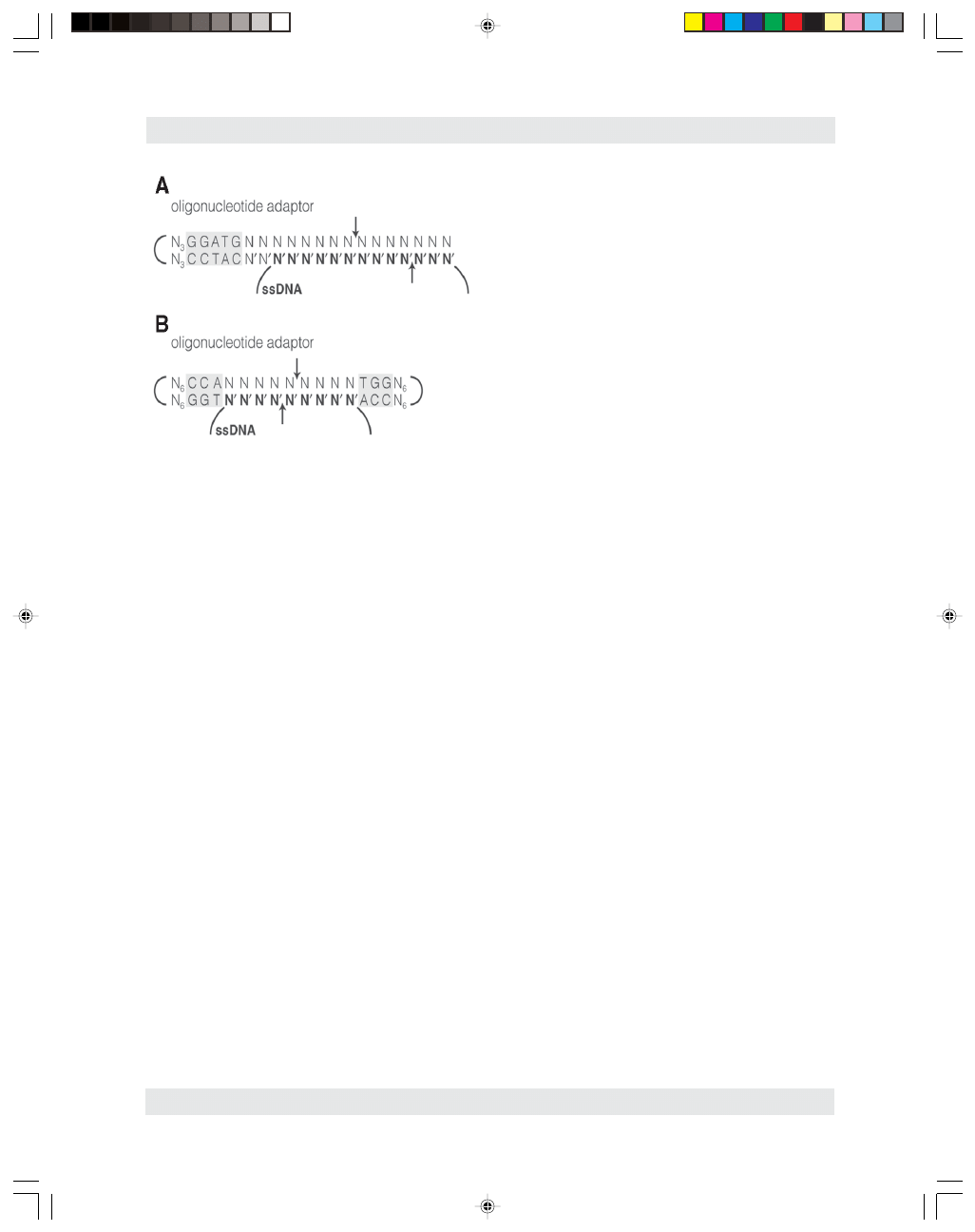
ing ssDNA uses an oligonucleotide adaptor and a
Type IIs enzyme where the recognition site and
cleavage site are significantly separated such as
FokI. The oligonucleotide contains a hairpin loop,
a double-stranded region with the recognition site
of the enzyme, and a single-stranded tail extend-
ing past the recognition site. This single-stranded
04/JW549/Williams/225-244
2/10/03, 9:54 AM
235
M
OLECULAR
B
IOTECHNOLOGY
Volume 23, 2003
236
Williams
region of the oligonucleotide protruding past the
recognition site is complementary to the ssDNA
substrate. The endonuclease is then able to recog-
nize its cognate site on the double-stranded region
of the oligonucleotide and cleave in the region
formed by the oligo:ssDNA hybrid as illustrated
in Fig. 1A. After cleavage, the oligonucleotide
fragment bound to the ssDNA can be heat dena-
tured. This method cannot be used to cleave
ssRNA, however (19). A similar approach for cut-
ting ssDNA uses the enzyme XcmI, which recog-
nizes the longest (9 nt) degenerative sequence
known (5'-CCANNNNN/NNNNTGG-3'). An
oligo is designed with two hairpin loops and adja-
cent inside regions of dsDNA containing the rec-
ognition nucleotides but leaving the center,
nonspecific nucleotides single-stranded. A com-
plementary ssDNA that hybridizes to these non-
specific nucleotides will be cleaved as illustrated
in Fig. 1B (65).
A small group of naturally occurring nickases
are now known that mimic restriction enzymes in
recognizing a double-stranded DNA site but
cleaving only one specific phosphate diester bond.
The two best characterized nickases, N.BstSE (66)
and N.BstNBI (67), are isoschizomers recognizing
the nonpalindromic sequence GAGTC and cleav-
ing the top strand four bases from the 3' end of the
recognition site. N.BstNBI has been cloned and
shows a high degree of sequence similarity with the
Type IIs enzymes PleI and MlyI, which also recog-
nize GAGTC. Furthermore, PleI cleaves the top
strand in the same position as N.BstNBI. Based
on gel filtration with bound DNA, DNA cleavage,
and mutational analysis, a structure and mecha-
nism similar to that of FokI without the need for
dimerization at the cleavage site has been pro-
posed for N.BstNBI (68).
4.3. Specificity Alteration
Through Protein Engineering
Despite the complete sequence and 3D struc-
tural data available, site-directed mutagenesis of
restriction enzymes to alter the DNA sequence
they recognize has generally resulted only in
relaxed specificity and/or decreased cleavage
rates. Simple mutations that easily alter the recog-
nition site for restriction would be lethal to the
host without the same alteration in methylase ac-
tivity; therefore, the endonuclease has evolved to
be highly specific and redundant in recognition.
Mutations to accept a modified base or increasing
specificity of the site so far have proven to be
easier. Mutants of EcoRV have been constructed
that cleave recognition sites with a uracil instead
of thymine by more than two orders of magnitude
over wild-type (N188Q) (69) or with a methyl-
phosphonate in one of the phosphate backbone
positions by three orders of magnitude over wild-
type (T94V) (70). In addition, EcoRV A181K and
A181E mutants preferentially cleave sites with a
purine or thymine, respectively, 5' to the recogni-
tion site (71). A directed evolution approach has
produced a N97T/S183A/T222S mutant with a
20-fold preference for an oligonucleotide with a
GC-rich flanking region and a K104N/A181T
mutant with a seven-fold preference for an AT-
rich flanking region (72). Heterodimers of EcoRV
containing a catalytically inactive mutant subunit
act as site-specific nickase (73). To facilitate addi-
tional enzyme engineering, more structural infor-
mation is needed regarding the large number of
Fig. 1. Use of oligonucleotide adaptors for cleaving
ssDNA with the endonucleases FokI (A) and XcmI (B).
The ssDNA appears in bold, the endonuclease recog-
nition site contained within the oligonucleotide is
highlighted, and the cleavage positions are indicated
the arrows.
04/JW549/Williams/225-244
2/10/03, 9:54 AM
236

M
OLECULAR
B
IOTECHNOLOGY
Volume 23, 2003
Restriction Endonucleases
237
enzyme–DNA contacts and intra/intermolecular
protein shifts. However, several other approaches
to achieve custom designed, sequence-specific
cleavage have also been investigated such as
metal-catalyzed cleavage, Achilles heel cleavage,
and fusion proteins.
4.4. Metal Catalyzed Cleavage
One approach to custom-designed specificity
and cleavage uses either an oligonucleotide
capable of forming a triple helix (74) or a DNA-
binding protein to provide the specificity.
Covalently attached to the oligonucleotide or pro-
tein is a metal complex, usually EDTA-iron or
o-phenanthroline-copper, which catalyzes phos-
phodiester cleavage in the presence of a reducing
agent. Proteins successfully used to supply speci-
ficity include Cro (75), the catabolite activator
protein “CAP” (76), and the Msx-1 homeodomain
(77). However, cleavage at more than one
phosphodiester bond in each strand results in a
mixed population of overhangs and cleavage does
not proceed to completion.
4.5. Achilles Heel Cleavage
A technique to achieve more precise and com-
plete cleavage but at a less frequent number of
sites than standard endonucleases is known as
“Achilles heel cleavage.” First, a target sequence
is protected by a bound RecA/oligo complex
(78,79) or triple helix formation (80). A methy-
lase modifies all sites except the protected target.
The methylase is removed by purification, fol-
lowed by the protecting group. The target
sequence is then specifically cleaved by a methyl
sensitive restriction enzyme.
4.6. Fusion Proteins
Hybrid enzymes can be constructed by fusing
recognition and cleavage domains from different
proteins. In one such example, the recognition
domain of the Type IIs enzyme AlwI was fused to
the catalytic domain of the nicking enzyme
N.BstNBI, which does not contain the dimeriza-
tion potential necessary for double stranded cleav-
age. The resultant chimeric enzyme, N.AlwI, cuts
only the top strand four bases downstream from
the AlwI recognition site, GGATC (81).
Using various spacers and constructs, the Type
IIs FokI catalytic domain has been combined with
DNA binding domains from the Drosophila Ubx
homeodomain (82), the zinc finger region of the
eukaryotic transcription factor Sp1, the designed
zinc finger consensus sequence protein QQR (83),
and the zinc finger region of the yeast transcrip-
tion factor Gal4 (84). Interestingly, the fusions
with Sp1 and QQR will also bind and cleave the
DNA strand of DNA–RNA hybrids. Since the rec-
ognition sequence of Gal4 is palindromic and the
protein dimerizes at the site, the strands are
cleaved on opposite sides of the recognition site.
This results in a
ⱖ24 base, 5' overhang, which
includes the recognition nucleotides. Sites for the
Ubx, Sp1, and QQR hybrids are nonpalindromic,
the fusion proteins may act as monomers, and both
strands are cleaved to one side of the recognition
site at high chimeric enzyme concentration
although generally at more than one position.
This approach has recently been further refined.
A new chimeric enzyme was created with the
cleavage domain of FokI fused to QNK, another
designed zinc finger protein. However, the cleav-
age domain of the fusion protein was again not
under the same level of allosteric regulation as in
the native FokI and therefore low levels of
hydrolysis occurs at nearby phosphates when
enzyme concentrations are sufficient to produce
double-stranded cuts (85). Cleavage efficiency
was greatly enhanced, and alternate site cleavage
reduced, by positioning two of the nonpalin-
dromic consensus sequences close together in a
tail-to-tail orientation. The greatest fidelity of
cleavage for QQR at a single phosphate bond
in each strand occurred when the intervening
sequence was 12 bp. In this manner, each strand
was cleaved eight bases from their respective rec-
ognition site, approximately one helical turn, and
a 5' overhang of four bases was generated as in
native FokI. In addition, the substrate DNA could
consist of one site each of QQR and QNK in simi-
lar tail-to-tail orientation, and requiring both
respective chimeric enzymes for cleavage. When
the dimerization-deficient mutants of the FokI
catalytic domain were used, cleavage did not
occur. Therefore, it appears likely that catalytic
04/JW549/Williams/225-244
2/10/03, 9:54 AM
237

M
OLECULAR
B
IOTECHNOLOGY
Volume 23, 2003
238
Williams
domain dimerization in the spacer DNA of
approximately one helical turn is necessary for
efficient and specific cleavage (86).
Taken a step further, a DNA containing appro-
priately spaced and orientated QQR sites was
injected into oocytes and allowed to assemble into
chromatin. Subsequent injection with the chimeric
QQR enzyme and incubation yielded nearly 100%
homologous recombination when the spacer was
8 bp in length. Significant homologous recombi-
nation was also observed when the hybrid QQR/
QNK substrate and both chimeric enzymes were
injected. Since each consensus sequence is 9
nucleotides in length, the predicted occurrence of
such an 18-bp site would be 4
18
or 6.9
× 10
10
. Use
of additional zinc fingers would expand the con-
sensus binding sequence. Given that the human
genome is approx 3
× 10
9
base pairs, continued
development of this technique may hold promise
for stimulating targeted homologous recombina-
tion in vivo (87).
5. Commercially Prepared Restriction
Endonucleases
5.1. Unit Definition and Application
to Other Substrates
Commercial vendors of restriction endonu-
cleases use standard assays for unit activity defi-
nition with only minor variations. The products of
digestion are generally separated by electrophore-
sis in agarose gels and detected by ethidium bro-
mide staining. An activity unit is defined as the
amount of enzyme necessary to completely digest
1
µg of the defined substrate, usually lambda
DNA, in a 50
µL reaction volume in 1 h at the
specified temperature. If the number of sites is
small (
ⱕ3), lambda pre-digested with another
enzyme (e.g., EcoRI) may be used to improve gel
resolution of the fragments resulting from diges-
tion with the enzyme in question. A different DNA
such as Adenovirus 2 is used if there is a single or
no sites in lambda.
The reaction conditions and enzyme units
needed to digest a given substrate must be chosen
carefully to ensure performance. The buffer sys-
tems provided with commercially obtained en-
zymes are designed to balance optimal individual
enzyme performance and limiting the number of
different buffers. Very few provided reaction buff-
ers are specifically optimized for a single enzyme,
nor is there a true “universal” buffer. As with any
group of similar enzymes, endonucleases are all
unique to some degree in their preferences for
buffer components and concentration such as
cation (Na
+
or K
⫹
), anion (Cl
⫺
or acetate), pH
(7.2–8.5), stabilizer (BSA, detergent, or spermi-
dine), and reducing agent. The storage buffer of
the enzyme may adversely affect use when it com-
prises an unusually large amount of the total reac-
tion volume (volume exclusion of glycerol
causing star activity, chelators for stability bind-
ing Mg in reaction, and so on).
As it may also constitute a large percentage of
the reaction volume, substrate preparation is criti-
cal for enzyme performance. Unit definitions are
generally given for high purity linear phage or
viral DNA, which is not necessarily the situation
in many applications. Enzymes vary in their resis-
tance to proteases, interference by DNA binding
proteins, competitive inhibition from RNA, and
tolerance for EDTA, PEG, SDS, CsCl, phenol,
chloroform, and alcohols. Extra caution should
also be used for cutting near the end of a DNA
substrate. Endonucleases require contact with the
DNA backbone for several bases adjacent to the
recognition sequence. In general, the recognition
site must lie 3 bp from the end to give good cleav-
age. Tables have been developed for a limited list
of enzymes based on cutting a short oligo (88),
cutting a PCR fragment near one end (89), and
double digests of adjacent sites in a polylinker
(90). One has to keep in mind the number of
pmoles of cut sites used to define a unit vs a sub-
strate of interest. The following table suggests the
theoretical enzyme units needed for complete cut-
ting with BamH I based strictly on the number of
cut sites and optimal conditions. Although no
other parameters are taken into account, this
approach can be a useful approximation.
5.2. Quality Control Assays
An overdigest or nonspecific exo- and endonu-
clease assay is performed in the same fashion as an
activity assay except that a large excess of enzyme
04/JW549/Williams/225-244
2/10/03, 9:54 AM
238

M
OLECULAR
B
IOTECHNOLOGY
Volume 23, 2003
Restriction Endonucleases
239
units is used (5–100 U) and incubation times are
long (generally 16 h). Often the result is reported
as (X-) fold-overdigestion (units
× hours) in spite
of the fact that the activity of the enzyme typically
diminishes significantly over this length of time at
the reaction temperature. Although star activity is
an inherent property of the enzyme itself and not a
separate, distinct contaminant, it does result in
cleavage at noncognate sites, which will interfere
with downstream applications, and therefore must
be considered in determining the endpoint of this
assay. Some suppliers only consider the absence of
discrete contaminants in their specifications, which
can be misleading.
The cut–ligate–recut assay is more sensitive
with regard to contaminating exonuclease and can
additionally detect phosphatase activity. DNA is
slightly overdigested, ligated with T4 DNA ligase,
and then recut. Dephosphorylated ends will not be
ligated and staggered ends that have been blunted
by single-stranded exonuclease activity will
exhibit less efficient ligation. Loss of any of the
original terminal nucleotides after cleavage will
almost always also result in the loss of the recog-
nition site for recutting even if the substrate
religated. Although ligation of a one base over-
hang is still efficient enough to be useful in T-vec-
tor cloning of PCR fragments, for unknown
reasons the efficiency of ligation, as indicated by
transformation efficiencies of plasmids, can be as
much as two orders of magnitude lower than that
observed for blunt end ligation. In contrast, a four-
base G-C overhang is stable enough to transform
well even without in vitro ligation.
Some vendors also test for the presence of
nickases by incubating the enzyme and a super-
coiled substrate (RF I form) that does not contain
a recognition site to determine the amount of sub-
strate converted to a relaxed, open circle (RF II
form). However, the impact of nicking activity on
the major applications of cloning and mapping are
minimal. One application that would be influ-
enced by nicking is the generation of nested dele-
tions with exonuclease III. However, improperly
purified DNA is a far more likely source of nicks
than contaminant activity present in the endonu-
clease. Current purification methods and the sen-
sitivity of other assays are such that rarely, if ever,
is a nickase test warranted.
Another test used for the detection of exonu-
cleases is to digest
3
H-ds and -ssDNA, TCA pre-
cipitate the remaining DNA, and detect released
nucleotides by scintillation counting of the super-
natant. Be aware that if the enzyme cleaves the
substrate within 30 bp of the DNA end, inefficient
precipitation of the resultant small fragments may
lead to an incorrect interpretation that suggests
exonuclease activity. Labeling the 5' or 3' ends of
DNA with
32
P is a sensitive way to detect con-
taminating phosphatase or exonuclease. However,
this method requires frequent preparation of
substrate.
The Blue/White assay combines excellent sen-
sitivity and a verification of performance. A clon-
ing plasmid is used that contains a multiple
cloning site flanked by RNA polymerase
promoter(s) within a coding sequence for the lacZ
gene
α-peptide and a separate selectable marker
such as ampicillin resistance. The plasmid is sev-
eral-fold overdigested with an enzyme having a
single site within the multiple cloning region. The
DNA is ligated (without insert) and then trans-
formed into cells lacking the
α-peptide region of
lacZ. An agarose gel of cut, ligated, and recut
DNA is also examined. If the integrity of the cut
ends is perfectly maintained, ligation will produce
mostly higher molecular weight concatamers and
a lesser amount of circularized monomer. Upon
transformation and
α-peptide expression, the
functional lacZ gene product
β-galactosidase is
produced through
α-complementation. When
plated in the presence of IPTG and X-Gal, blue
colonies will result. Expected transformation effi-
ciency will be 1–2 orders of magnitude lower than
Enzyme
Base
Picomoles Cut sites Picomoles
units
DNA
pairs
in 1
µg* (BamHI) cut sites needed
Unit Definition
(ex. lambda)
48,502
0.0312
5
0.156
1
Plasmid
3,000
0.5
1
0.5
3–4**
PCR Fragment
700
2.16
1
2.16
12–15
Oligonucleotide 25
60.6
1
60.6
400–380
*Based on 660 Daltons per bp of DNA.
**Enzymes differ in their ability to cut supercoiled vs linear-
ized substrates.
04/JW549/Williams/225-244
2/10/03, 9:54 AM
239

M
OLECULAR
B
IOTECHNOLOGY
Volume 23, 2003
240
Williams
with control supercoiled plasmid. Both phos-
phatase and exonuclease contamination will lower
ligation efficiency and thereby decrease transfor-
mation efficiency. More importantly, loss of
nucleotides at the cut site, even if ligatable, yields
a mixed population of clones containing frame
shifts and codon deletions in the lacZ gene
α-pep-
tide. These cause white colonies, i.e., false posi-
tives in a cloning experiment with an insert (91).
A special case is an as yet unidentified contami-
nant that is found in native and cloned prepara-
tions of endonuclease that removes a single 3'
nucleotide from the end of DNA. The resultant
colonies are then able to use an alternative start
codon that shifts to become in-frame. However,
it produces weak translation initiation and/or
improper complementation for fully active
β-galac-
tosidase and the colonies develop a faint blue
color, which is easily mistaken for white. This is
especially problematic with blunt end cutting
enzymes. Not all commercial vendors specifi-
cally assay for and remove this contaminant (92).
Acknowledgment
The author would like to thank Michael Slater,
Mark Klekamp, Terri Sundquist, and Isobel
Maciver for their review of the manuscript and
many helpful suggestions.
References
1. Roberts, R. J. and Halford, S. E. (1993), Type II
Restriction Endonucleases, in Nucleases, 2nd Edition
(Linn S. M., Lloyd, S. R., and Roberts, R. J. eds.), Cold
Spring Harbor Laboratory Press, Cold Spring Harbor,
NY, pp. 35–88.
2. Stein, D.C., Gunn, J.S., Radlinska, M., and
Piekarowicz, A. (1995) Restriction and modification
systems of Neisseria gonorrhoeae. Gene 157, 19–22.
3. Lin, L-F. Posfai, J., Roberts, R. J., and Kong, H.
(2001) Comparative genomics of the restriction-modi-
fication systems in Helicobacter pylori. Nuc. Acids
Res. 98, 2740–2745.
4. Rocha, E. P. C., Danchin, A., and Viari, A. (2001)
Evolutionary Role of Restriction/Modification Sys-
tems as Revealed by Comparative Genome Analysis.
Genome Res. 11, 946–958.
5. Nobusato, A., Uchiyama, I., and Kobayashi, I. (2000)
Diversity of restriction-modification gene homologues
in Helicobacter pylori Gene 259, 89–98.
6. Smith, H. O. and Nathans, D. (1973) A suggested
nomenclature for bacterial host modification and re-
striction systems and their enzymes. J. Mol. Biol. 81,
419–423.
7. Wilson, G. G. and Murray, N. E. (1991) Restriction and
Modification Systems. Annu. Rev. Genet. 25, 585–627.
8. Stahl, F., Wende, W., Jeltsch, A., and Pingoud, A.
(1998) The Mechanism of DNA Cleavage by the Type
II Restriction Enzyme EcoRV: Asp36 Is Not Directly
Involved in DNA Cleavage but Serves to Couple Indi-
rect Readout to Catalysis. Biol. Chem. 379, 467–473.
9. Smith, H. O., Annau, T. M., and Chandrasegaran, S. (1990)
Finding sequence motifs in groups of functionally related
proteins. Proc. Natl. Acad. Sci. USA 87, 826–830.
10. Szomolanyi, E., Kiss, A., and Venetianer, P. (1980)
Cloning the modification methylase gene of Bacillus
sphaericus R in Escherichia coli. Gene 10, 219–225.
11. Tao, T., Bourne, J. C., and Blumenthal, R. M. (1991)
A Family of Regulatory Genes Associated with Type
II Restriction-Modification Systems. J. Bact. 173,
1367–1375.
12. Ives, C. L., Sohail, A., and Brooks, J. E. (1995) The
Regulatory C Proteins from Different Restriction-
Modification Systems Can Cross-Complement. J.
Bact. 177, 6313–6315.
13. Bickle, T. A. (1993) The ATP-dependent Restriction
Enzymes, in Nucleases, 2nd ed. (Linn S. M., Lloyd, S.
R., and Roberts, R. J. eds.), Cold Spring Harbor Labo-
ratory Press, Cold Spring Harbor, NY, pp. 35–88.
14. Murray, N. E. (2000) Type I restriction systems:
sophisticated molecular machines. Microbiol. Mol.
Biol. Revs. 64, 412–434.
15. Pingoud, A. and Jeltsch, A. (1997) Recognition and
cleavage of DNA by type-II restriction endonucleases.
Eur. J. Biochem. 246, 1–22.
16. Pingoud, A., Jeltsch, A. (2001) Structure and function
of type II restriction endonucleases. Nuc. Acids Res.
29, 3705–3727.
17. Reuter, M., Kupper, D., Pein, C. D., Petrusyte, M.,
Siksnys, V., Frey, B., and Kruger, D. H. (1993) Use of
Specific Oligonucleotide Duplexes to Stimulate
Cleavage of Refractory DNA Sites by Restriction
Endonucleases. Anal. Biochem. 209, 232–237.
18. Oller, A. R., Broek, W. V., Conrad, M., and Topal, M.
(1991) Ability of DNA and Spermidine To Affect the
Activity of Restriction Endonucleases from Several
Bacterial Species. Biochemistry 30, 2543–2549.
19. Szybalski, W., Kim, S. C., Hasan, N., and Podhajska,
A. J. (1991) Class-IIS restriction enzymes - a review.
Gene 100, 13–26.
20. Kong, H. (1998) Analyzing the Functional Organiza-
tion of a Novel Restriction Modification System, the
BcgI System. J. Mol. Biol. 279, 823–832.
21. Sears, L. E., Zhou, B., Aliotta, J. M., Morgan, R. D., and
Kong, H. (1996) BaeI, another unusual BcgI-like restric-
tion endonuclease. Nucleic Acids Res. 24, 3590–3592.
22. Belfort, M. and Roberts, R. J. (1997) Homing endonu-
cleases: keeping the house in order. Nuc. Acids Res.
25, 3379–3388.
04/JW549/Williams/225-244
2/10/03, 9:54 AM
240

M
OLECULAR
B
IOTECHNOLOGY
Volume 23, 2003
Restriction Endonucleases
241
23. Meisel, A., Mackeldanz, P., Bickle, T. A., Kruger, D.
H., and Schroeder, C. (1995) Type III restriction
endonucleases translocate DNA in a reaction driven
by recognition site-specific ATP hydrolysis. EMBO J.
14, 2958–2966.
24. Kruger, D. H., Kupper, D., Meisel, A., Reuter, M., and
Schroeder, C. (1995) The significance of distance and
orientation of restriction endonuclease recognition
sites in viral DNA genomes. FEMS Microbiol. Rev.
17, 177–184.
25. Janulaitis, A., Petrusyte, M., Maneliene, Z.,
Klimasauskas, S., and Butkus, V. (1992) Purification
and properties of the Eco57I restriction endonuclease
and methylase — prototypes of a new class (type IV).
Nucl. Acids Res. 20, 6043–6049.
26. Williams, R. J. (2001), Restriction Endonucleases and
Their Uses, in The Nucleases (Schein, C. H., ed.),
Humana Press, New York, NY, pp. 409–429.
27. Mise, K. and Nakajima, K. (1985) Purification of a
new restriction endonuclease, StyI, from Escherichia
coli carrying the hsd
⫹ minplasmid. Gene 33,
357–361.
28. Jeltsch, A., and Pingoud, A. (1998) Kinetic Character-
ization of Linear Diffusion of the Restriction Endonu-
clease EcoRV on DNA. Biochemistry 37, 2160–2169.
29. Engler, L. E., Sapienza, P., Dorner, L. F., Kucera, R.,
Schildkraut, I., and Jen-Jacobson, L. (2001) The Ener-
getics of the Interaction of BamHI Endonuclease with its
Recognition Site GGATCC. J. Mol. Biol. 307, 619–636.
30. Horton, N. C. and Perona, J. J. (2000) Crystallographic
snapshots along a protein-induced DNA-bending path-
way. Proc. Natl. Acad. Sci. 97, 5729–5734.
31. Kovall, R. A. and Matthews, B. W. (1998) Structural,
functional, and evolutionary relationships between
λ-
exonuclease and the type II restriction endonucleases.
Proc. Natl. Acad. Sci. 95, 7893–7897.
32. Kovall, R. A. and Matthews, B. W. (1999) Type II
restriction endonucleases: structural, functional and
evolutionary relationships. Curr. Opin. Chem. Biol. 3,
578–583.
33. Kubareva, E. A., Thole, H., Karyagina, A. S.,
Oretskaya, T. S., Pingoud, A., and Pingoud, V. (2000)
Identification of a base-specific contact between the
restriction endonuclease SsoII and its recognition
sequence by photocross-linking. Nucl. Acids Res. 28,
1085–1091.
34. Lukacs, C. M., Kucera, R., Schildkraut, I., and
Aggarwal, A. K. (2000) Understanding the immuta-
bility of restriction enzymes: crystal structure of BglII
and its DNA substrate at 1.5 Å resolution. Nature
Struc. Biol. 7, 134–140.
35. Huai, Q., Colandene, J. D., Chen, Y., Luo, F., Zhao,
Y., Topal, M. D., and Ke, H. (2000) Crystal structure
of NaeI-an evolutionary bridge between DNA endo-
nuclease and topoisomerase. EMBO J. 19, 3110–3118.
36. Reuter, M., Kupper, D., Meisel, A., Schroeder, C., and
Kruger, D. H. (1998) Cooperative Binding Properties
of Restriction Endonuclease EcoRII with DNA Rec-
ognition Sites. J. Biol. Chem. 273, 8294–8300.
37. Pein, C. D., Reuter, M., Meisel, A., Cech D., and
Kruger, D. H. (1991) Activation of restriction endo-
nuclease EcoRII does not depend on the cleavage of
stimulator DNA. Nuc. Acids Res. 19, 5139–5142.
38. Conrad, M., and Topal, M. (1992) Modified DNA
fragments activate NaeI cleavage of refractory DNA
sites. Nuc. Acids Res. 20, 5127–5130.
39. Senesac, J. H. and Allen, J. R. (1995) Oligonucleaotide
Activation of the Type IIe Restriction Enzyme NaeI
for Digestion of Refractory Sites. BioTechniques 19,
990–993.
40. Jo, K. and Topal, M. D. (1995) DNA Topoisomerase
and Recombinase Activities in NaeI Restriciton
Endonuclease. Science 267, 1817–1820.
41. Jo, K. and Topal, M. D. (1998) Step-wise DNA relax-
ation and decatenation by NaeI-43K. Nucleic Acids
Res. 26, 2380–2384.
42. Colandene, J. D. and Topal, M. D. (2000) Evidence
for Mutations That Break Communication between the
Endo and Topo Domains in NaeI Endonuclease/
Topoisomerase. Biochemistry 39, 13703–13707.
43. Wah, D. A., Hirsch, J. A., Dorner, L. F., Schildkraut,
I., and Aggarwal, A. K. (1997) Structure of the
multimodular endonuclease FokI bound to DNA.
Nature 388, 97–100.
44. Bitinaite, J., Wah, D. A., Aggarwal, A. K., and
Schildkraut, I. (1998) FokI dimerization is required for
DNA cleavage. Proc. Natl. Acad. Sci. 95, 10570–10575.
45. Scheuring, E. S., Santagata, S., and Aggarwal, A. K.
(2001) FokI Requires Two Specific DNA Sites for
Cleavage. J. Mol. Biol. 309, 69–78.
46. Wentzell, L. M., Nobbs, T. J. and Halford, S. E. (1995)
The SfiI restriction endonuclease makes a 4-strand
DNA break at two copies of its recognition sequence.
J. Mol. Biol. 248, 581–595.
47. Sato, H., Suzuki, T., and Yamada, Y. (1990) Purifica-
tion of Restriction Endonuclease from Acetobacter
aceti IFO 3281 (AatII) and its Properties. Agric. Biol.
Chem. 54, 3319–3325.
48. Siksnys, V., Skirgaila, R., Sasnauskas, G., Urbanke,
C., Cherny, D., Grazulis, S. (1999) The Cfr10I restric-
tion enzyme is functional as a tetramer. J. Mol. Biol.
291, 1105–1118.
49. Deibert, M., Grazulis, S., Sasnauskas, G., Siksnys, V.,
and Huber, R. (2000) Struture of the tetrameric restric-
tion endonuclease NgoMIV in complex with cleaved
DNA. Nature Struct. Biol. 7, 792–799.
50. Nobbs, T. J., Williams, S. A., Connolly, B. A., and
Halford, S. E. (1998) Phosphorothioate Substrates for the
SfiI Restriction Endonuclease. Biol. Chem. 379, 599–604.
51. Nobbs, T. J. and Halford, S. E. (1995) DNA cleavage
at two recognition sites by the SfiI restriction endonu-
clease: salt dependence of cis and trans interactions
between distand DNA sites. J. Mol. Biol. 252,
399–411.
04/JW549/Williams/225-244
2/10/03, 9:54 AM
241

M
OLECULAR
B
IOTECHNOLOGY
Volume 23, 2003
242
Williams
52. Embleton, M. L., Sidsnys, V., and Halford, S. E.
(2001) DNA Cleavage Reactions by Type II Restric-
tion Enzymes that Require Two Copies of their Rec-
ognition Sites. J. Mol. Biol. 311, 503–514.
53. Williams, S. A. and Halford, S. E. (2001) SfiI endonu-
clease activity is strongly influenced by the non-spe-
cific sequence in the middle of its recognition site.
Nucl. Acids Res. 29, 1476–1483.
54. Stankevicius, K., Lubys, A., Timinskas, A.,
Vaitkevicius, D., and Janulaitis, A. (1998) Cloning and
analysis of the four genes coding for Bpu10I restriction-
modification enzymes. Nucl. Acids Res. 26, 1084–1091.
55. Hsieh, P.-C., Xiao, J.-P., O’Loane, D., and Xu, S.-Y.
(2000) Cloning, Expression, and Purification of a
Thermostable Nonhomodimeric Restriction Enzyme,
BslI. J. Bact. 182, 949–955.
56. Kong, H. and Smith, C. L. (1998) Does BcgI, a Unique
Restriction Endonuclease, Require Two Recognition
Sites for Cleavage? Biol. Chem. 379, 605–609.
57. Kong, H. and Smith, C. L. (1997) Substrate DNA and
cofactor regulate the activities of a multi-functional
restriction-modification enzyme, Bcg I. Nucl. Acids
Res. 25, 3687–3692.
58. Galburt, E. A., Chevalier, B., Tang, W., Jurica, M. S.,
Flick, K. E., Monnat, R. J., and Stoddard, B. L. (1999)
A novel endonuclease mechanism directly visualized
for I-PpoI. Nature Struct. Biol. 6, 1096–1099.
59. Robinson, C. R. and Sligar, S. G. (1998) Changes in
solvation during DNA binding and cleavage are criti-
cal to altered specificity of the EcoRI endonuclease.
Proc. Natl. Acad. Sci. USA 95, 2186–2191.
60. Lesser, D. R., Kurpiewski, M. R., and Jen-Jacobson,
L. (1990) The Energetic Basis of Specificity in the
EcoRI Endonuclease-DNA Interaction. Science 250,
776–786.
61. Sidorova, N. Y. and Rau, D. C. (2001) Linkage of
EcoRI Dissociation from its Specific DNA Recogni-
tion Site to Water Activity, Salt Concentration, and
pH: Separating their Roles in Specific and Non-spe-
cific Binding. J. Mol. Biol. 310, 801–816.
62. Vermote, C. L. M. and Halford, S. E. (1992) EcoRV
restriction endonuclease: Communication between
catalytic metal ions and DNA recognition. Biochemis-
try 31, 6082–6089.
63. Yoo, O. J. and Agarwal, K. L. (1980) Cleavage of
single strand oligonucleotides and bacteriophage
phiX174 DNA by Msp I endonuclease. J. Biol. Chem.
255, 10559–10562.
64. Blakesley, R. W., Dodgson, J. B., Nes, I. F., Wells, R.
D. (1977) Duplex regions in “single-stranded”
phiX174 DNA are cleaved by a restriction endonu-
clease from Haemophilus aegypius. J. Biol. Chem.
252, 7300–7306.
65. Shaw, P. C. and Mok, Y. K. (1993) XcmI as a univer-
sal restriction enzyme for single-stranded DNA. Gene
133, 85–89.
66. Abdurashitov, M. A., Belichenko, O. A., Shevchenko,
A. V. and Degtyarev, S. K. (1996) N.BstSE-site-spe-
cific nuclease from Bacillus stearothermophilus
SE-589-restriction endonuclease production. Mol.
Biol. (Mosk.) 30, 1261–1267.
67. Morgan, R. D., Calvet, C., Demeter, M., Agra, R. and
Kong, H. (2000) Characterization of the specific DNA
nicking activity of restriction endonuclease N.BstNBI.
Biol. Chem. 381, 1123–1125.
68. Higgins, L. S., Besnier, C. and Kong, H. (2001) The
nicking endonuclease N.BstNBI is closely related to
Type IIs restriction endonucleases MlyI and PleI.
Nucl. Acids Res. 29, 2492–2501.
69. Wenz, C., Selent, U., Wende, W., Jeltsch, A., Wolfes,
H., and Pingoud, A. (1994) Protein engineering
of the restriction endonuclease EcoRV: replacement
of an amino acid residue in the DNA binding site
leads to an altered selectivity towards unmodified
and modified substrates. Biochim. Biophys. Acta.
1219, 73–80.
70. Lanio, T., Selent, U., Wnez, C., Wende, W., Schulz,
A., Adiraj, M., Katti, S. B., and Pingoud, A. (1996)
EcoRV-T94V: a mutant restriction endonuclease with
an altered substrate specificity towards modified
oligodeoxynucleotides. Protein Eng. 9, 1005–1010.
71. Schottler, S., Wenz, C., Lanio, A., Jeltsch, A.,
and Pingoud, A. (1998) Protein engineering of the
restriction endonuclease EcoRV: Structure-guided
design of enzyme variants that recognize the base pairs
flanking the recognition site. Eur. J. Biochem. 258,
184–191.
72. Lanio, T., Jeltsch, A., and Pingoud, A. (1998) Towards
the Design of Rare Cutting Restriction Endonucleases:
Using Directed Evolution to Generate Variants of
EcoRV Differing in Their Substrate Specificity by
Two Orders of Magnitude. J. Mol. Biol. 283, 59–69.
73. Stahl, F., Wende, W., Jeltsch, A., and Pingoud, A.
(1996) Introduction of asymmetry in the naturally
symmetric restriction endonuclease EcoRV to investi-
gate intersubunit communication in the homodimeric
protein. Proc. Natl. Acad. Sci. USA 93, 6175–6180.
74. Dervan, P. B. (1992) Reagents for the site-specific
cleavage of megabase DNA. Nature 359, 87–88.
75. Ebright, Y. W., Chen, Y., Pendergrast P. S., and
Ebright, R. H. (1992) Incorporation of an EDTA-metal
complex at a rationally selected site within a protein:
application to EDTA-iron DNA affinity cleaving with
catabolite gene activator protein (CAP) and Cro. Bio-
chemistry 31, 10664–10670.
76. Pendergrast, P. S., Ebright, Y. W., and Ebright, R. H.
(1994) High-Specificity DNA Cleavage Agent:
Design and Application to Kilobase and Megabase
DNA Substrates. Science 265, 959–962.
77. Shang, Z., Ebright, Y. W., Iler, N., et al. (1994) DNA
affinity cleaving analysis of homeodomain-DNA
interaction: Identification of homeodomain consensus
04/JW549/Williams/225-244
2/10/03, 9:54 AM
242

M
OLECULAR
B
IOTECHNOLOGY
Volume 23, 2003
Restriction Endonucleases
243
sites in genomic DNA. Proc. Natl. Acad. Sci. USA 91,
118–122.
78. Koob, M., Burkiewicz, A., Kur J., Szybalski, W.
(1992) RecA-AC: single-site cleavage of plasmids and
chromosones at any predetermined restriction site.
Nucl. Acids. Res. 20, 5831–5836.
79. Schoenfeld, T., Harper, T., and Slater, M. (1995) RecA
Cleavage and Protection for Genomic Mapping and
Subcloning. Promega Notes 50, 9–14.
80. Strobel, S. A. and Dervan, P. B. (1991) Single-site
enzymatic cleavage of yeast genomic DNA mediated
by triple helix formation. Nature 350, 172–174.
81. Xu, Y., Lunnen, K. D., and Kong, H. (2001) Engineer-
ing a nicking endonuclease N.AlwI by domain swap-
ping. Proc. Natl. Acad. Sci. 98, 12990–12995.
82. Kim, Y. G. and Chandrasegaran, S. (1994) Chimeric
restriction endonuclease. Proc. Natl. Acad. Sci. USA.
91, 883–887.
83. Kim, Y. G., Shi, Y., Berg, J. M., and Chandrasegaran,
S. (1997) Site-specific cleavage of DNA-RNA hybrids
by zinc finger/FokI cleavage domain fusions. Gene
203, 43–49.
84. Kim, Y. G., Smith, J., Durgesha, M., and
Chandrasegaran, S. (1998) Chimeric Restriction
Enzyme: Gal4 Fusion to FokI Cleavage Domain. Biol.
Chem. 379, 489–495.
85. Smith, J., Berg, J. M., Chandrasegaran, S. (1999) A
detailed study of the substrate specificity of a chimeric
restriction enzyme. Nucl. Acids Res. 27, 674–681.
86. Smith, J., Bibikova, M., Whitby, F. G., Reddy, A. R.,
Chandrasegaran, S. and Carroll, D. (2000) Require-
ments for double-strand cleavage by chimeric restric-
tion enzymes with zinc finger DNA-recognition
domains. Nucl. Acids Res. 28, 3361–3369.
87. Bibikova, M., Carroll, D., Segal, D. J., Trautman, J.
K., Smith, J., Kim, Y-G. and Chandrasegaran, S.
(2001) Stimulation of Homologous Recombination
through Targeted Cleavage by Chimeric Nucleases.
Mol. and Cell. Biol. 21, 289–297.
88. New England BioLabs 2000·01 Catalog (2000). New
England BioLabs, Inc., 210–211.
89. Dallas-Yang, Q., Jiang, G., and Sladek, F. M. (1998)
Digestion of Terminal Restriction Endonuclease Rec-
ognition Sites on PCR Products. BioTechniques 24,
582–584.
90. Moreira, R. F. and Noren, C. J. (1995) Minimum
Duplex Requirements for Restriction Enzyme Cleav-
age Near the Termini of Linear DNA Fragments.
BioTechniques 19, 57–59.
91. Hung, L., Murray, E., Murray, W., Bandziulis, R.,
Lowery, R., Williams, R., Noble, R. (1991) A Blue/
White Cloning Assay for Quality Control of DNA Re-
striction and Modifying Enzymes. Promega Notes 33,
12–13.
92. Murray, E., Singer, K., Cash, K., and Williams, R.
(1993) Cloning-qualified blunt end restriction
enzymes: Causes and cures for light blue colonies.
Promega Notes 41, 1–5.
04/JW549/Williams/225-244
2/10/03, 9:54 AM
243
Wyszukiwarka
Podobne podstrony:
Skrypt - Rozdzial 3 - endonukleazy restrykcyjne, materiały medycyna SUM, biochemia, Kolokwium I
Kardiomiopatia restrykcyjna
Restrykcja
enzymy restrykcyjne
Komórka prokariotyczna a eukariotyczn restryktazy
Enzymy restrykcyjne referat
Charakterystyka enzymów restrykcyjnych
ENZYMY RESTRYKCYJNe
restrykcje
Przewlekłe restrykcyjne choroby płuc
1001562-enzymy restrykcyjne, semestr IV, genetyka, Genetyka
Inżynieria genetyczna enzymy restrykcyjne
Analiza restrykcyjna?kteriofaga ƛx
enzymy restrykcyjne-stud, Studia, Inżynieria genetyczna
Przykład restrykcji zapisywanych w rejestrze (4)
enzymy restrykcyjne-stud, Biologia molekularna
wykłady Gabryś, Temat 10, Natomiast wskazuje, że korzystna może być restrykcyjna polityka pieniężna,
Analiza restrykcyjna plazmidowego DNA
więcej podobnych podstron