CHAPTER 17
Notochordal Tumours
Notochordal tumours arise from remnants of the notochord and
hence occur exclusively along the midline. Tumours which occur
elsewhere may resemble chordomas.
The majority of the tumours occur in the sacrum or in the clivus.
Involvement of the remainder of the spine is unusual. One of the
characteristic histological features of chordoma is a lobulated
growth pattern.
Chondroid chordomas occur exclusively in the base of the skull
and show features of both low grade chondrosarcoma and chor-
doma. Some studies have indicated a better prognosis for this
subtype.
bb5_25.qxd 13.9.2006 13:47 Page 315
Chordoma
J.M. Mirra
C. Della Rocca
S.D. Nelson
F. Mertens
Definition
Chordoma is a low to intermediate grade
malignant tumour that recapitulates noto-
chord.
ICD-O codes
Chordoma NOS
9370/3
Chondroid chordoma
9371/3
"Dedifferentiated" chordoma
9372/3
Epidemiology
Chordomas account for 1-4% of all pri-
mary malignant bone tumours. Chor-
doma most commonly presents after age
30, with the most common decade being
the sixth (30% of patients). It is very rare
under age 20 (1%). Male:female ratio is
1.8:1.
Sites of involvement
Axial spine (sacral 60%; spheno-occipi-
tal/nasal 25%; cervical 10%; & thoraco-
lumbar 5%).
Clinical features / Imaging
The clinical features are related to the
location and spread of the neoplasm.
Being a slow-growing mass chordoma
usually produces non specific symptoms
for months to years before the diagnosis
is made.
In the sacrococcygeal presentation pain
is the most frequent symptom. It is usual-
ly referred to the lower back or tip of the
spinal column. Constipation due to
obstruction may develop. Almost all
these neoplasms spread in the pre-
sacral area allowing physical detection
by rectal examination. Nerve dysfunc-
tions, such as anesthesia and paresthe-
sia, are unusual and late manifestations.
Those located in the spheno-occipital
region are often associated with a chron-
ic headache and symptoms due to com-
pression of a cranial nerve. Ocular nerve
involvement is the most frequent; com-
pression and destruction of the pituitary
gland may lead to endocrine distur-
bances; if spread is lateral a cerebello-
pontine angle tumour symptomatology
can be evident. In case of spread inferi-
orly nasal obstruction, bleeding and
even a nasal mass may appear.
Chordomas arising in the cervical,
thoracic and lumbar spine usually pro-
duce symptoms related to nerve roots
or spinal cord compression and / or a
palpable mass can be present. Charac-
teristically cervical chordoma may cli-
nically manifest as a parapharyngeal
mass. Clinically, most patients experi-
ence progressive pain, swelling and/or
neurological deficits that may ultimately
be incapacitating.
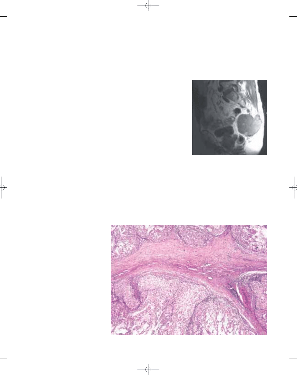
Radiologically, chordomas are typically
solitary, central, lytic, destructive lesions
of the axial skeleton {2058} They are
almost always associated with a soft tis-
sue mass and shards of bony detritus.
Intratumoural calcification may be pres-
ent particularly in sacral tumours. In the
sacral area they tend to displace the
bowel and/or bladder {1302}. MRI stud-
ies best visualise soft tissue extension
and its relationship to anatomic struc-
tures. On MRI, T-1 weighted images are
hypo- or isointense {418}, while T-2
weighted images are of high signal inten-
sity {418,1551}.
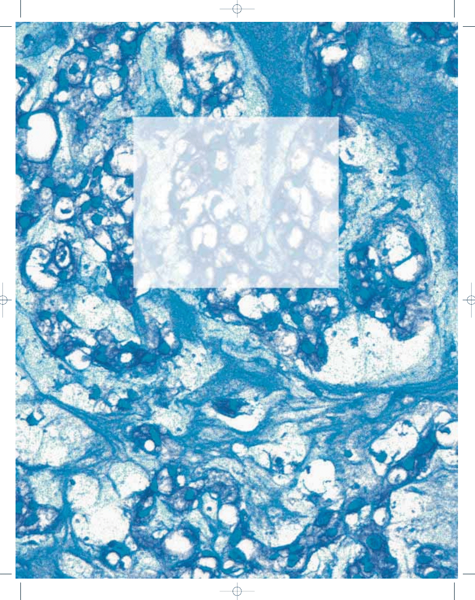
Macroscopy
Chordoma is a lobulated, glistening,
greyish tan to bluish white, muco-gelati-
nous to friable, dark-red haemorrhagic
tumour, generally from 5 to 15 cm. In
most cases it is associated with exten-
sion beyond the contours of the bone into
the surrounding soft tissues {418, 1468}.
Fig. 17.01 Chordoma. T2 MRI showing a dark, lobu-
lated, destructive mass in the sacrococcygeal
region.
316
Notochordal tumours
Fig. 17.02 Chordoma. Bands of fibrosis and lobularity typify this neoplasm on low power.
bb5_25.qxd 13.9.2006 13:47 Page 316
Histopathology
Chordomas are lobulated tumors, with
individual lobules being separated by
fibrous bands. The tumour cells are
arranged in sheets, cords or float singly
within an abundant myxoid stroma.
They typically have an abundant pale
vacuolated cytoplasm (the classic
"physaliphorous cells"). They show mild
to moderate nuclear atypia. There may
be considerable variability in the
appearance of the tumour from area
to area. Mitoses are infrequent {1468}.
In the chondroid variants, there are
areas that may mimic hyaline or myxoid
cartilage {925}. Chordoma associated
with a high grade sarcoma is called
a "dedifferentiated" chordoma {1398}
or sarcomatoid chordoma {1506}.
They account for less than 5% of all
chordomas.
Immunophenotype
Chordomas are reactive with antibodies
against S100 protein, pan-keratin, low
molecular cytokeratins and Epithelial
Membrane Antigen (EMA).
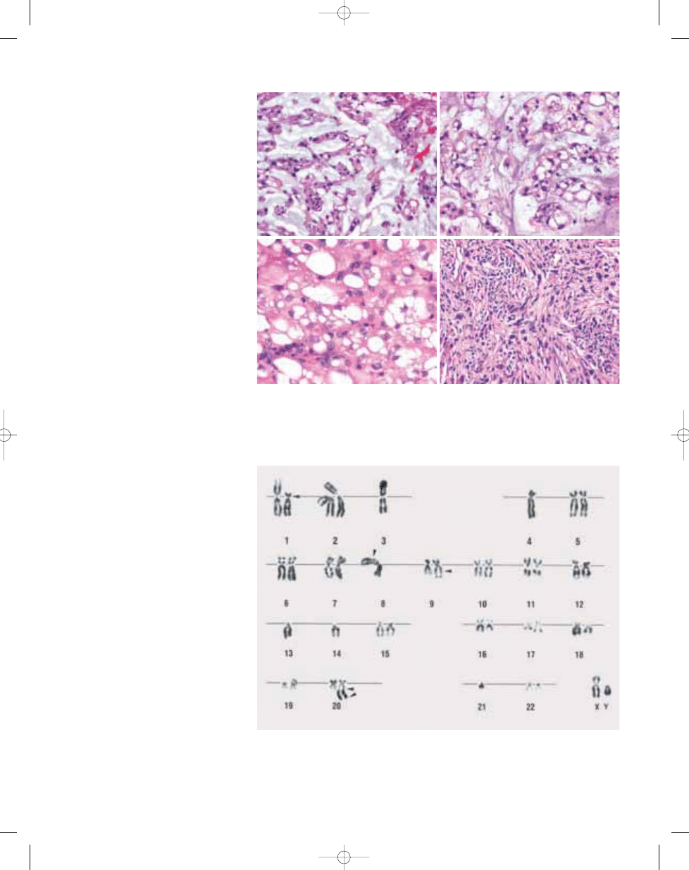
Genetics
Clonal chromosome aberrations have
been detected in 16 cases {1477,
2082}. Nine of them had a hypodiploid
stemline, with a chromosome number
ranging from 33 to 44. Frequent numer-
ical changes include loss of chromo-
somes 3, 4, 10, and 13, and the most
commonly (half of the cases) deleted
segments are 1p31-pter, 3p21-pter,
3q21-qter, 9p24-pter, and 17q11-qter.
These results are in agreement with
data obtained by comparative genomic
hybridisation (CGH) {1880}. By CGH,
also gains of chromosome arms 5q and
7q and chromosome 20 are frequently
seen. The possibility of a tumour sup-
pressor locus of significance for chor-
doma development at distal 1p is fur-
ther strengthened by the finding of loss
of heterozygosity at band 1p36 in spo-
radic as well as familial chordomas
{1465}.
Prognostic factors
Prognosis has improved considerably
with modern surgical techniques of
resection especially with tumours of the
sacrum {1051,2027} and even of mobile
spine {210}. The chondroid variant has
been reported to be associated with a
better prognosis {925} although this
experience is not universal. Metastases
to lung, bone, soft tissue, lymph node
and skin occur, and are more frequent
in patients with advanced disease.
B
A
D
C
Fig. 17.03 Chordoma. A,B Chords of tumour cells in a myxoid background. Note occasional cells displaying a
bubbly cytoplasm. C Some of the classic physaliphorous cells contain multiple intracytoplasmic bubbles that
may cause nuclear indentations similar to those seen in lipoblasts.
D Sarcomatoid, or "dedifferentiated" chon-
droma displaying prominent storiform architecture. Note the large, pleomorphic nuclei and the rather solid
arrangement of cells without a prominent myxoid background.
Fig. 17.04 Chordoma with complex karyotype, including the characteristic loss of chromosomes 3, 4 and 13.
Arrowheads indicate breakpoints in structural rearrangements.
317
Chordoma
bb5_25.qxd 13.9.2006 13:47 Page 317
Wyszukiwarka
Podobne podstrony:
bb5 chap3
bb5 chap8
bb5 chap1
BB5 BOX
bb5 chap16
bb5 chap15
bb5 contents
bb5 chap12
bb5 chap4
bb5 references
bb5 chap6
bb5 chap20
bb5 chap5
Lista wszystkich dostępnych polskich Product Code dla telefonów platformy BB5
bb5 chap21
bb5 source
bb5 chap13
bb5 chap19
więcej podobnych podstron