
53
Prace poglądowe
Chirurgia Polska 2005, 7, 1, 53–58
ISSN 1507–5524
Copyright © 2005 by Via Medica
Guz ziarnistokomórkowy (guz Abrikossoffa)
— przegląd piśmiennictwa
Granular cell tumor (Abrikossoff tumour) — a review of literature
Lidia Mazur-Psonka
1
, Daria Wziątek–Kuczmik
1
, Jacek Pająk
2
, Jan Drugacz
1
, Marcin Mazur
1
1
Katedra i Klinika Chirurgii Szczękowo-Twarzowej Śląskiej Akademii Medycznej, Katowice (Department of Maxillofacial Surgery,
Silesian Medical University, Katowice, Poland)
2
Histologiczna Pracownia Diagnostyczna Katedry i Zakładu Patomorfologii Śląskiej Akademii Medycznej, Katowice (Histology Laboratory,
Department of Pathomorphology, Silesian Medical University, Katowice, Poland)
Streszczenie
Guz ziarnistokomórkowy odkryty po raz pierwszy przez Abrikossoffa to nowotwór najczęściej łagodny,
o niewyjaśnionej do końca histogenezie. Pojawia się rzadko, zwłaszcza u osób dorosłych, i lokalizuje się
najczęściej na bocznej ścianie trzonu języka, wargach, dziąsłach, błonie śluzowej policzka, przełyku i gar-
dle. Jest to guz mały, gładki, niebolesny, elastyczno-twardy, rośnie powoli i bezobjawowo. Często o roz-
poznaniu decyduje badanie histopatologiczne. W niniejszej pracy przedstawiono przegląd piśmiennictwa
oraz 4 przypadki rozpoznanego histopatologicznie guza ziarnistokomórkowego w obrębie trzonu języka.
Słowa kluczowe: guz ziarnistokomórkowy, diagnostyka, leczenie
Abstract
Granular cell tumour (GCT), first described by Abrikosoff typically develops as a benign lesion of yet
unclear histogenesis. It occurs rarely, mostly in adults, and is localised on the lateral aspect of the tongue,
lips, gingiva, buccal mucosa, and in the pharynx and oesophagus. GCT tends to be a small, smooth,
painless, elastic-firm mass which arises slowly and asymptomatically. Diagnosis is established based on
histopathological examination. The present paper comprises a review of the literature on the subject and
a brief report on four cases of lingual GCT.
Key words: granular cell tumour, diagnostic, treatment
Guz ziarnistokomórkowy (guz Abrikossoffa, GCT, gra-
nular cell tumor) jest najczęściej łagodnym nowotworem
zbudowanym z ziarnistych kwasochłonnych komórek
o niewyjaśnionej do końca histogenezie. Po raz pierw-
szy został opisany w 1926 roku przez rosyjskiego patolo-
ga Abrikossoffa, który jednocześnie zasugerował mięś-
niowe pochodzenie guza [1]. Zgodnie z tą koncepcją przez
pewien okres nowotwór funkcjonował pod nazwą gra-
nular cell myoblastoma. Późniejsze doniesienia poparte
wynikami badań ultrastrukturalnych oraz immunohisto-
chemicznych udowodniły związek komórek guza
z komórkami Schwanna lub ich prekursorami [2–8]. Pu-
blikacje te znalazły odzwierciedlenie w nomenklaturze.
Granular cell tumour (Abrikosoff tumour, GCT) com-
posed of granular eosinophilic cells, most often presents
as a benign tumour of yet unclear histogenesis. It was first
described in 1926 by a Russian pathologist Abrikosoff,
who believed the tumour was derived from myoblasts [1].
Thus, the neoplasm used to be referred to as granular
cell myoblastoma. However, subsequent reports from
ultrastructural and immunohistochemical studies re-
vealed an association between tumour cells and Schwann
cells or their precursors [2–8]. Hence, granular cell tu-
mour has been referred to as granular cell schwannoma,
granular cell neurofibroma, neurogenic granular cell tu-
mour [9]. Some authors would negate the neuronal

Lidia Mazur-Psonka
et al.
Granular cell tumor
Polish Surgery 2005, 7, 1
54
Guz ziarnistokomórkowy funkcjonował pod nazwami:
ziarnistokomórkowa schwannoma, ziarnistokomórkowa
neurofibroma, neurogenny guz ziarnistokomórkowy [9].
Niektórzy autorzy negowali nerwowe pochodzenie ko-
mórek ziarnistych [10]. Kolejne badania wykazały, że ko-
mórki ziarniste identyczne jak w guzie ziarnistokomórko-
wym występują również w nowotworach pochodzenia
mięśniowego i szkliwiakach [11, 12]. Część autorów uwa-
ża, że komórki GCT są zwyrodniałym lub reaktywnym
odpowiednikiem komórek Schwanna oraz innych komó-
rek, które mogą być prekursorami łagodnych i złośliwych
guzów o różnicowaniu nerwowym, mięśniowym i naczy-
niowym [13–17].
Guz ziarnistokomórkowy występuje u osób w każdym
wieku, ale rzadko u dzieci. Najczęściej spotykany jest mię-
dzy 4. a 6. lub według innych autorów między 5. a 7. de-
kadą życia [9, 18–20]. Zdecydowanie dominuje u kobiet,
chociaż w niektórych publikacjach opisuje się jego
większą częstość u mężczyzn [9, 21–24]. Na ogół ujaw-
nia się on jako guz pojedynczy. Postacie mnogie, syn-
chroniczne lub asynchroniczne obejmują 5–16% przypad-
ków [6, 8, 18, 20–22, 25]. Opisano również rodzinne wy-
stępowanie GCT. Najczęściej guz lokalizuje się w skórze,
w tkankach podskórnych oraz mięśniach głowy, szyi,
kończyn i tułowia [9, 24, 26]. Około 10–25% GCT umiej-
scowionych jest na języku [24].
Guz ziarnistokomórkowy jest często wykrywany w prze-
wodzie pokarmowym [27–29], przełyku [22], jelicie grubym
i w okolicach odbytu [22, 28], rzadziej w żołądku [22], wy-
rostku robaczkowym i jelicie cienkim [28]. Rzadko GST
można spotkać w drogach żółciowych [28, 30, 31], pę-
cherzyku żółciowym [32], trzustce [30], pęcherzu moczo-
wym [33], mózgu [34], przysadce mózgowej [35], sro-
mie [22, 36], macicy [37], gruczole piersiowym [10, 38],
drogach oddechowych i węzłach chłonnych [20, 39–41].
W gruczole piersiowym GST rozwija się w miąższu na-
rządu lub, częściej, w pokrywającej gruczoł skórze i tkance
podskórnej [22, 24, 38]. W drogach oddechowych guzki
najczęściej lokalizują się w krtani w okolicy fałdów gło-
sowych, najrzadziej zajęta jest tchawica [9, 22, 39–41].
Pierwotny GCT w węzłach jest rzadki i wymaga różnico-
wania z przerzutem złośliwego GCT [20].
W jamie ustnej GCT może występować zarówno
w postaci pojedynczej, jak i mnogiej [42, 43]. Guz ziarni-
stokomórkowy w obrębie jamy ustnej pojawia się u cho-
rych w różnym wieku i zarówno u kobiet, jak i u męż-
czyzn czasami występuje rodzinnie [9, 18–24]. Pojawia się
rzadko, zlokalizowany jest najczęściej na bocznej ścianie
trzonu lub końcu języka, choć może również być obecny
na wargach, dziąsłach, błonie śluzowej policzka, przeły-
ku i w gardle [9, 18, 21, 22, 39–41, 43, 44]. W języku guz
może rozwijać się w błonie śluzowej lub śródmięśnio-
wo. Nabłonek płaski nad guzem zazwyczaj wykazuje ce-
chy znacznego rozrostu. Garlick i wsp. [45] opisali rzadki
desmoplastyczny wariant GCT w policzku. Kobiety cho-
rują prawie dwukrotnie częściej niż mężczyźni [18]. No-
wotwór ten przeważnie uwypukla się nad powierzchnię
języka, jest małym, pojedynczym, gładkim i elastyczno-
twardym guzem, pokrytym błoną śluzową, ale spotyka
origin of granular cells [10]. Other investigations showed
that identical granular cells had been found in adamantino-
mas and neoplasms originating from the muscle [11, 12].
Some others would consider GCT-cells as degenerated
or a reactive change corresponding to Schwann cells or
other cells, which could possibly act as precursors of
benign and malignant neoplasms of neural, muscular,
and vascular differentiation [13–17].
A GCT may occur at any age, most frequently be-
tween the fourth and sixth decade; according to other
authors between the fifth and seventh decade [9, 18–20].
Pediatric cases are quite rarely observed. The tumours are
twice as common in women as in men although some re-
ports seem to demonstrate a male predilection [9, 21–24].
Although a GCT generally grows as a solitary tumour;
multifocal (synchronous or asynchronous) granular cell
tumours will develop in 5–16% of patients [6, 8, 18, 20,
21, 22, 25]. Familial cases have also been described. The
lesions often arise in the skin, subcutaneous tissues, and
the muscles of the head, neck, limbs and trunk [9, 24, 26].
Around 10–25% of granular cell tumours are found on
the tongue [24].
Frequent sites of involvement include the digestive tract
[27–29], the oesophagus [22], the large intestine and anal
region [22, 28]; also, although more rarely, the stomach
[22], the appendix, and the small intestine [28]. Although
infrequently, GCT has also been diagnosed in the biliary
tree [28, 30, 31], gall bladder [32], pancreas [30], urinary
bladder [33], the brain [34], hypophysis [35], vulva [22, 36],
uterus [37], breast [10, 38], the respiratory tract and lymph
nodes [20, 39–41]. In the breast the tumour usually devel-
ops within the parenchyma, or, even more frequently, in
the overlying skin or subcutaneous tissue [22, 24, 38]. The
tumours of the respiratory tract are most often localised in
the larynx in the area of vocal folds; tracheal involvement
is extremely uncommon [9, 22, 39–41]. Primary GCT of
the lymph nodes is a rare entity and should be differenti-
ated with malignant GCT metastasis [20].
In the oral cavity both solitary and multifocal tumours
have been reported [42, 43], and in male and female
patients of different ages; sometimes also as a familial
disease [9, 18–24]. The lesion arises on the lateral as-
pect or anterior part of the tongue (intramuscularly or
in the mucous membrane), and possibly also on the
lips, gingiva, and buccal, oesophageal, and pharyngeal
mucosa [9, 18, 21, 22, 39–41, 43, 44]. The squamous
epithelium underlying the tumour tends to show fea-
tures of marked hyperplasia. Garlick [45] described
a rare desmoplastic granular cell tumour of the buccal
mucosa. Women outnumber men by as much as 2
to 1 [18]. The tumour usually bulges over the lingual
dorsum, and is a small, solitary, sessile, hard nodule of
smooth surface covered with mucous membrane; how-
ever, multiplicity has also been observed [42, 43]. Thick-
ening of the overlying epithelium is often found, with
the tumour extending into the immediately adjacent tis-
sues and giving an impression of pseudocancerous
hyperplasia. The nodules are immovable, and well cir-
cumscribed [9, 22, 24, 26, 38]; they arise slowly,
Lidia Mazur-Psonka i wsp.
Chirurgia Polska 2005, 7, 1
Guz ziarnistokomórkowy
55
się też postacie mnogie [42, 43]. Często stwierdza się
pogrubienie nabłonka pokrywającego guz, który wpukla
się w podścielisko, tworząc rozrosty rzekomorakowe.
Rzadziej umiejscawia się głębiej w tkankach. Guzek jest
nieruchomy, dobrze odgraniczony od otoczenia [9, 22,
24, 26, 38]. Rośnie powoli i bezobjawowo, osiągając
kształty płaskie do wypukłych i średnicę do około 2 cm,
nie ulega owrzodzeniu i jeśli nie ulegnie zezłośliwieniu,
nie osiąga dużych rozmiarów [20, 22]. W jamie ustnej
guzy te występują sporadycznie [46]. Guz ziarnistokomór-
kowy często stanowi przypadkowe odkrycie kliniczne
i decyduje o tym badanie histopatologiczne. Na przekro-
jach GCT jest szaro-biały, szaro-żółty lub żółty. Obraz
mikroskopowy jest charakterystyczny. Guz zbudowany
jest z płatów, gniazd i pasm okrągłych, wielobocznych
lub wydłużonych, komórek o obfitej ziarnistej, kwaso-
chłonnej cytoplazmie. W badaniach ultrastrukturalnych
wykazano, że cytoplazmatyczne ziarnistości kwasochłon-
ne odpowiadają dużym pleomorficznym, wtórnym lizo-
somom [9]. Między grupami komórek znajdują się pa-
sma tkanki łącznej, w starszych guzkach czasem grube
z licznymi włóknami kolagenowymi [9, 20, 47]. Komórki
guza mają wyraźną błonę jądrową. Jądra są małe, okrą-
głe, mocno barwiące się, z drobnymi jąderkami. Poje-
dyncze jądra mogą wykazywać zmiany zwyrodnieniowe
i pozorną atypię. Sporadycznie pojawiają się komórki
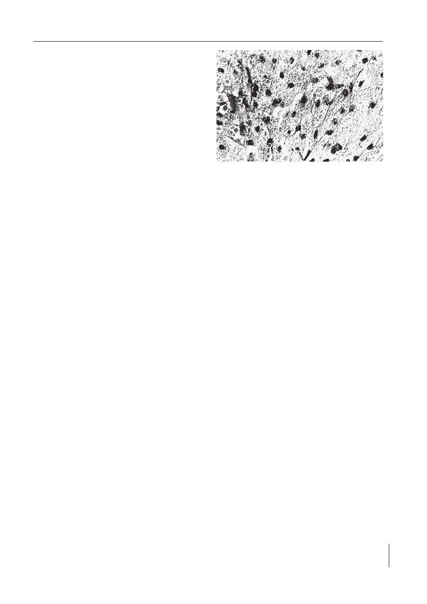
wielojądrowe. Figury podziałowe są rzadkie. W cytoplaz-
mie niektórych komórek oprócz ziarnistości mogą znaj-
dować się kwasochłonne kule otoczone jasną obwódką
(ryc. 1) [9, 20]. W podścielisku guza znajdują się charak-
terystyczne makrofagi zawierające duże łukowate ciałka
kwasochłonne odpowiadające lizosomom [17, 48]. Ziar-
nistości cytoplazmatyczne w komórkach guza, kule i ciałka
kwasochłonne są PAS-pozytywne i odporne na trawie-
nie diastazą. W utkaniu GCT lub w jego sąsiedztwie spo-
tyka się drobne gałązki nerwów obwodowych, rzadziej
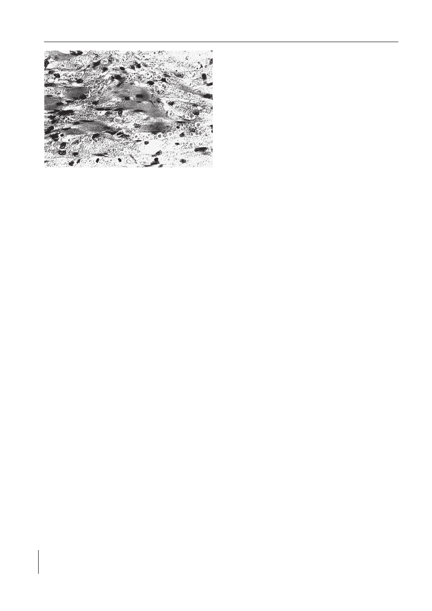
duże pnie nerwowe. Guz może rosnąć wzdłuż osłonek
nerwów obwodowych i wnikać między włókna mięśnio-
we (ryc. 2) [9, 20, 49]. W skórze i błonach śluzowych na-
błonek płaski pokrywający guz często wykazuje cechy
rozrostu i można pomylić, szczególnie w małych biop-
sjach z bardzo dojrzałym rakiem płaskonabłonkowym
[9, 19, 20, 36]. W diagnostyce histopatologicznej GST nie
sprawia na ogół większych problemów. Wymaga różni-
cowania głównie ze złośliwą postacią GCT, tak zwanym
„wrodzonym GCT” lub „wrodzonym nadziąślakiem” wy-
stępującym u niemowląt [50, 51], mięśniakiem prążko-
wanokomórkowym, ziarnistokomórkowym wariantem
mięśniaka gładkomórkowego oraz z zimowiakiem [9, 20].
Sporadycznie w różnicowaniu należy uwzględnić niektóre
postacie histologiczne raka przy lokalizacji GCT w gru-
czole piersiowym oraz gwiaździaki z komórkami ziarni-
stymi w przypadku GCT w ośrodkowym układzie nerwo-
wym [52, 53]. Decydujące znaczenie dla prawidłowej dia-
gnozy ma wykonanie odpowiednich różnicujących bar-
wień immunohistochemicznych.
Obraz kliniczny złośliwego GCT jest zbliżony do po-
staci łagodnej. Na ogół guz ma większą średnicę, od
4 do 14 cm [54], chociaż opisano złośliwe guzy o średni-
asymptomatically as flat or convex masses of 2 cm in
diameter; ulceration has not been noted. If malignant
transformation does not occur, the lesions do not grow
very large [20, 22]. Location deep in the tissues has not
been commonly observed. The tongue is the most fre-
quently involved structure of those within the head and
the neck [46]. The diagnosis is often established acci-
dentally, based on histopathological examination. GCT
sections appear grayish-white, grayish-yellow or yellow-
ish. The microscopic picture is quite characteristic: the
tumour is composed of lobules, nests and ribbons of
round, polygonal, and elongated cells with abundant
eosinophilic cytoplasm. The ultrastructural appearance
of cytoplasmatic eosinophilic granules has been con-
firmed as compatible with large pleomorphic second-
ary lysosomes [9]. Between cell clusters, fibrous con-
nective tissue is seen, sometimes thick with multiple
collagen fibres [9, 20, 47]. Tumour cells have distinct
nuclear membranes; the nuclei are small and intensively
staining, with tiny nucleoli. Some nuclei may demon-
strate degenerative changes and apparent atypia. Poly-
nuclear cells appear only sporadically; mitotic figures
are sparse. Apart from granules, eosinophilic globules
with a surrounding halo may be observed (Fig. 1) [9,
20]. Tumour adjacent tissues may demonstrate the pres-
ence of macrophages containing large arciform eosi-
nophilic bodies, whose structure is consistent with that
of lysosomes [17, 48]. Cytoplasmatic granules in tumor
cells, eosinophilic globules and granules are PAS-posi-
tive, and resistant to diastase digestion. In both a GCT
and its surrounding areas, tiny branches of peripheral
nerves, more rarely large nerve trunks, can be observed.
The tumour may arise along the peripheral nerves my-
elin sheaths and penetrate between myofibrils (Fig. 2)
[9, 20, 49]. A squamous epithelium covering the tumour
tends to show features of hyperplasia and pathologists
must be cautious about misdiagnosing this as well-dif-
ferentiated squamous cell carcinoma [9, 19, 20, 36]. GCT
Rycina 1. Obraz mikroskopowy przedstawia utkanie guza ziar-
nistokomórkowego. W cytoplazmie niektórych ko-
mórek kwasochłonne kule otoczone jasną obwódką.
Barwienie HE. Pow. ok. 250 ×
Figure 1. Microscopic structure of granular cell tumour. Eosi-
nophilic globules with surrounding halo seen in the
cytoplasm of some cells. (HE) staining. Magnifica-
tion × 250
Lidia Mazur-Psonka
et al.
Granular cell tumor
Polish Surgery 2005, 7, 1
56
cy 1 cm [55]. W skórze i błonach śluzowych często jest
owrzodziały. W wywiadzie klinicznym często udaje się
wychwycić moment gwałtownego wzrostu wcześniej ist-
niejącego guza, co może odpowiadać transformacji zło-
śliwej łagodnego GCT.
Wyróżniono dwie postacie złośliwego GCT. Do pierw-
szej grupy, obejmującej 90% przypadków, należą guzy
dające przerzuty odległe i niewykazujące znaczących róż-
nic w obrazie histologicznym z łagodnymi GCT [9].
W guzach tych stwierdza się czasami nieznaczne pod-
wyższenie indeksu mitotycznego oraz mierny polimor-
fizm jąder komórkowych. W grupie drugiej mieszczą się
nowotwory z cechami złośliwości histopatologicznej [9].
Morfologiczne kryteria złośliwego GCT opracowano
stosunkowo niedawno. Złośliwy GCT szybko daje wzno-
wy po usunięciu chirurgicznym. Przerzuty następują na-
czyniami krwionośnymi i limfatycznymi do węzłów chłon-
nych, płuc, wątroby, kości [9, 20]. W obrazie mikrosko-
powym obecność co najmniej trzech z następujących
cech histologicznych: martwicy, pęcherzykowych jąder
z jąderkami, wydłużonych i wielokształtnych komórek,
wysokiego indeksu mitotycznego powyżej 2/10 HPF oraz
wysokiego stosunku jądrowocytoplazmatycznego po-
zwala na rozpoznanie złośliwego GCT w tej grupie gu-
zów. Guzy, w których utkaniu spotyka się tylko jedną lub
dwie powyższe cechy, nazywane są atypowymi GCT
i mają bardzo dobre rokowanie [20].
Złośliwy GCT wymaga różnicowania w obrazie histo-
patologicznym z mięsakami tkanek miękkich — przede
wszystkim z mięsakiem z mięśni gładkich i prążkowanych,
złośliwym guzem fibrohistiocytarnym oraz mięsakiem na-
czyniowym. W barwieniach immunohistochemicznych
reaguje z tymi samymi przeciwciałami, co łagodny GCT,
jednak w niektórych przypadkach reakcja może być ujem-
na. Odmiana złośliwa tego guza jest rzadka.
Złośliwy GCT obejmuje mniej niż 2% wszystkich GCT.
Ravich i wsp. po raz pierwszy opisali złośliwą odmianę
GCT u 31-letniej pacjentki w pęcherzu moczowym [33].
histolopathology generally does not pose problems. The
following enter into the differentials: malignant GCT,
congenital GCT, congenital epulis as diagnosed in in-
fants [50, 51], rhabdomyoma, granular cell leiomyoma
and hibernoma [9, 20]. When a GCT is localised in the
breast, some histological forms of cancer should also
be considered; with central nervous system involve-
ment — astrocytoma with granular cells [52, 53]. Im-
munohistochemical differential staining is crucial to ar-
rive at a correct diagnosis.
Clinical presentation of malignant granular cell tumour
resembles that of a benign GCT. Generally, malignancy
results in a larger lesion of 4 to 14 cm in diameter [54]
although masses as small as 1 cm have also been de-
scribed [55]. Ulceration has frequently been found in skin
and mucous membrane involvement. The clinical his-
tory often reveals the approximate moment of rapid
growth which might signal malignant transformation of
a benign GCT.
Two subtypes of malignant GCT have been discussed
in the literature. The first (90% of cases) includes tumours
which metastasize late, and whose histopathology does
not substantially differ from that of a benign GCT [9].
A slightly increased mitotic index and mild polymorphism
of the cell nuclei are characteristic. The other variant en-
compasses tumours with distinct features of histopatho-
logical malignancy [9].
The morphological markers for malignancy have been
only recently developed. Malignant GCTs may recur soon
after surgical excision, and spread via blood and lymphatic
vessels to lymph nodes, lungs, liver and bones [9, 20].
Microscopic examination may reveal the following his-
tological markers: necrosis, a vesicular nucleus with
nucleoli, elongated and polymorphic cells, mitotic index
over 2/10HPF, and elevated nucleocytoplasmatic index.
When three of these features are found, a diagnosis of
malignant GCT is established. Tumours, whose structure
shows only one or two of the above mentioned charac-
teristics are referred to as atypical GCT and the progno-
sis is very favourable [20].
Malignant GCT should enter into histopathological
differentiation with soft tissue sarcomas, and especially
with leiomyosarcoma and rhabdomyosarcoma, fibro-
histiocytary malignancy, and angiosarcoma. The tumour
cells reveal the same positive immunohistochemical
staining as benignity; however in some cases the reac-
tion may prove negative.
Malignant GCT occurs rarely; the incidence is as low
as 2% of all granular cell tumours. Ravich et al. [33] first
described malignant GCT masses in the urinary bladder
of a 31-year old female patient.
In the case of benign GCTs, the treatment of choice
is surgical excision. Typically, the tumour is removed with
some margin of healthy tissue. If unencapsulated, it is
excised with the overlying mucous membrane, and a rela-
tively small margin of unaffected tissue. Following com-
plete excision, recurrence is seen in 2–8% of cases. With
incomplete surgery the figures tend to be much higher,
i.e., 21–50% [56].
Rycina 2. Obraz mikroskopowy przedstawia komórki kwaso-
chłonne guza wnikające pomiędzy włókna mięśni ję-
zyka. Barwienie HE. Pow. ok. 250 ×
Figure 2. Microscopic picture of eosinophilic cells of the tu-
mor migrating between the muscle fibres of the
tongue. (HE) staining. Magnification × 250

Lidia Mazur-Psonka i wsp.
Chirurgia Polska 2005, 7, 1
Guz ziarnistokomórkowy
57
Leczenie z wyboru łagodnych GCT polega na chirur-
gicznym usunięciu. Wykonuje się wycięcie guza z mar-
ginesem tkanek zdrowych, zwłaszcza gdy jest on nieotor-
biony, usuwa się guz z pokrywającą błoną śluzową i nie-
wielkim marginesem tkanek otaczających. Nawroty
towarzyszące całkowitej resekcji występują w 2–8% przy-
padków. U pacjentów z niecałkowitą resekcją guza czę-
stość nawrotów jest zdecydowanie wyższa (21–50%) [56].
W Klinice Chirurgii Szczękowo-Twarzowej w Katowi-
cach w latach 1991–2004 operowano 4 chorych (2 kobie-
ty i 2 mężczyzn) z powodu niebolesnych guzów języka,
które rozpoznano u osób o średniej wieku około 40 lat.
U 2 chorych były one zlokalizowane na bocznej powierzch-
ni języka po stronie prawej, a u pozostałych 2 pacjentóww
trzonie języka w linii pośrodkowej. Guzki były wielkości
od 0,7 do 2,0 cm. Chorych bez współistniejących scho-
rzeń ogólnych operowano ambulatoryjnie, a pacjentów
z obciążonym wywiadem ogólnym operowano w warun-
kach szpitalnych. U wszystkich operowanych chorych guz-
ki języka wycinano z marginesem zdrowych tkanek. Powi-
kłań śródoperacyjnych, pooperacyjnych i wznów nie stwier-
dzono. W badaniu histopatologicznym materiału poopera-
cyjnego u wszystkich chorych rozpoznano GCT.
Piśmiennictwo (References)
1. Abrikossoff AI. Über Myome, ausgehend von der quergestreif-
ten willkürlichen Musculatur. Virchows Arch Pathol Anat
Histopathol. 1926; 260: 215–233.
2. Bedetti CD, Martinez AJ, Beckford NS et al. Granular cell tumor
arising in myelinated peripheral nerves. Light and electron mi-
croscopy and immunoperoxidase study. Virchows Arch. 1983;
402: 175–183.
3. Budzilovich GN. Granular cell „myoblastoma” of vagus nerve.
Acta Neuropathol. 1968; 10: 162–169.
4. Fischer ER, Wechsler H. Granular cell myoblastoma — a mis-
nomer. Electron microscopic and histochemical evidence con-
cerning its Schwann cell derivation and nature (granular cell
schwannoma). Cancer 1962; 15: 936–957.
5. Maiorano E, Favia G, Napoli A et al. Cellular heterogeneity of
granular cell tumours: a clue to their nature? J Oral Pathol Med.
2000; 29: 244–290.
6. Moscovic EA, Azar HA. Multiple granular cell tumors („myobla-
stomas”). Case report with electron microscopic observations
and review of the literature. Cancer 1967; 20: 2032–2047.
7. Rao TV, Puri R, Reddy GN. Intracranial trigeminal nerve granular
cell myoblastoma. Case report. J Neurosurg. 1983; 59: 706–709.
8. Seo IS, Azzarelli B, Warner TF et al. Multiple visceral a cutaneous
granular cell tumors. Ultrastructural and immunocytochemical
evidence of Schwann cell origin. Cancer 1984; 53: 2104–2110.
9. Scheithauer BW, Woodruff JM, Erlandson RA. Miscellaneous be-
nign neurogenic tumors. Primary malignant tumors of periphe-
ral nerve. In: Atlas of tumor pathology. Tumors of the peripheral
nervous system. Washington: AFIP, 1999: 248–258, 358–372.
Scheithauer BW, Woodruff JM, Erlandson RA. Tumors of the
peripheral nervous system atlas of tumor pathology. Third Se-
ries, Fascicle 24. Bethesda, Maryland: Armed Forces Institute
of Pathology 1999.
10. Le Boit PE, Barr RJ, Burall S et al. Primitive polypoid granular-
cell tumor and other cutaneous granular-cell neoplasms of ap-
parent nonneural origin. Am J Surg Pathol. 1991; 15: 48–58.
11. Christ ML, Ozzello L. Myogenous origin of a granular cell tumor
of the urinary bladder. Am J Clin Pathol. 1971; 56: 736–749.
12. Navarrette AR, Smith M. Ultrastructure of granular cell amelo-
blastoma. Cancer 1971; 27: 948–955.
13. Finkel G, Lane B. Granular cell variant of neurofibromatosis. Ul-
trastructure of benign and malignant tumors. Hum Pathol. 1982;
13: 959–963.
14. McWilliam LJ, Harris M. Granular cell angiosarcoma of the skin.
Histology, electron microscopy and immunohistoochemistry of
a newly recognized tumor. Histopathology 1985; 9: 1205–1216.
15. Miettinen M, Lehtonen E, Lehtola H et al. Histogenesis of gra-
nular cell tumor. An immunohistochemical and ultrastructural
study. J Pathol. 1984; 142: 221–229.
16. Nistal M, Paniagua R, Picazo ML et al. Granular changes in va-
scular leiomyosarcoma. Virchows Arch. 1980; 386: 239–248.
17. Sobel HJ, Marquet E. Granular cell and granular cell lesions.
Pathol Ann. 1974; 9: 43–79.
18. Baden E, Divaris M, Quillard J. A light microscopic and immu-
nohistochemical study of a multiple granular cell tumor
and review of the literature. J Oral Maxillofac Surg. 1990; 48:
1093–1099.
19. Strong EW, McDivitt RW, Brasfield RD. Granular cell myobla-
stoma. Cancer 1970; 25: 415–422.
20. Weiss SW, Goldblum IR. Rozdział 20. In: Weiss SW, Goldblum
JR. Enzinger and Weiss´s Soft Tissues Tumours. 4th ed. St.
Louis, Mosby, 2001: 1178–1187.
Weiss SW, Goldblum JR. Benign tumors of peripheral nerves.
In: Weiss SW, Goldblum JR. Enzinger and Weiss´s Soft Tissues
Tumours. St. Louis, 4th ed. Mosby 2001: 1171–1173.
21. Billeret Lebranchu V. Granular cell tumor. Epidemiology of 263
cases. Arch Anat Cytol Pathol. 1999; 47: 26–30.
22. Lack EE, Worsham GF, Callihan MD et al. Granular cell tumor.
A clinicopathologic study of 110 patients. J Surg Oncol. 1980;
13: 301–316.
23. Mazur MT, Schultz JJ, Myers JL. Granular cell tumor. Immuno-
histochemical analysis of 21 benign tumors and one malignant
tumor. Arch Pathol Lab Med. 1990; 114: 692–696.
24. Peterson LJ. Granular cell tumor. Review of the literature
and report of a case. Oral Surg Oral Med Oral Pathol. 1974; 37:
724–735.
25. Martin RW, Nelder KH, Boyd AS et al. Multiple cutaneous gra-
nular cell tumors and neurofibromatosis in childhood. Arch Der-
matol. 1990; 126: 1051–1056.
In the years 1991 through 2004, four patients were
operated on in the Department of Maxillofacial Surgery
for asymptomatic tumours located on the tongue. There
were two male and two female patients with a mean age
of around 40 years. In two patients the tumours were
localised on the right lateral lingual surface, in the other
two in the median line of the lingual dorsum. Tumor size
was between 0.7–2.0 cm. Patients with no concomitant
disease were operated on in the outpatient clinic while
those with medical abnormalities were hospitalized. All
tumours were excised with a surgical margin of healthy
tissue. Intra- and postoperative complications were not
noted. Recurrence did not occur in any of the patients.
A histopathology of intraoperative biopsies revealed
a benign granular cell tumour.

Lidia Mazur-Psonka
et al.
Granular cell tumor
Polish Surgery 2005, 7, 1
58
26. Vance SF, Hudson RP. Granular cell myoblastoma. Clinicopatho-
logic study of 42 patients. Am J Clin Pathol. 1969; 52: 208–211.
27. Brady PG, Nord HJ, Connor RG. Granular cell tumor of the eso-
phagus: natural history, diagnosis and therapy. Dig Dis Sci.
1988; 33: 1329–1333.
28. Johnston J, Helwig EB. Granular cell tumours of the gastrointe-
stinal tract and perianal region. A study of 74 cases. Dig Dis Sci.
1981; 26: 807–816.
29. Rickert RR, Larkey IG, Kantor EB. Granular-cell tumors (myobla-
stoma) of the anal region. Dis Colon Rectum 1978; 21: 417–423.
30. Chandrasoma P, Fitzgibbons P. Granular cell tumor of the intra-
pancreatic common bile duct. Cancer 1984; 53: 2178–2182.
31. Eisen RN, Kirby WM, O’Quinn IL. Granular cell tumor of the bi-
liary tree. A report of two cases and a review of the literature.
Am J Surg Path. 1991; 15,: 460–465.
32. Yamaguchi K, Kuroki S, Daimaru Y et al. Granular cell tumor of
the gallbladder. Report of a case. Acta Pathol Jpn. 1985; 35:
687–691.
33. Ravich A, Stout AT, Ravich RA. Malignant granular cell myoblasto-
ma involving the urinary bladder. Ann Surg. 1945; 121: 361–372.
34. Sakurama N, Matsukado Y, Marubayashi T et al. Granular cell
tumour of the brain and its cellular identity. Acta Neurochir.
1981; 56: 81–94.
35. Schlachter LB, Tindall GT, Pearl GS. Granular cell tumor of the
pituitary gland associated with diabetes insipidus. Neurosurge-
ry 1980; 6: 418–421.
36. Wolber RA, Talerman A, Wilkinson EJ et al. Vulvar granular cell
tumors with pseudocarcinomatous hyperplasia. A comparati-
ve analysis with well-differentiated squamous carcinoma. Int
J Gynecol Pathol. 1991; 10: 59–66.
37. Copas P, Dyer M, Hall DJ et al. Granular cell myoblastoma of
the uterine cervix. Diagn Gynecol Obstet. 1981; 3: 251–254.
38. DeMay RM, Kay S. Granular cell tumor of the breast. Pathol
Ann. 1984; 19: 121–148.
39. Compagno J, Hyams VJ, Ste-Marie P. Benign granular cell tu-
mors of the larynx: a review of 36 cases with clinicopathologic
data. Ann Otol Rhinol Laryngol. 1975; 84: 308–314.
40. Hamid AM, Alshaikly A. Granular cell tumour of the larynx in an
eight-year-old girl. J Laryngol Otol. 1993; 107: 940–941.
41. Solomons NB. Extensive granular cell tumour of the larynx and
trachea. J Laryngol Otol. 1988; 102: 658–660.
42. Collins BM, Jones AC. Multiple granular cell tumors of the oral
cavity: report of a case and review of the literature. J Oral Ma-
xillofac Surg. 1995; 53: 707–711.
43. Worsaae N, Schwartz O, Pindborg JJ. Follow-up study of 14
oral granular cell tumors. Int J Oral Surg. 1979; 8: 133–139.
44. Zahid L, Cariappa KM. Granular cell tumor of buccal mucosa
— a case report. Int J Oral Maxillofac Surg. 1996; 25: 134–135.
45. Garlick JA, Dayan D, Buchner A. A desmoplastic granular cell
tumour of the oral cavity: report of a case. Br J Oral Maxillofac
Surg. 1992; 30: 119–121.
46. Wetzel W, Leipzig B, Grunow W et al. Malignant granular cell
tumor of the tongue. Arch Otolaryngol. 1982; 108: 603–605.
47. McMahon JN, Rigby HS, Davis JD. Elastosis in granular cell
tumours: prevalence and distribution. Histopathology 1990; 16:
37–41.
48. Dingemans KP, Mooi WJ, van den Bergh Weeman MA. Angula-
te lysosomes. Ultrastruct Pathol. 1983; 5: 113–122.
49. Ophir D, Lifschitz-Mercer B, Czernobilsky B. Expression of in-
termediate filaments and stromal proteins in granular cell tu-
mor of the tongue. Oral Surg Oral Med Oral Pathol. 1988; 66:
689–696.
50. Kay S, Elzay RP, Wilson MA. Ultrastructural observations on
a gingival granular cell tumor (congenital epulis). Cancer 1971;
27: 674–680.
51. Tucker MC, Rusnock EJ, Azumi N et al. Gingival granular cell
tumors of the newborn. An ultrastructural and immunohisto-
chemical study. Arch Pathol Lab Med. 1990; 114: 895–898.
52. Melaragno MJ, Prayson RA, Murphy MA et al. Anaplastic astro-
cytoma with granular cell differentiation: case report and re-
view of the literature. Hum Pathol. 1993; 24: 805–808.
53. Townsend MC, Stellato TA. Granular cell myoblastoma of the
breast: a report of five cases and a review. Breast 1985; 11:
12–21.
54. Al Sharraf M, Loud AV, Vaitkevicius VK. Malignant granular cell
tumor. Histochemical and electron microscopic study. Arch
Pathol. 1971; 91: 550–558.
55. Fanburg-Smith JC, Meis-Kindblom JM, Fante R et al. Malignant
granular cell tumor of soft tissue. Diagnostic criteria and clini-
copathologic correlation. Am J Surg Pathol. 1998; 22: 779–794.
56. Alessi DM, Zimmermann MC. Granular cell tumors of the head
and neck. Laryngoscope 1988; 98: 810–814.
Adres do korespondencji (Address for correspondence):
Dr med. Daria Wziątek-Kuczmik
Katedra i Klinika Chirurgii Szczękowo-Twarzowej Śląskiej Akademii Medycznej
ul. Francuska 20/24
40–027 Katowice
tel. kom. 0600–380–458
Praca wpłynęła do Redakcji: 14.09.2004 r.
Wyszukiwarka
Podobne podstrony:
ziarnica zlosliwa
chloniak ziarniczy
Guz mózgu - pielegnowanie pacjenta, Magisterka materiały, Neurochirurgia
derma-ziarniniak grzybiasty, IV rok Lekarski CM UMK, Dermatologia, Historie choroby
Guz Pancoasta, studia pielęgniarstwo
Mikrobiologia opracowanie na podstawie części II Skryptu WAM wersja ostateczna wreszcie kurna!!! , Z
Protokoły, rozpoznanie 3 karolina, guz pęcherza moczowego - tumor vesicae urinariae
ziarnistosc
guz wilmsa
ziarnica kurs
1 Guz chromochłonny nadnerczy
Biochemia, Oznaczanie aktywnościi amylazy metodą Noeltinga i Bernfelda w ziarnie pszenicy
Buleczki ziarniste Thinness
OPRACOWANIE ZIARNIAKOW EXTRA, II ROK STOMATOLOGIA SUM ZABRZE, MIKROBIOLOGIA, MIKROBIOLOGIA, mikroby
CHOROBY ZIARNINIAKOWE SKÓRY i wirusowe, KOSMETYKA, anatomia
więcej podobnych podstron