
Journal of Basic Microbiology 2011, 51, 205 – 214
205
© 2011 WILEY-VCH Verlag GmbH & Co. KGaA, Weinheim
www.jbm-journal.com
Research Paper
Production, characterization, cloning and sequence analysis
of a monofunctional catalase from Serratia marcescens
SYBC08
Hua-Wei Zeng
1
, Yu-Jie Cai
1
, Xiang-Ru Liao
1
, Feng Zhang
1
and Da-Bing Zhang
2
1
The Key Laboratory of Industrial Biotechnology, Ministry of Education, School of Biotechnology,
Jiangnan University, Wuxi, Jiangsu, China
2
Hanbon Science& Technology Co.Ltd., Huaian 223001, China
A monofunctional catalase from Serratia marcescens SYBC08 produced by liquid state fermen-
tation in 7 liter fermenter was isolated and purified by ammonium sulfate precipitation (ASP),
ion exchange chromatography (IEC), and gel filtration (GF) and characterized. Its sequence
was analyzed by LC-MS/MS technique and gene cloning. The highest catalase production
(20,289 U · ml
–1
) was achieved after incubation for 40 h. The purified catalase had an estimated
molecular mass of 230 kDa, consisting of four identical subunits of 58 kDa. High specific acti-
vity of the catalase (199,584 U · mg
–1
protein) was 3.44 times higher than that of Halomonas sp.
Sk1 catalase (57,900 U · mg
–1
protein). The enzyme without peroxidase activity was found to be
an atypical electronic spectrum of monofunctional catalase. The apparent K
m
and V
max
were
78 mM and 188, 212 per µM H
2
O
2
µM heme
–1
s
–1
, respectivly. The enzyme displayed a broad
pH activity range (pH 5.0–11.0), with optimal pH range of 7.0–9.0: It was most active at 20 °C
and had 78% activity at 0 °C. Its thermo stability was slightly higher compared to that of
commercial catalase from bovine liver. LC–MS/MS analysis confirmed that the deduced amino
acid sequence of cloning gene was the catalase sequence from Serratia marcescens SYBC08. The
sequence was compared with that of 23 related catalases. Although most of active site residues,
NADPH-binding residues, proximal residues of the heme, distal residues of the heme and
residues interacting with a water molecule in the enzyme were well conserved in 23 related
catalases, weakly conserved residues were found. Its sequence was closely related with that of
catalases from pathogenic bacterium in the family Enterobacteriaceae. This result imply that
the enzyme with high specific activity plays a significant role in preventing those micro-
organisms of the family Enterobacteriaceae against hydrogen peroxide resulted in cellular
damage. Calalase yield by Serratia marcescens SYBC08 has potential industrial application in
scavenging hydrogen peroxide.
Keywords: Monofunctional catalase / Gene coning and sequencing / ESI-Q-TOF MS/MS / Serratia marcescens
Received: April 14, 2010; accepted: July 18, 2010
DOI 10.1002/jobm.201000147
Introduction
*
Aerobic organisms use molecular oxygen (O
2
) for respi-
ration or oxidation of nutrients to obtain their energy.
Reactive oxygen species (ROS) including superoxide an-
Correspondence: Xiang-Ru Liao, Key Laboratory of Industrial Biotech-
nology, Ministry of Education, School of Biotechnology, Jiangnan Uni-
versity, Lihu Road 1800, Wuxi 214122, Jiangsu Province, China
E-mail: liaoxiangru@163.com; yu_jie_cai@yahoo.com.cn
Phone: 86 0510 85916372
ion radical (O
2
), hydrogen peroxide (H
2
O
2
), and the
highly reactive hydroxyl radicals (
·
OH) are generally
generated by the leakage of single electrons from cellu-
lar respiratory chain. The biological targets for these
highly ROS are DNA, RNA, proteins, and lipids. Much of
the damage is caused by hydroxyl radicals generated
from H
2
O
2
[1]. Catalase is one of the central components
of the detoxification pathways that prevent the forma-
tion of highly reactive hydroxyl radical by catalyzing
the decomposition of H
2
O
2
into water and dioxygen by

206 H.-W.
Zeng
et al.
Journal of Basic Microbiology 2011, 51, 205 – 214
© 2011 WILEY-VCH Verlag GmbH & Co. KGaA, Weinheim
www.jbm-journal.com
two-electron transfer [2], and exist in aerobic, faculta-
tive aerobic and anaerobic microorganisms [3–5], it is
widely used in several industrial fields such as textiles,
pulp and paper. Commercially available catalase is pre-
pared from bovine livers or microorganisms, an ap-
proach limited by low yield [6].
Based on enzymological properties, catalases can be
classified into one of their three types, heme-containing
monofunctional catalases, heme-containing bifunctio-
nal catalase-peroxidases, and non-heme-containing or
Mn-containing catalases [7]. Monofunctional catalases
have a two stage mechanism for the degradation of
H
2
O
2
in which one hydrogen peroxide molecule oxi-
dizes the heme to an oxyferryl species in catalytic sites
and hydrogen peroxide molecule is utilized as a reduc-
tant of compound I to regenerate the resting-state en-
zyme, water and oxygen [8]. Some amino acid residues
of monofunctional catalase such as in NADPH binding
sites and catalytic sites play a very important role in the
reaction [8, 9], in this way, amino acid sequence analy-
sis has a better understanding of its catalytic behavior
in the process of decomposing H
2
O
2
.
In the present study, a catalase of Serratia marcescens
SYBC08 from sludge with hydrogen peroxide was puri-
fied and characterized as a
monofunctional enzyme. Its
amino acid sequence was analyzed by LC-MS/MS tech-
nology and gene cloning.
Materials and methods
Strains and culture conditions
A strain with highest catalase production among 104
catalase-producing microorganisms was isolated from
sludge with hydrogen peroxide in bleaching workshop
of textile factory. The strain was identified and desig-
nated as Serratia marcescens SYBC08 by 16S rDNA se-
quence (Genbank Accession no. GU188473). It was sub-
sequently conserved in China General Microbiological
Culture Collection Center (Preserved no. CGMCC 3449).
Prior to use, the strain was recovered from 10% glyc-
erol stocks stored at –70 °C. For seed preparation, the
microorganism was inoculated into 50 ml seed medium
(glucose 20 g · l
–1
, peptone 10 g · l
–1
, beef 5 g · l
–1
extract
(NaCl 5 g · l
–1
, pH 7.2) in 250 ml flasks and cultivated at
30 °C on a rotary shaker at 200 rpm for 12 h. Seed with
4% size of inoculation (V · V
–1
) was inoculated into the
optimized fermentation medium (corn steep liquor
powder 33.8 g · l
–1
, citric acid 30 g · l
–1
, initial pH 5.91).
Batch fermentation was carried out in a 7 l fermentor
with a working volume of 5 l. The aeration rate was
1.5 V · V
–1
· min
–1
, and agitation speed was 400 rpm.
The temperature and pH were controlled at 32.8 °C and
7.0, respectively.
Preparation of crude enzyme and determination
The broth was centrifuged at 4 °C and 10,800 × g for
15 min. The precipitation was dried at 105 °C to con-
stant weight followed by weighting with electrical level
to determine the biomass or was disrupt by supersonic
instrument at 0 °C for 20 min to prepare crude enzyme
extract. The cell debris was removed by centrifugation
at 4 °C and 18,000 × g for 15 min, and the supernatants
were pooled as crude enzyme extract.
Catalase activity was measured spectrophotometri-
cally by monitoring the decrease in absorbance at
240 nm caused by the decomposition of hydrogen per-
oxide [10]. The
ε
of H
2
O
2
at 240 nm was 43.6 mM
–1
cm
–1
.
The reaction mixture contained suitable amount of en-
zyme solution, 30 mM H
2
O
2
, and 50 mM Na
2
HPO
4
–
NaH
2
PO
4
buffer (pH 7.0) in a total volume of 4 ml. The
linear range of the reaction (30 s) was used to calculate
the rate of the reaction, and one unit of catalase activ-
ity was defined as the amount of enzyme that required
to transform 1 µmol of hydrogen peroxide to water and
oxygen per min [11]. Catalase activity was determined
for three times for each sample. The catalase activity
was calculated and analyzed by SPSS 11.5 software.
Peroxidase activity was measured spectrophotomet-
rically by monitoring the increasing in absorbance
at 470 nm. The reaction mixture contained suitable
amount of enzyme solution, 50 mM Na
2
HPO
4
-citrate
buffer (pH 6.0), 10 mM H
2
O
2
, and 10 mM guaiacol in a
total volum of 4 ml. One unit of peroxidase was ex-
pressed as the enzyme amount required for producing
1 µmol guaiacol oxidants [12].
The protein concentration was determined by the
method according to Bradford [13] with bovine serum
albumin as the standard. Residual citric acid was de-
tected according to the method described by Cen et al.
[14].
Catalase purification
The pooled crude enzyme extract was firstly precipi-
tated by using 40% (w ⋅ v
–1
) ammonium sulfate satura-
tion. The pellets were removed, and the supernatants
with catalase activity were collected by centrifugation
at 4 °C and 10,800 × g for 20 min. After the precipita-
tion by using 60% (w ⋅ v
–1
) ammonium sulfate satura-
tion, the precipitates were collected by centrifugation
at 4 °C and 12,000 × g for 20 min. The precipitates were
fully dissolved in a small amount of 50 mM Na
2
HPO
4
-
NaH
2
PO
4
buffer (pH 8.0) followed by centrifugation
(4 °C and 17,300 × g for 20 min) to discard the undissol-

Journal of Basic Microbiology 2011, 51, 205 – 214
Sequence analysis of a monofunctional catalase from Serratia marcescens 207
© 2011 WILEY-VCH Verlag GmbH & Co. KGaA, Weinheim
www.jbm-journal.com
ved fractions. The supernatants were dialyzed against
the same buffer for 12 h and then subjected to a DEAE
(1.0 cm × 10.0 cm) column which had been equilibrated
with the same buffer. The adsorbed enzyme was eluted
with a linear gradient of NaCl from 0 to 0.7 M in 50 mM
Na
2
HPO
4
–NaH
2
PO
4
buffer (pH 8.0) at the flow rate of
1 ml × min
–1
. The fraction with catalase activity was
pooled and 2 ml of the active fraction was loaded on a
Sephacryl Tm S-200 column (16 mm × 60 cm) which
had been equilibrated with the same buffer. The en-
zyme was eluted with the same buffer at the flow rate
of 1 ml × min
–1
, and the eluted catalase fractions were
collected. The solution in each purified step was col-
lected for assaying catalase activity and protein con-
tent.
The molecular masses of the catalase subunits and
holoenzyme were determined by 12% (w × v
–1
) SDS-
PAGE according to the method of Laemmli [15] and GF,
respectively. The gels were silver staining according to
the Ref. [16].
Spectrophotometric analysis
The absorption spectrum of the purified catalase was
recorded at each 2 nm at room temperature between
280 and 700 nm using double beam UV-Vis spectropho-
tometer.
Effect of pH and temperature on the activity
and stability of the purified catalase
Effect of catalytic pH value on catalase activity was
determined by incubating the purified enzyme in
50 mM Na
2
HPO
4
-citric acid buffer (pH 4.0 ~ 6.0), 50 mM
NaH
2
PO
4
–Na
2
HPO
4
buffer (pH 7.0 ~ 8.0), and 50 mM
Na
2
CO
3
–NaHCO
3
buffer (pH 9.0 ~ 11.0) at 30 °C, and the
highest catalase activity was defined as 100%. The ef-
fect of the pH range from 5.0 to 11.0 on catalase stabil-
ity was investigated by incubating the enzyme at 30 °C
for 180 min, and its initial activity was expressed as
100%. The effect of temperature on catalase activity
was measured at the temperature from 0 to 70 °C at pH
7.0, and the highest activity was regarded as 100%. For
thermal stability determination, the enzyme was incu-
bated at 60 °C, 65 °C and 70 °C at pH 7.0 followed by
periodical measurement at 30 °C, and initial activities
of the sample at corresponding temperatures were cal-
culated as 100%.
Kinetic parameters (V
max
and K
m
)
The effect of H
2
O
2
concentration (7.5, 10, 12.5, 15, 20,
25, 30 mM) on catalase activity was evaluated in 50 mM
NaH
2
PO
4
–Na
2
HPO
4
buffer (pH 7.0) at 20 °C. The kinetic
parameters (Michaelis-Menten constant, Km, and maxi-
mal reaction velocity, V
max
) were estimated by linear re-
gression from double-reciprocal plots according to
Lineweaver and Burk [17].
Amino acid sequence analysis using ESI-Q-TOF
MS/MS
The purified enzyme partially digested with trypsin
(Sigma-Aldrich, Germany). Mass spectrometry analysis
was carried out in electrospray ionization quadrupole
time-of-flight mass spectrometr (ESI-Q-TOF-MS/MS).
MS/MS data were investigated using MASCOT searching
tool (Matrix Science Ltd., London, UK)
DNA preparation and gene cloning
DNA was extracted according to the method descri-
bed by Tao et al. [18]. Primers for PCR amplification
were designed according to the DNA sequence of
serratia proteamaculans 568 catalase (Genbank Acces-
sion no. CP000826.1). It included upstream primer
(ACCGGAATTCATGAGCAAGAAAGGACTG) and down-
stream primer (ACCGGCGGCCGCTTATTTCAGACCTAA
CGCC). The reaction system included genomic DNA, the
PCR reaction buffer, and two units of Taq polymerase
were mixed and performed PCR amplification in the
condition which was an initially denatured step at
95 °C
for 4 min, followed by 35 cycles of a three-stages
program with 1 min at 94 °C, 1 min at 52 °C for renatu-
ration, then 1.5 min at 72 °C, and a final elongation
step runed for 6 min at 72 °C. The PCR products were
then recovered with Agarose Gel DNA Purification Kit
Ver. 2.0 (TaKaRa). The purified products were ligated
into pET 28 vectors and transformed into E. coli DH5α.
The transformants were selected on Luria-Bertani (LB)
broth containing 100 µg · ml
–1
ampicillin. The positive
clone was screened by H
2
O
2
bubbling test. Plasmid DNA
from positive colonies was extracted from E. coli using a
Plasmid DNA Extraction Kit (Takar) for sequence analy-
sis using an automated DNA sequencer ABI3700.
Amino acid sequence analysis
The deduced amino acid sequence of Serratia marces-
cens SYBC08 was blasted in NCBI database (http://www.
ncbi.nlm.nih.gov/Database/). Twenty four amino acid
sequences of related catalases from Serratia marces-
cens SYBC08 (accession no.ADI55329.1), Yersinia entero-
colitica subsp. enterocolitica 8081 (accession no.
YP_001005695.1), Vibrio fischeri MJ11 (accession no.
YP_002157580), Syntrophobacter fumaroxidans MPOB (ac-
cession no. YP_845843.1), Serratia proteamaculans 568
(accession no. YP_001479504.1), Saccharomonospora viri-
dis DSM 43017 (accession no. YP_003133951.1), Rhodo-
coccus jostii RHA1 (accession no. YP_705771.1), Ralstonia
208 H.-W.
Zeng
et al.
Journal of Basic Microbiology 2011, 51, 205 – 214
© 2011 WILEY-VCH Verlag GmbH & Co. KGaA, Weinheim
www.jbm-journal.com
eutropha H16 (accession no. YP_727552.1), Pseudovibrio
sp. JE062 (accession no. ZP_05083841.1), Pseudomonas
aeruginosa pao1 (accession no. NP_252926.1), Providencia
rustigianii DSM 4541 (accession no.ZP_05973780.2), Pro-
teus Mirabilis Pr (accession no. 2CAH_A), Polaromonas
naphthalenivorans CJ2 (accession no. YP_982885.1),
Photorhabdus luminescens subsp. laumondii tto1 (acces-
sion no. NP_930300.1), Pelobacter propionicus DSM 2379
(accession no. YP_901599.1), Nitrosomonas sp. al212 (ac-
cession no. ZP_05316133.1), Moritella sp. PE36 (accession
no. ZP_01899777.1), Desulfovibrio vulgaris str. 'Miyazaki
F'(accession no. YP_002435660), Cupriavidus metallidurans
CH34 (accession no. YP_587727.1), Colwellia psychreryth-
raea 34H (accession no. YP_269157.1), Arsenophonus na-
soniae (accession no. CBA76514.1), Aromatoleum aromati-
cum EBn1(accession no. YP_158186.1), Aliivibrio salmo-
nicida LFI1238 (accession no. YP_002264567 ) and Bovine
liver (accession no. NP_001030463.1) were chosen for
alignment by DNAMAN Version.v5.2.2. The phyloge-
netic relationships of the 24 sequences were generated
by using CLUSTALX version 1.8 and the software pack-
ages MEGA version 4.1. Unrooted phylogenetic trees
were constructed by using the neighbour joining [19].
Minimum evolution and maximum parsimony methods
was carried out according to the reference [20], and
they were evaluated by bootstrap resampling (1000 rep-
lications).
Results
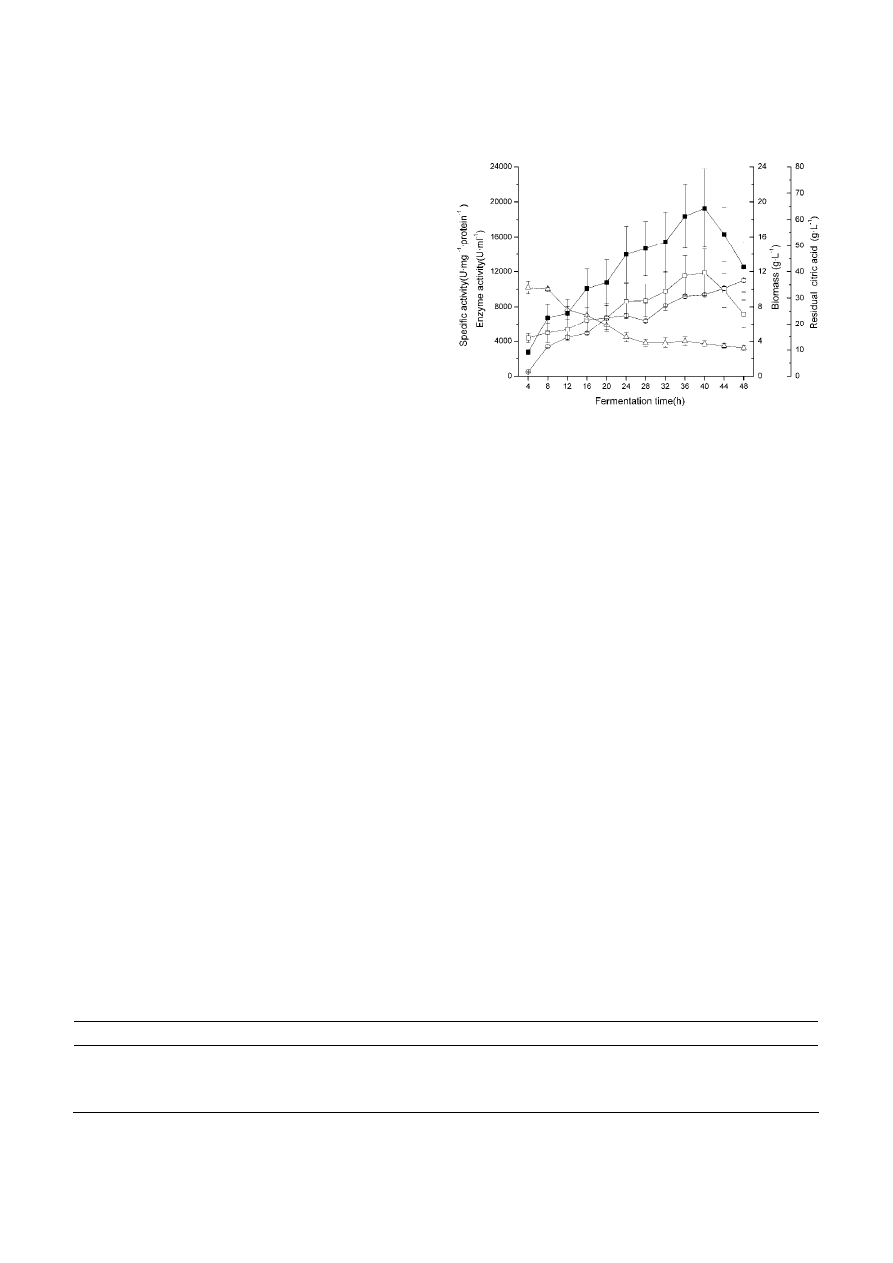
Catalase production in 7 l fermenter
The fermentation time course for catalase produc-
tion by Serratia marcescens SYBC08 in a 7 l fermenter
was presented in Fig. 1, which reveals the relation-
ship between the biomass, specific activity, and cat-
lase production. The maximum catalase production
(20,289 U ⋅ ml
–1
) and specific activity (11,863 U ⋅ mg
–1
of
protein) was achieved at 40 h after incubation, while
biomass constantly increased at all the time course.
Specific activity and catalase production were signi-
ficantly associated (r 0.97). Biomass was closely asso-
ciated with catalase production (r 0.86).
Figure 1. Time course of production of catalase from Serratia
marcescens SYBC08 under optimized medium in 7 l fermenter. The
aeration rate was 1.5 V · V
–1
· min
–1
, agitation speed was 400 rpm,
and pH was 7.0. Values given are the means of at least triplicate
experiments, and error bars represent the SD. Catalase activity (
䊏
),
Specific activity (
ⵧ
), Biomass (
䊊
), Residual citric acid (
䉭
).
Catalase purification
Serratia marcescens SYBC08 catalase was purified by ASP,
IEC and GF. The purification procedure was summa-
rized in Table 1.
Catalase
from
Serratia marcescens SYBC08 was purified
1.6-fold after ASP. During IEC, five protein peaks were
appeared and the fifth peak contained catalase activity
(data not shown). The catalase was purified 5.4-fold
after this process. The protein solution from GF was
separated and appeared six peaks, and only the sixth
peaks contained catalase activity (data not shown).
The enzyme was purified 13.8-fold with a recovery
of 22% after this procedure. It displays high specific ac-
tivity of 99,584 U ⋅ mg
–1
protein, 3.44 time higher than
that Halomonas sp. Sk1 catalase (57,900U ⋅ mg
–1
protein)
[21].
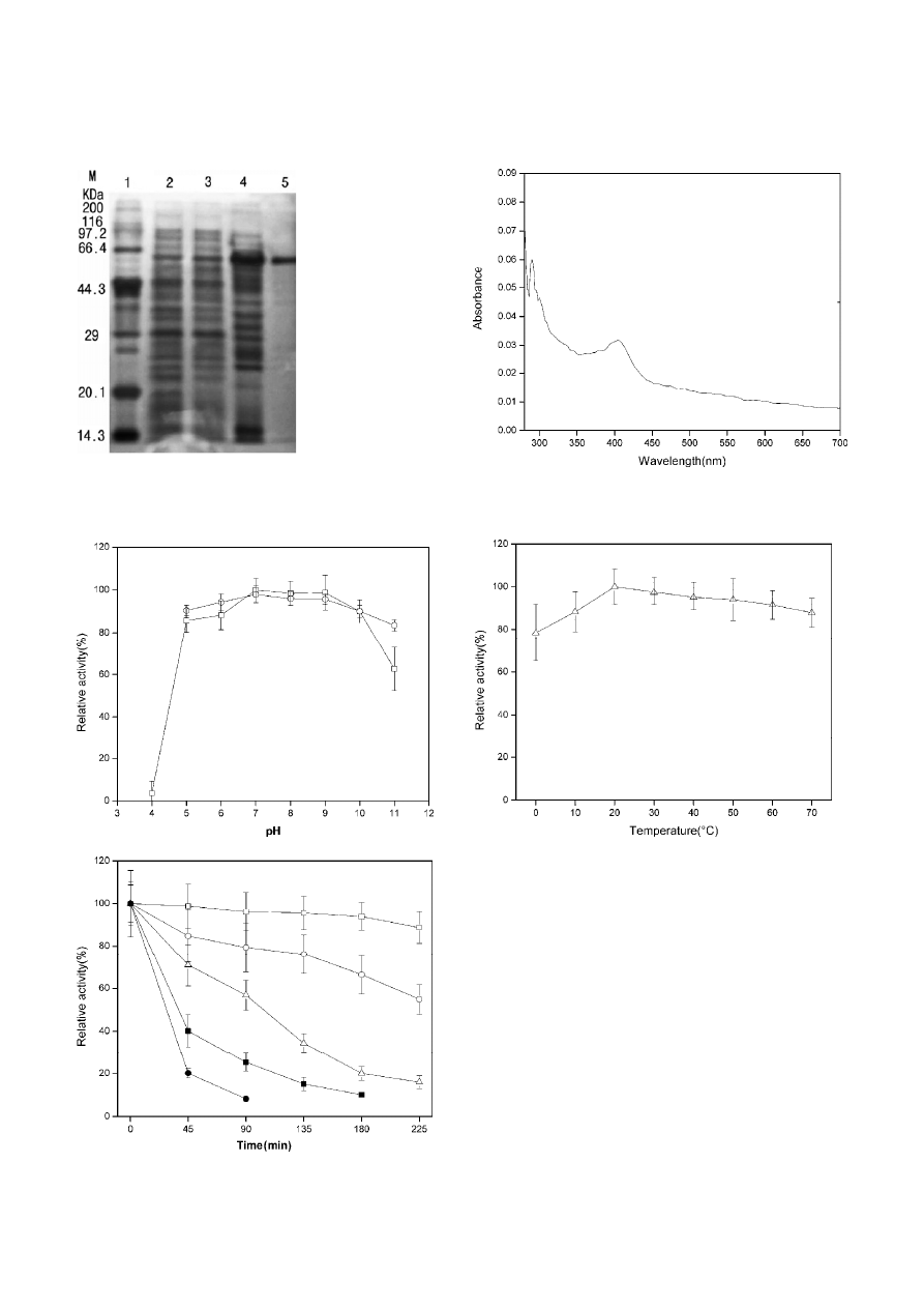
Samples from each procedure were analyzed by SDS-
PAGE, and the result is present in Fig. 2. A single band
from GF was revealed by silver staining and suggested
that the purified catalase was obtained, and the mo-
lecular mass of the subunit was 58 kDa. Molecular mass
of the purified catalase was estimate to be 230 kDa by
GF (data not shown). Thus, we proposed that the puri-
fied catalase was tetramer consisted of 4 homosubunits.
Table 1. Summary of the purification of catalase from Serratia marcescens SYBC 08.
Step
Total activity (U)
Total protein (mg) Specific activity (U · mg
–1
protein) Yield (%) Purification (fold)
Crude extract
5,790,186
399
14,487
100
1.0
ASP
2,569,373
112
22,863
44
1.6
IEC
1,742,347
22.1
78,683
30
5.4
GF 1,325,786
6.6 199,585
22
13.8
Journal of Basic Microbiology 2011, 51, 205 – 214
Sequence analysis of a monofunctional catalase from Serratia marcescens 209
© 2011 WILEY-VCH Verlag GmbH & Co. KGaA, Weinheim
www.jbm-journal.com
Figure 2. Electrophoretic analysis of catalase from Serratia marces-
cens SYBC08 by SDS-PAGE with silver staining. Lane 1, Marker.
Lane 2, crude extract; Lane 3, ASP; Lane 4, IEC; Lane 5, GF.
Figure 3. Spectrum analysis of the purified catalase from Serratia
marcescens SYBC08. The spectra of the enzyme were recorded
against a blank of identical buffer.
(a)
(b)
(c)
Figure 4. (a) Effect of pH on catalase activity from Serratia mar-
cescens SYBC08. Values given are the means of at least triplicate
experiments, and error bars represent the SD. pH catalytic activity
(
ⵧ
); pH stabilition (
䊊
). (b) Effect of temperture on catalase catalytic
activity from Serratia marcescens SYBC08.
Values given are the
means of at least triplicate experiments, and error bars represent
the SD. (c) The thermal stability of catalase at various tempera-
tures. Values given are the means of at least triplicate experiments,
and error bars represent the SD. Serratia marcescens SYBC08,
60
°C (
ⵧ
); 65
°C (
䊊
); 70
°C (
䉭
); bovine liver, 60 °C (
䊏
); 65
°C (
䊉
).
210 H.-W.
Zeng
et al.
Journal of Basic Microbiology 2011, 51, 205 – 214
© 2011 WILEY-VCH Verlag GmbH & Co. KGaA, Weinheim
www.jbm-journal.com
Spectroscopic analysis
Spectroscopic analysis of the purified catalase was per-
formed, and the result was displayed in Fig. 3. The two
maxima at 405 nm (Soret peak) and 280 nm (protein
maximum) was obviously appeared, and the Rz value
(A405/A280) of 0.42 ± 0.041 was calculated.
Effect of pH and temperature on catalase activity
and stability
The catalytic activity of the purified catalase under dif-
ferent pH values is presented in Fig. 4a, and had a
wide pH range of 5.0–11.0. The enzyme was also highly
stable in a broad pH range of 5.0–11.0 (Fig. 4a). Al-
though the enzyme had a slight dependence of tem-
perature, it still showed optimum temperature of 20 °C,
and it maintained 78% of the maximal activity at 0 °C
(Fig. 4b). The temperature stability was determined by
incubating the purified enzyme at 60 °C, 65 °C and
70 °C at pH 7.0, respectively (Fig. 4c). At 60 °C, the en-
zyme from Serratia marcescens SYBC08 was stable for
240 min, while bovine liver catalase only retained 40%
activity for 45 min. At 65 °C, the enzyme from Serratia
marcescens SYBC08 retained more than 55% of its activ-
ity by incubating the enzyme at pH 7.0 after 225 min,
while bovine liver catalase only retained 20% activity
for 45 min. At 70 °C, the enzyme could retain 57% of
its initial activity after incubating at pH 7.0 for 90 min.
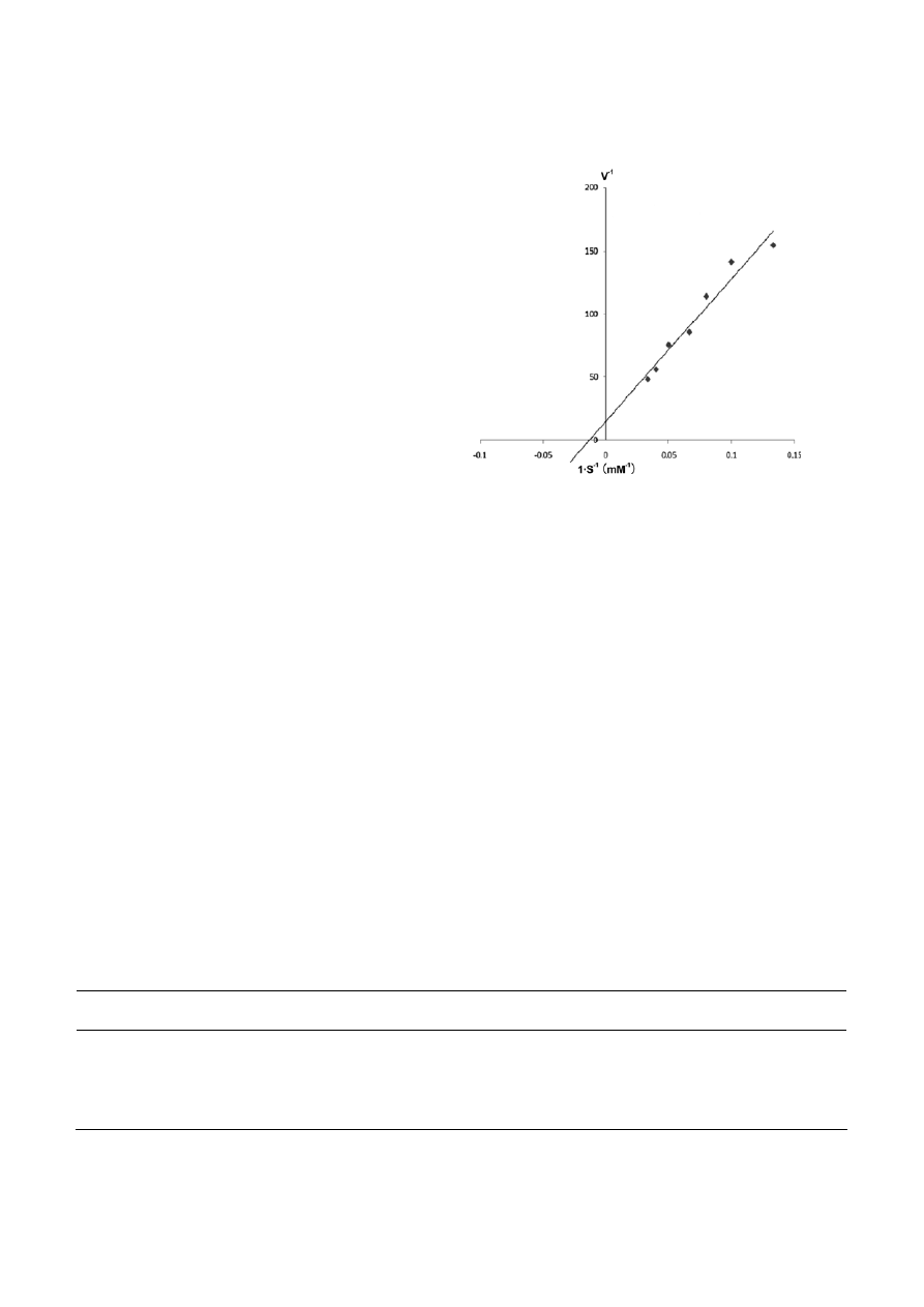
Kinetic analysis
The kinetic parameters of the purified catalase were
analyzed by Lineweaver–Burk plot (Fig. 5). The K
m
and
V
max
for the enzyme at 20 °C was 78 mM and 188,212
per µM H
2
O
2
µM heme
–1
s
–1
, respectively.
MS peptide sequence
The peptides mass fingerprint from LC-MS/MS were
used as a query against the NCBI Protein database
(MASCOT search), and the result is represent in Table 2.
Four peptide sequences was identical with the se-
quences of the two catalases from Yersinia enterocolitica
subsp. enterocolitica 8081 (gi|123441711) and Serratia
Figure 5. Lineweave-Burk plot of the catalase from Serratia mar-
cescens SYBC08.
proteamaculans 568 (gi|157371515), but two peptide se-
quences did not completely matched that of the two
catalases, respectively.
Gene cloning and sequence analysis
A encoding gene was cloned by using two PCR primers
which was designed according to highly homologized
gene sequence from Serratia proteamaculans 568 catalase
under LC-MS/MS analysis, and it was deposited in the
GenBank under the accession number HM 068611. The
deduced 479 amino acid sequence according to an open
reading frame of 1437bp completely matched mass
spectrometric sequence in Table 3. Comparative analy-
sis of those amino acid squences revealed it had high
homology with the sequences of other catalases from
Serratia proteamaculans 568 (94% amino acid sequence
identity), Yersinia enterocolitica subsp. enterocolitica 8081
(91%), and other sources (the range of 53%–85%).
Multiple alignments of 24 catalases were performed
(data not shown). The amino acid residues of the
active site are very important in preservation of en-
zyme functions and much research revealed it was
Table 2. Identification of tryptic peptides of catalase from Serratia marcescens SYBC 08.
Observed ion (m/z)
Expected
molecular mass
Calculated
molecular mass
Delta Sequence
Matched
organism
494,5961 1480,7665
1480,7786
–0.0122 LAHFDREVIPER
1,2
741,3950 1480,7754
1480,7786
–0.0032 LAHFDREVIPER
1,2
761,4196 1520,8246
1520,8351
–0.0105 DPLKFPDLNHVVK 1,2
644,2604 1286,5062
1286,5051
0.0011 EDDDYYSQPR
1,2
841,9160 1681,8174
1681,8060
0.0114 GSGAYGTFTVTHDITR
1
784,4251 1566,8356
1480,7786
–0.0032 IAGELSQVPEQIQR 2
1 or 2 present Yersinia enterocolitica subsp. enterocolitica 8081 (gi|123441711) or Serratia proteamaculans 568 (gi|157371515).
Journal of Basic Microbiology 2011, 51, 205 – 214
Sequence analysis of a monofunctional catalase from Serratia marcescens 211
© 2011 WILEY-VCH Verlag GmbH & Co. KGaA, Weinheim
www.jbm-journal.com
Table 3. Weakly conservative amino acid residuals from important functional sites.
Organism M53
S93
H341
H284
K216
F194
Serratia marcescens SYBC08
M
S
H
H
R
F
Proteus mirabilis PR
M
S
H
H
R
F
Providencia rustigianii DSM 4541
M
S
Q
H R F
Photorhabdus luminescens subsp. laumondii TTO1
M
S
Q
H R F
Arsenophonus nasoniae
M S H H R F
Serratia proteamaculans 568
M
S
H
H
R
F
Yersinia enterocolitica subsp. enterocolitica 8081
M
S
A
H R F
Pseudomonas aeruginosa PAO1 M
S
H
H
K
F
Aliivibrio salmonicida LFI1238
M
S
Q
H V F
Vibrio fischeri MJ11
M
T Q H
K
F
Moritella sp. PE36
M
T Q H
K
F
Colwellia psychrerythraea 34H
M
T Q H
E
F
Nitrosomonas sp. AL212
M
T Q H
K
F
Rhodococcus jostii RHA1
M
S
Q K R F
Saccharomonospora viridis DSM 43017
M
S
Q K R F
Aromatoleum aromaticum EBN1
M
S
Q
H
E
F
Pelobacter propionicus DSM 2379
M
S
Q K R F
Syntrophobacter fumaroxidans MPOB
M
S
Q K R F
Desulfovibrio vulgaris str. ‘Miyazaki F’
M
S
H
H
R
F
Cupriavidus metallidurans CH34
M
S
A K V F
Pseudovibrio sp. JE062
M
S
A K E F
Polaromonas naphthalenivorans CJ2 M
S
Q
H
V
F
Ralstonia eutropha H16
V
S
Q
H
K
F
Bovine liver H16
V
S H H K Y
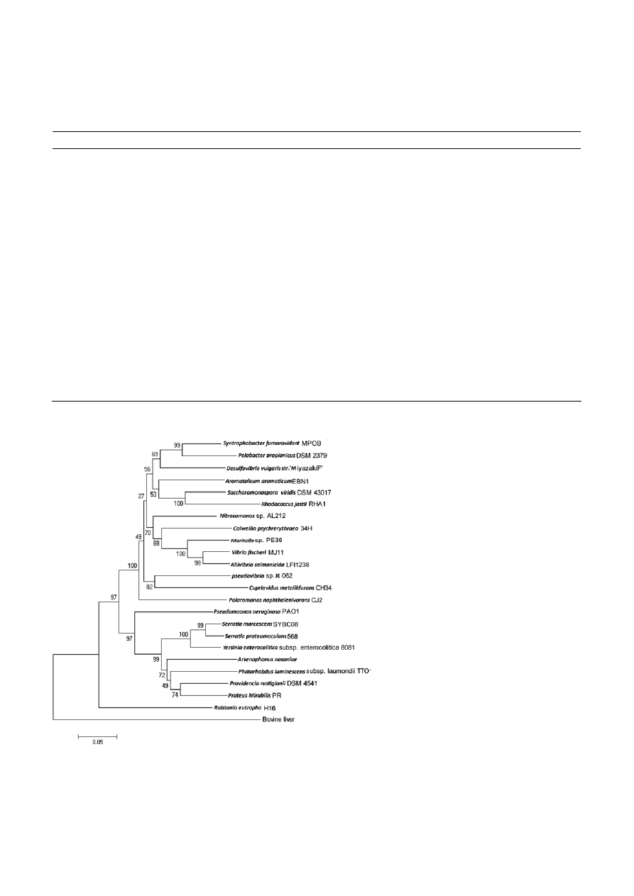
Figure 6. The phylogenetic relationship of the catalase from Serratia marcescens SYBC08 with other 23 related catalase sequences. The
dendrogram was constructed from a matrix of pairwise genetic distances by the neighbor-joining method using the MEGA 4.1 software.
Numbers above branches indicate a bootstrap values (1000 replicates). The scale bar represents five per substitutions 1000 amino acid
positions.

212 H.-W.
Zeng
et al.
Journal of Basic Microbiology 2011, 51, 205 – 214
© 2011 WILEY-VCH Verlag GmbH & Co. KGaA, Weinheim
www.jbm-journal.com
rather conserved in catalases [22, 23]. According to the
amino acid residues of bovine liver catalase site and
alignment analysis of amino acid sequence, the active
sites of the catalase from Serratia marcescens SYBC08
were consisted of H54, N127, and S93. Its sites of
proximal side of heme were composed of P301, R333,
Y337, M329, and H341, and sites of the distal side of the
heme contained M53, N127, F132, and F140. The Func-
tion of catalase-bound NADPH in bovine and human
catalase was that both prevents and reverses the accu-
mulation of compound II, an inactive form of catalase
that is generated slowly when catalaseis exposed to hy-
drogen peroxide [24]. Amino acid residues involved in
NADPH binding sites of the catalase were H173, S180,
R182, and H284. In the bovine catalase, a water mole-
cule has been considered as possibly involved in a redox
mechanism of NADPH [25]. The amino acid residues in-
volved in such procedure were K216, Y194, and H214. A
phylogenetic relation of 24 catalases was presented in
Fig 6. From this figure, it was found that the catalase of
Serratia marcescens SYBC08 had closest relationship with
Serratia proteamaculans 568 and yersinia enterocolitica
subsp. enterocolitica 8081, and it was distincted from
the bovine liver catalases. Those catalases from arseno-
phonus nasoniae, photorhabdus luminescens subsp. laumon-
dii tto1, proteus mirabilis pr and providencia rustigianii
dsm 4541 belonging to the family Enterobacteriaceae
were located on a small branch.
Discussion
The highest catalase yield (20,289 U ⋅ ml
–1
) was achieved
in 7 l fermenter after incubation of 40 h. Many literatu-
res showed catalase productions by microorganisms did
not exceed 5,000 Um ⋅ ml
–1
[6, 26–29]. Nakayama et al.
[11] reported that Micrococcus luteus strain showed rather
high catalase production of 34,601 U ⋅ ml
–1
. Although,
in our study, the yield of catalase from Serratia marces-
cens SYBC08 was slightly lower compared to the report
of Nakayama et al. [11], it had a great rising space by
adding some suitable inducers such as H
2
O
2
. There-
fore, the enzyme yield had a good attraction in applica-
tion.
The 13.8-fold purification achieved in this study
was lower than most of reports. Literature survey re-
vealed that main ranges of purification fold was
54.1-fold for a catalase from Vibrio rumoiensis S-1
T
[30]
to 1,538-fold for a catalase from Methanosarcina barkeri
[5]. Since one of the goals of our study was to evaluate
its industrial applications, low fold purification ob-
tained from the above procedure meant high ratio of
catalase production and helped to reduce its purifica-
tion cost.
The protein had a molecular mass of 230 kDa and a
subunit size of approximately 58 kDa. Thus, we pro-
posed that the purified monofunctional catalase was
tetramer consisted of 4 homosubunits. The subunit
number and native enzyme sizes for this monofunc-
tional enzyme were similar to those of bacteria (i.e., Vi-
brio rumoiensis S-1
T
with 57.3 kDa and 230 kDa [30],
Halophilic bacterium Halobacterium halobium with 68
and 240 kDa [31], Deinococcus radiodurans with 65 kDa
and 240 kDa subunit and native molecular mass [32],
Vibrio salmonicida with 57 kDa and 235 kDa [33], respec-
tively).
The Rz value of the purified catalase from Serratia
marcescens SYBC08 (0.042 ± 0.041) was lower than that
of monofunctional catalase which usually exhibit
ratios of approximately 1. Thus, the observed spectrum
would be considered as atypical electronic spectrum of
monofunctional catalase. Similar atypical spectra
were found in catalases from other bacteria such as
Methanosarcina barkeri (0.48) [5] and Rhodobacter sphaer-
oides ATH 2.4.1(0.513) [34]. Shima et al. and Terzenbach
et al. explained the phenomenon, which is caused by
partially loss of the heme in this purified procedure
[5, 33].
The purified enzyme of Serratia marcescens SYBC08
showed maximum catalase activity in the pH range
from 7.0 to 9.0. The broad pH optimum range is a
common feature of monofunctional catalases, but cata-
lase-peroxidases have narrow pH optimum range [12,
30, and 31]. Our catalase from Serratia marcescens
SYBC08 was found to be stable in the broad pH range
from pH 5.0 to 11.0. This result was similar to the re-
port of the monofunctional catalase of Yumoto et al.
[30]. It displayed high relative activity at wide tempera-
ture range from 0 to 70 °C. The temperature depen-
dence of catalase activity was poor. The phenomenon
was also observed in other monofunctional catalases
[30, 34]. The heat stability of the catalase from Serratia
marcescens SYBC08 was higher than that of commercial
bovine liver catalase. From an industrial application of
view, those property of pH and temperature could meet
the demands of waste water treatment under wide pH
or temperature conditions.
Generally, monofunctional catalses have high K
m
val-
ues of about over 50 mM, while catalase-peroxidases
have low K
m
values of 1–20 mM [5, 34, 35]. Catalase
from Serratia marcescens SYBC08 exhibited a K
m
of
78 mM which is very similar to other monofunctional
enzyme, but its K
m
values was much lower than that of
other Serratia marcescens catalase (228) [36]. This indi-

Journal of Basic Microbiology 2011, 51, 205 – 214
Sequence analysis of a monofunctional catalase from Serratia marcescens 213
© 2011 WILEY-VCH Verlag GmbH & Co. KGaA, Weinheim
www.jbm-journal.com
cated that the catalase is more efficient with regard to
the scavenging of hydrogen peroxide than the Serratia
marcescen catalase in literature [36].
Rapid advances in protein analytical technologies,
fuelled by the addition of MS and sequence data-
bases, have made it possible for protein chemists to
identify new proteins and designed primers for gene
coining [37, 38]. Two peptide sequences did not com-
pletely matched catalase sequence of Yersinia enterocoli-
tica subsp. enterocolitica 8081 (gi|123441711) or Ser-
ratia proteamaculans 568 (gi|157371515 ). Therefore, the
catalase was regarded as a new protein. 479 amino
acid residues encoded by the open reading frame
matched the LC-MS/MS sequences. This confirmed that
the enzyme gene was obtained. The above study re-
presents a excellent application of LC–MS/MS technol-
ogy.
Catalase primarily responsible for the metabolism of
hydrogen peroxide, is an essential antioxidant enzyme
that is present throughout phylogeny, from bacteria to
animal [39]. Amino acid residues of Serratia marcescens
SYBC08 catalase in the active sites (H54 and N127),
NADPH binding sites (H173, S180, and R182), proximal
sites of heme (P301, R333, Y337, and M329), distal sites
of the heme (N127, F132, and F140) and sites inter-
acting with a water molecule (K216 and Y194) were
well conserved in 23 catalases. Met changed to Val in
bovine liver and ralstonia eutropha h16 (Table 3). The
Met in Proteus mirabilis PR could produce some steric
hindrance impairing the accessibility of large substra-
tes or inhibitors to the iron of the active site, this
result of Met replacing with Val caused significantly
greater sensitivity to aminotriazole of a specific in-
hibitor of catalases than P. mirabilis PR [40]. Some other
replacements of the residues from Serratia marcescens
SYBC08 catalase (Table 3) might further supported
some degree of specificity in their catalysis behaviors.
The catalase of high specific activity was closely related
with the enzyme from pathogenic bacterium in the
family Enterobacteriaceae which developed the ability
to survive in host against the presence of H
2
O
2
. This sup-
ported the growth environment of Serratia marcescens
SYBC08.
As a summary, in the study, a high catalase produc-
tion was obtained by Serratia marcescens SYBC08. The
purified catalase was characterized as a monofunc-
tional enzyme. LC–MS/MS technology confirmed that
the cloning gene was the encoding gene of the mono-
functional enzyme. Amino acid sequences analyses
suggest the enzyme from Serratia marcescens SYBC08 has
highly conserved catalysis behaviors in the microorgan-
isms from the family Enterobacteriaceae.
Acknowledgements
This work was financially supported by the National
High Technology and Development Program of China
(863 Program; grant humber 2010AA101501).
References
[1] Cabiscol, E., Tamarit, J., Ros, J., 2000. Oxidative stress in
bacteria and protein damage by reactive oxygen species.
Int. Microbiol., 3, 3–8.
[2] Shin, D.H., Choi, Y.S., Cho, Y.H., 2008. Unusual properties
of catalase A (KatA) of Pseudomonas aeruginosa PA14 are
associated with its biofilm peroxide resistance. J. Bacte-
riol., 190, 2663–2670.
[3] Chagas, R.F., Bailao, A.M., Fernandes, K.F., Winters, M.S.,
Pereira, M., Soares, C.M., 2009. Purification of Paracocci-
dioides brasiliensis catalase P; subsequent kinetic and
stability studies. J. Biochem., 147, 345–351.
[4] Rio, R.V., Anderegg, M., Graf, J., 2007. Characterization of
a catalase gene from Aeromonas veronii, the digestive-tract
symbiont of the medicinal leech. Microbiology., 153,
1897–1906.
[5] Shima, S., Netrusov, A., Sordel, M., Wicke, M., Hartmann,
G.C., Thauer, R.K., 1999. Purification, characterization,
and primary structure of a monofunctional catalase from
Methanosarcina barkeri. Arch. Microbiol., 171, 317–323.
[6] Shi, X.L., Feng, M.Q., Zhao, Y.J., Guo, X., Zhou, P., 2008.
Overexpression, purification and characterization of a
recombinant secretary catalase from Bacillus subtilis. Bio-
technol. Lett., 30, 181–186.
[7] Jones, P., Wilson, I., 1978. Catalases and iron complexes
with catalase-like properties. 7, Marcel Dekker., New York.
[8] Chelikani, P., Fita, I., Loewen, P.C., 2004. Diversity of
structures and properties among catalases. Cell. Mol. Life.
Sci., 61, 192–208.
[9] Buzy, A., Bracchi, V., Sterjiades, R., Chroboczek, J., Thi-
bault, P., Gagnon, J., Jouve H.M., Hudry-Clergeon, G.,
1995. Complete amino acid sequence of Proteus mirabilis
PR catalase. occurrence of a methionine sulfone in the
close proximity of the active site. J. Protein. Chem., 14,
59–72.
[10] Beers, R., Jr., F., Sizer, I.W., 1952. A spectrophotometric
method for measuring the breakdown of hydrogen per-
oxide by catalase. J. Biol. Chem., 195, 133–140.
[11] Nakayama, M., Nakajima-Kambe, T., Katayama, H.,
Higuchi, K., Kawasaki, Y., Fuji, R., 2008. High catalase
production by Rhizobium radiobacter strain 2-1. J. Biosci.
Bioeng., 106, 554–558.
[12] Brown-Peterson, N.J., Salin, M.L., 1993. Purification of a
catalase-peroxidase from Halobacterium halobium: characte-
rization of some unique properties of the halophilic en-
zyme. J. Bacteriol., 175, 4197–4202.
[13] Bradford, M.M., 1976. A rapid and sensitive method for
the quantitation of microgram quantities of protein
utilizing the principle of protein-dye binding. Anal. Bio-
chem., 72, 248–254.

214 H.-W.
Zeng
et al.
Journal of Basic Microbiology 2011, 51, 205 – 214
© 2011 WILEY-VCH Verlag GmbH & Co. KGaA, Weinheim
www.jbm-journal.com
[14] Cen, H.Y., He, Y., Mang, H., Feng, F.Q., 2007. Rapid
measurement of citric acids in orange juice using visible
and near infrared reflectance spectroscopy. Spectrosc.
Spect. Anal., 27, 1747–1750 (in chinese).
[15] Laemmli, U.K., 1970. Cleavage of structural proteins
during the assembly of the head of bacteriophage T4.
Nature, 227, 680–685.
[16] Merril, C.R., 1990. Silver staining of proteins and DNA.
Nature, 343, 779–780.
[17] Lineaweaver, H., Burk, D., 1934. The determination of
enzyme dissociation constants. J. Am. Chem. Soc., 56,
658–666.
[18] Tao, T.S., Yang, R.Y., Zhu, D.X., 2007. Procaryote biosyste-
matics, CN: Chemical Industry pressm, pp. 1–586.
[19] Saitou, N., Nei, M., 1987. The neighbor-joining method: a
new method for reconstructing phylogenetic trees. Mol.
Biol. Evol., 4, 406–425.
[20] Takahashi, K., Nei, M., 2000. Efficiencies of fast algo-
rithms of phylogenetic inference under the criteria of
maximum parsimony, minimum evolution, and maxi-
mum likelihood when a large number of sequences are
used. Mol. Biol. Evol., 17, 1251–1258.
[21] Phucharoen, K., Hoshino, K., Takenaka, Y., Shinozawa, T.,
2002. Purification, characterization, and gene sequencing
of a catalase from an alkali- and halo-tolerant bacterium,
Halomonas sp. Sk1. Biosci. Biotechnol. Biochem., 66, 955–
962.
[22] Lee, D.H., Oh, D.C., Oh, Y.S., Malinverni, J.C., Kukor, J.J.,
Kahng, H.Y., 2007. Cloning and characterization of mono-
functional catalase from photosynthetic bacterium Rho-
dospirillum rubrum S1. J. Microbiol .Biotechnol., 17, 1460–
1468.
[23] Barriere, C., Bruckner, R., Centeno, D., Talon, R., 2002.
Characterization of the katA gene encoding a catalase and
evidence for at least a second catalase activity in Staphylo-
coccus xylosus, bacteria used in food fermentation. FEMS
Microbiol. Lett., 216, 277–283.
[24] Kirkman, H.N., Galiano, S., Gaetani, G.F., 1987. The func-
tion of catalase-bound NADPH. J. Biol. Chem., 262, 660–
666.
[25] Fita, I., Rossmann, M.G., 1985. The NADPH binding site
on beef liver catalase. Proc. Natl. Acad. Sci., 82, 1604–
1608.
[26] Petruccioli, M., Fenice, M., Piccioni, P., Federici, F., 1995.
Effect of stirrer speed and buffering agents on the
production of glucose oxidase and catalase by Penicillium
variable (P16) in benchtop bioreactor. Enzyme, Microb.
Technol., 17, 336–339.
[27] Venkateshwaran, G., Somashekar, D., Prakash, M.H., Ba-
sappa, S.C., Richard, J., 1999. Production and utilisation of
catalase using Saccharomyces cerevisiae. Process. Biochem.,
34, 187–191.
[28] Gromada, A., Fiedurek, J., 1997. Optimization of catalase
biosynthesis in submerged cultures of Aspergillus niger
mutant. J. Basic. Microbiol., 37, 85–91.
[29] Caridis, K.-A., Christakopoulos, P., Macris, B.J., 1991. Si-
multaneous production of glucose oxidase and catalase by
Alternaria alternata. Appl. Micobiol. Biotechnol., 34, 794–
797.
[30] Yumoto, I., Ichihashi, D., Iwata, H., Istokovics, A., Ichise,
N., Matsuyama, H., Okuyama, H., Kawasaki, K., 2000.
Purification and characterization of a catalase from the
facultatively psychrophilic bacterium Vibrio rumoiensis
S-1(T) exhibiting high catalase activity. J. Bacteriol., 182,
1903–1909.
[31] Brown-Peterson, N.J., Salin, M.L., 1995. Purification and
characterization of a mesohalic catalase from the halo-
philic bacterium Halobacterium halobium. J .Bacteriol., 177,
378–384.
[32] Kobayashi, I., Tamura, T., Sghaier, H., Narumi, I., Yama-
guchi, S., Umeda, K., Inagaki, K., 2006. Characterization
of monofunctional catalase KatA from radioresistant bac-
terium Deinococcus radiodurans. J. Biosc. Bioengin., 101,
315–321.
[33] Lorentzen, M.S., Moe, E.H., Jouve, M., Willassen, N.P.,
2006. Cold adapted features of Vibrio Salmonicida catalase:
characterisation and comparison to the mesophilic coun-
terpart from Proteus mirabilis. Extremophiles, 10, 427–
440.
[34] Terzenbach, D.P., Blaut, M., 1998. Purification and charac-
terization of a catalase from the nonsulfur phototrophic
bacterium Rhodobacter sphaeroides ATH 2.4.1 and its role in
the oxidative stress response. Arch. Microbiol., 169, 503–
508.
[35] Singh, R., Wiseman, B., Deemagarn, T., Jha, V., Switala, J.,
Loewen, P.C., 2008. Comparative study of catalase-per-
oxidases (KatGs). Arch. Biochem. Biophys., 471, 207–214.
[36] Switala, J., Loewen, P.C., 2002. Diversity of properties
among catalases. Arch. Biochem.Biophys., 401, 145–154.
[37] Sun, M.Z.,Liu, S,Q., Yang, F., Greenaway, F.T., Xu, Y.F.,
2009. A novel phospholipase A2 from Agkistrodon blom-
hoffii ussurensis venom: purification, proteomic, functional
and structural characterizations. Biochimie., 91, 558–
567.
[38] Vafiadi, C., Topakas, E., Biely, P., Christakopoulos, P.,
2009. Purification, characterization and mass spectro-
metric sequencing of a thermophilic glucuronoyl esterase
from Sporotrichum thermophile. FEMS. Microbiol. Lett., 296,
178–184.
[39] Gerhard, G.S., Kauffman, E.J., Grundy, M.A., 2000. Mole-
cular cloning and sequence analysis of the Danio rerio
catalase gene. Comp. Biochem. Physiol. B Biochem. Mol.
Biol., 127, 447–457.
[40] Jouve, H.M., Lasauniere, C., Pelmont, J., 1983. Properties
of a catalase from a peroxide-resistant mutant of Proteus
mirabilis. Can. J. Biochem. Cell. Biol., 61, 1219–1227.
((Funded by
• Program for Changjiang Scholars and Innovative Re-
search Team in University, Ministry of Science and
Technology, P. R. China; grant number: IRT0532))
Wyszukiwarka
Podobne podstrony:
jobm 201000013
jobm 201000298
jobm 201000191
jobm 201000321
jobm 201000018
jobm 201000214
jobm 201000067
jobm 201000037
jobm 201000074
jobm 201000280
jobm 201000385
jobm 201000198
jobm 201000458
jobm 201000520
jobm 201000327
jobm 201000342
jobm 201000420
jobm 201000364
jobm 201000317
więcej podobnych podstron